
Abstract
Introduction
PDT is based on the interaction between visible light and a photosensitizing agent that, under photoactivation, generates cytotoxic species “in situ.” When stimulated, the photosensitizing agent is promoted to the singlet and triplet forms, leading to species that could react with molecular oxygen or other biologic macromolecules present in the medium. This process results in the production of radical species and hydrogen peroxide, which react with the biologic system by destroying cellular constituents, such as organelles, proteins, and nucleic acids, thereby leading to cell death. 9
Aggregatibacter actinomycetemcomitans is the main pathogen related to aggressive periodontitis, characterized by rapid destruction of the tooth supportive tissues, leading to tooth loss. The use of antibiotics as an adjuvant factor in the mechanical treatment of periodontitis may lead to bacterial drug resistance. Therefore, the use of PDT to inactivate microorganisms responsible for periodontal disease seems to be an easy and low-cost alternative protocol for the treatment of this disease. 11
Many dyes have been used for PDT, including methylene blue (MB) and erythrosine (ERY). For more than a century, MB has been used in histology for surgical identification, at fairly high concentrations, normally 1% wt/vol (26.7 mmol/L), without causing human toxicity. 3,12 Many PDT studies focusing on the treatment of bacterial diseases have used MB as a photosensitizing agent because it is very effective against pathogenic organisms, including viruses, bacteria and yeasts. 13
Among the many classes of photosensitizing agents, those derived from xanthenes, merocyanines, phthalocyanines, and hematoporphyrins have been the most frequently used, and some have already been approved by the FDA for cancer treatment. 14 ERY belongs to a class of cyclic compounds called xanthenes (subgroup phlorone), together with eosin and rose bengal dyes. 15,16 These were the first photosensitizers to be used in the treatment of diseases such as versicolor pytyriasis, contagious molluscum, syphilis, lupus vulgaris, and skin cancer. 17 –19 Now ERY has been used to inactivate many gram-positive and gram-negative bacteria. 7
The possibility of different responses to dye photosensitivity when one varies the wavelength, light intensity, the presence or absence of oxygen in the medium, and in the chemical structure of the compounds used as photosensitizing agents has certainly broadened the versatility of their use for both biologic and medical applications. 20
The present study aimed to compare the effect of PDT application in dentistry by combining ERY or MB (as a dye for ROS generation) and light emitted by an odontologic resin photopolymerizer against A. actinomycetemcomitans grown as planktonic or biofilm cultures.
Materials and Methods
Photosensitizer and light source
Stock solutions of methylene blue and erythrosinee (2 mmol/L) were prepared in 2 mmol/L saline phosphate buffer (PBS), pH 7.4 (Na2HPO4 8.1 mmol/L, KH2PO4 1.47 mmol/L, NaCl 0.13 mol/L, and KCl 2.7 mmol/L). This solution was filter-sterilized (0.22 μm) and stored at −20°C.
A hand-held photopolymerizer or dental photopolymerizer (HHP) was purchased from Dabi Atlante SA (Ribeirão Preto SP, Brazil) and used as light source in all the experiments. The HHP had the following characteristics: continuous output of 350–500 mW/cm2 of potency from a halogen light isolated from an inner filter that selected the wavelength range of 400–500 nm. The energy output was measured by the use of a radiometer from Continuns company, model Field Master A, with a CW L-3M head. HHP was used as light source, which allowed photosensitivity of the dyes, as previously described. 16
Bacterial culture
A. actinomycetemcomitans JP2 was cultivated in Tryptic Soy Broth (TSB- soybean-casein digest medium) (Acumedia, Lansing, MI) or Tryptic Soy Agar (TSA-soybean-casein digest agar) from Difco (Sparks, MD), by using the candle jar technique, as described by Goulart et al. 16
Cytotoxic effects of MB and ERY on planktonic growing A. actinomycetemcomitans in the presence and absence of light
Before incubation with different concentrations of dyes (MB or ERY), the cells (A600nm = 0.5) were serially diluted in TSB medium, to obtain a 103 CFU/mL count. The cells were distributed (1 mL) into assay tubes (125 × 15 mm). One set of tubes was submitted to a control-cell experiment, to evaluate the dye toxicity per se in the dark and the light toxicity per se without any dye (in the concentration range of 0.1–10.0 μmol/L).
Light exposure was performed by using an HHP tip at an 11-cm distance from the cell suspension. First, the light toxicity per se was evaluated by irradiating a cell suspension (1 mL), not incubated with the dye, with a light dose of 0.65 J/cm2 (1 min of continuous HHP light). After that, dye-treated cells were submitted to different times of light exposure ranging from 1 to 3 min (∼2 J/cm2) under a stable irradiation power, to promote bacterial inactivation. The contact time with the dyes before irradiation, which varied from 10 to 30 min, was also studied. Assays to evaluate dye toxicity per se (in the dark) were performed in the same way, without light exposure. The whole process was performed under gentle magnetic stirring.
Cells were grown by dropping and spreading 50 μL of previously diluted cell suspension directly onto TSA and incubating it (protected from light) in a candle jar for 72 h at 37°C. After this period, the CFU/milliliter was calculated. 16
Cytotoxic effects of MB and ERY on biofilm growing A. actinomycetemcomitans in the presence and absence of light
The biofilm (A. actinomycetemcomitans cellular aggregates) was grown after inoculating a 24-well cell-culture cluster (Corning Costar 3524, flat bottom) (1 mL/well) with 15 μL of a 109 CFU/mL cell suspension, followed by 24-h incubation in a candle jar. After this period, the medium was removed, and the wells were washed 3 times with PBS, pH 7.4. Each well containing biofilm was then incubated with 1 mL MB or ERY solution, 0.5 and 1.0 μmol/L, respectively, for 30 min. Next, each well was irradiated (1 or 3 min, 0.65 to ∼2 J/cm2) with an HHP; the dye solution was replaced with 1 mL tryptic soy broth (TSB) medium, and the cell-culture cluster was incubated for 24 h in a candle jar. Then each well was washed with PBS, dried at room temperature, and photographed with a Leica DC 300 F digital camera associated with a Leica DMLB light microscope (Leica, Bensheim, Germany; 50 × magnification).
The remaining cells present in the biofilm after the PDT process (or without PDT, as control) were determined by using crystal violet, as described by Kaplan et al. 21 , by measuring the optical density at 550 nm of the ethanol-dye solution in each well.
Statistical analysis
Data are reported as the mean of triplicate measurement of three different preparations; statistical significance was set at p ≤ 0.05.
Results
Figure 1 shows that both MB and ERY may be photoactivated by HHP, because its light emission ranges from 300 to 800 nm, and the absorption spectrum of ERY and MB are 536 nm and 665 nm, respectively. Although we must explain that using a wide bandpass filter, in the range of 400–570 nm, the light emission presents a 100% transmission, which is appropriate for dyes having an absorption center in this range, such as ERY. In the red region ≥600, a reduction of light transmission (∼70% at 665 nm) must be considered in the choice of the dye to be activated.
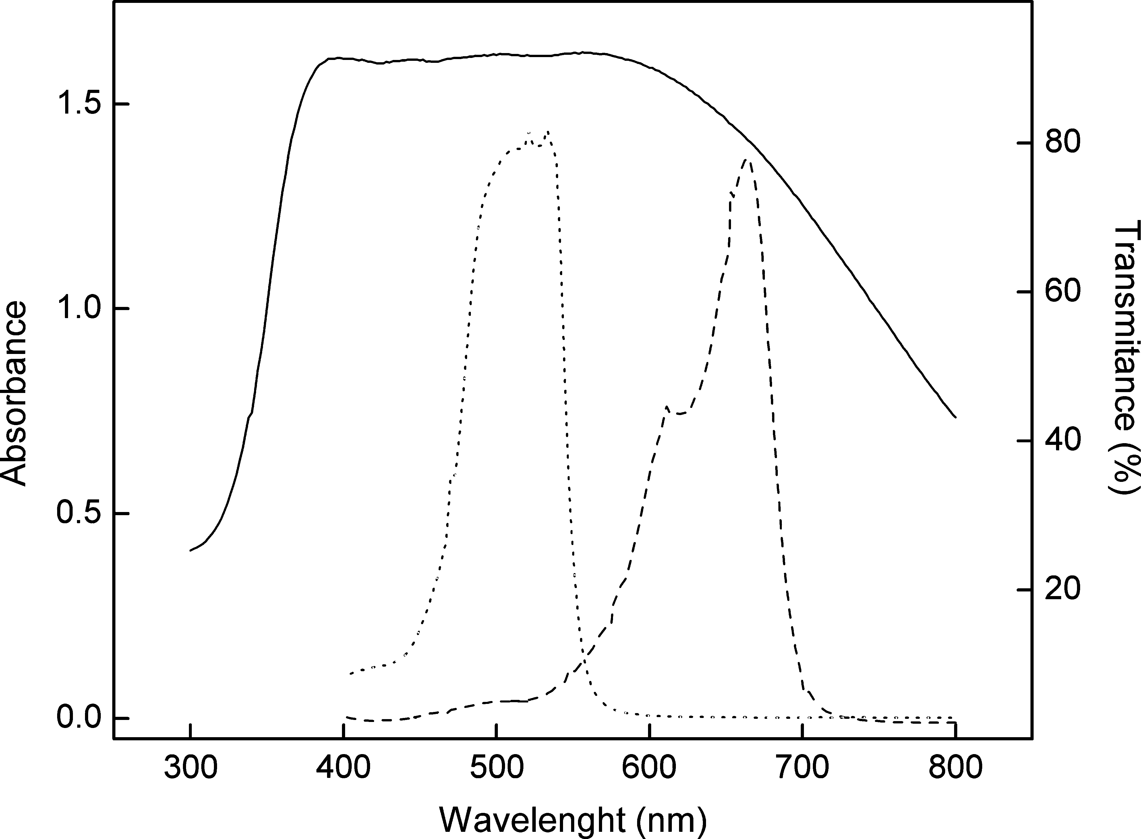
Absorption spectra of 26 μmol/L of methylene blue (dashed line) and 50 μmol/L of erythosin (dotted line) in water and transmittance spectra of the HHP filter (solid line).
Figure 2 depicts the dark toxicity of MB and ERY. As can be observed in Fig. 2A, MB does not show toxicity in concentrations ≤0.1 μmol/L, either with 10 or 30 min of previous incubation with a suspension of A. actinomycetemcomitans cells. At greater than this concentration, MB has increasing toxicity, reaching ∼50% cell death at a dye concentration of 20 μmol/L. As for ERY, a higher linearity of the dose–effect curve is observed for both 10- and 30-min preincubation, with a progressive decrease of bacterial viability ranging from 0.1 to 10 μmol/L, with values of 40–50% cell death being reached (Fig. 2B).

The effect of methylene blue (
When MB incubation and light exposure were associated, a dose-dependent effect of dye concentration on A. actinomycetemcomitans death was observed for planktonic cultures. Preincubations for 30 min in concentrations of ∼0.5 μmol/L were enough to cause 15% cell death, whereas 1 μmol/L (which gives low dark toxicity) caused 25% cell death after PDT (Fig. 3A).
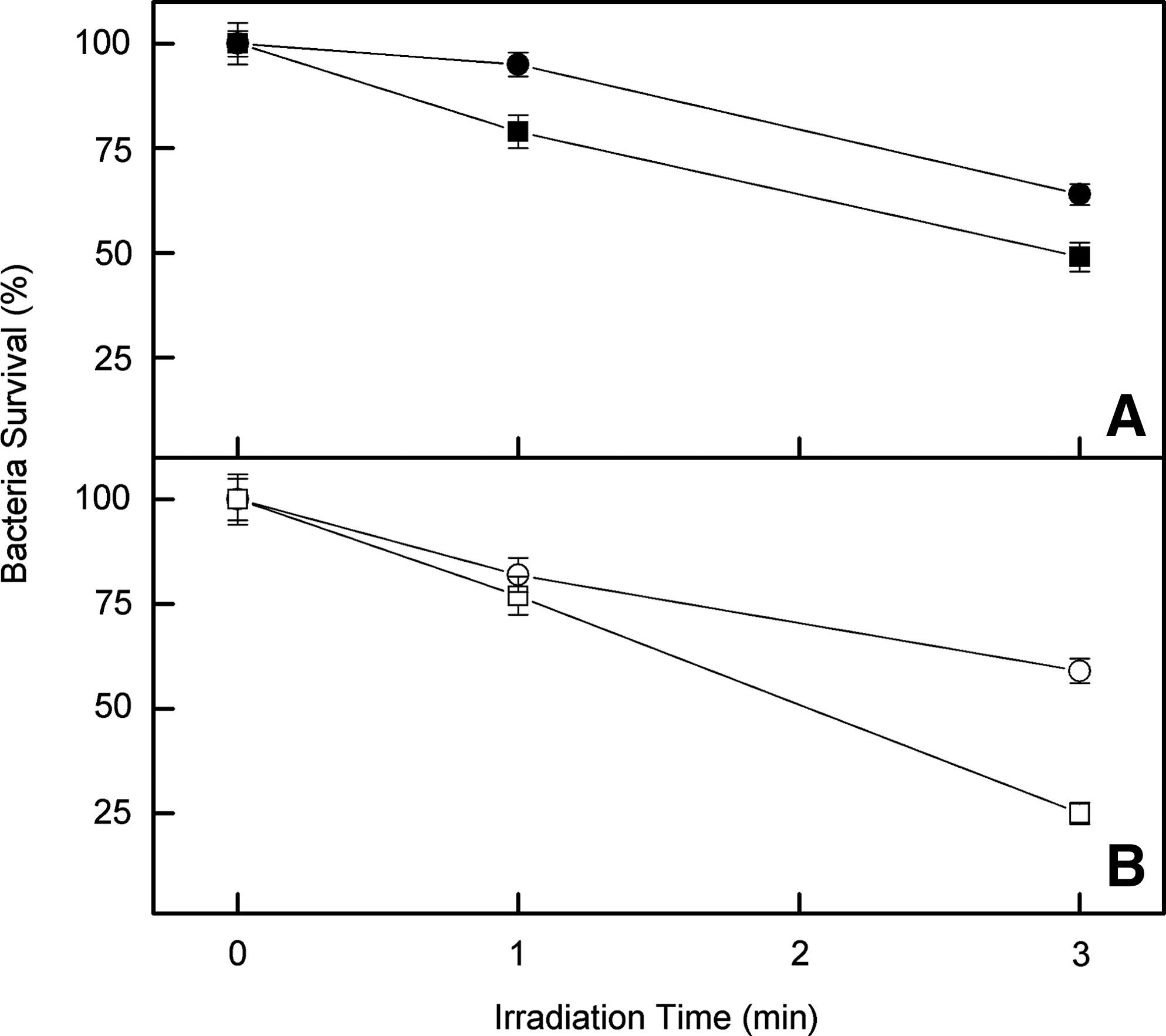
The effect of irradiation time of planktonic cultures of Aggregatibacter actinomycetemcomitans, treated with methylene blue (
The association between ERY and PDT showed behavior similar to that observed for MB. Nevertheless, it was more efficient at killing bacteria. Preincubation for 30 min of A. actinomycetemcomitans with ERY (0.5 μmol/L) and followed by 1-min irradiation, led to 20% cell deaths, whereas irradiation for 3 min resulted in 41% bacterial death. With 1.0 μmol/L ERY, bacterial deaths of ∼25% and 75% with 1- and 3-min irradiation were achieved, respectively (Fig. 3B). It was also observed that higher cell death occurred after 30 min of preincubation with the dye before light exposure in the case of plankton cultures, by using either MB or ERY. With prolonged times (60 min), little additional cell death was found for all the tested dye concentrations (data not shown).
PDT effects on the cellular aggregate (biofilm) formed by A. actinomycetemcomitans only were verified. The bacteria were grown in cell culture, to evaluate whether growth in the compact form could reduce the efficiency of dye action compared with bacteria in solution (planktonic). It is noteworthy that the effects of toxicity per se and PDT were not reduced by using the same experimental conditions. Quantification of the bacteria in the biofilm after treatments with dyes by using the crystal violet method (concentration range from 0.5 to 1.0 μmol/L) led to ∼54% and 77% maximum bacterial death by using MB and ERY, respectively (Figs. 4 and 5).
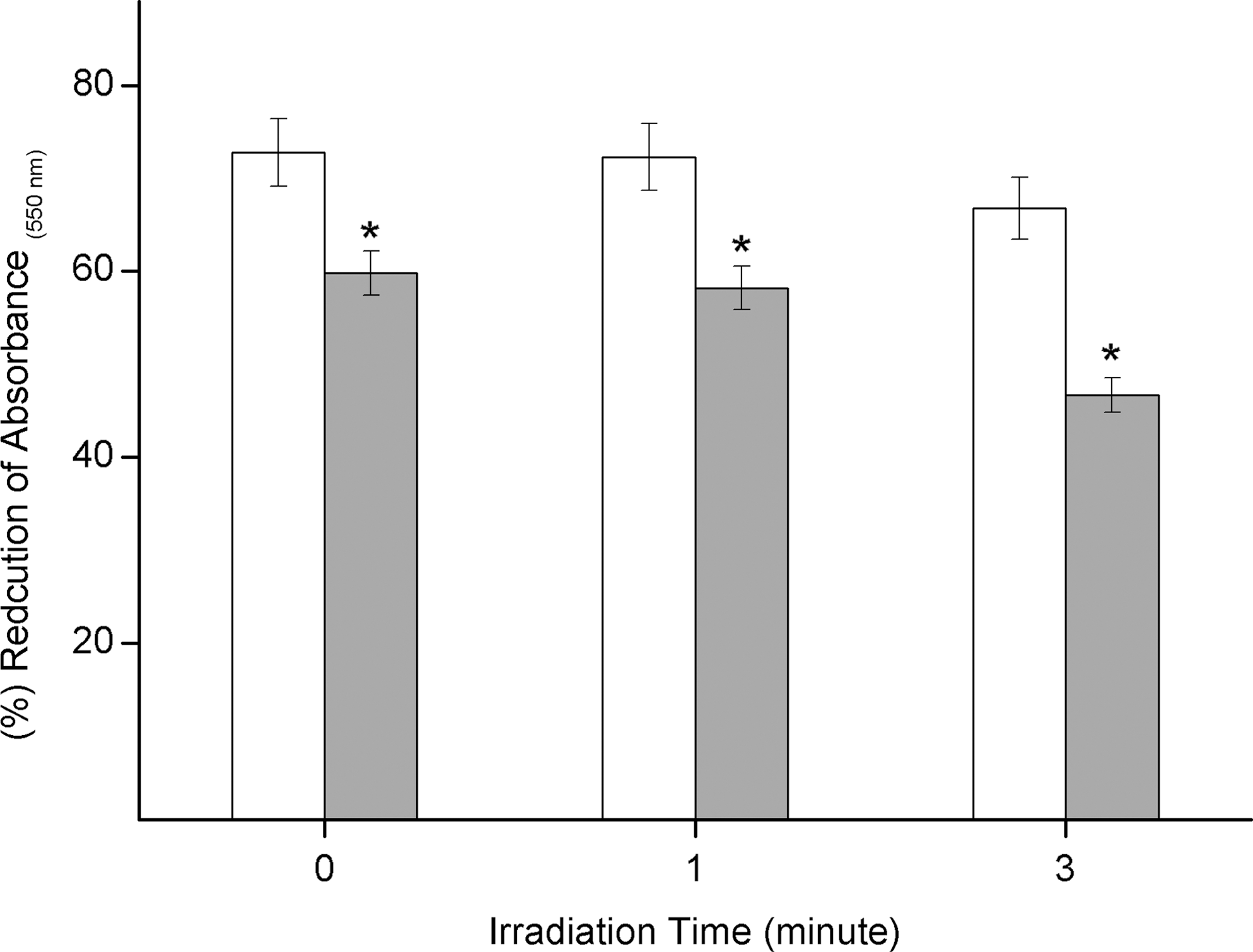
The effect of PDT on A. actinomycetemcomitans biofilms treated with methylene blue. The previously formed biofilm (24 h) was incubated with 0.5 μmol/L (open bars) or 1.0 μmol/L (solid bars) methylene blue for 30 min, and light was irradiated for 1 or 3 min. The plates were incubated at 37°C in a candle jar for 24 h, to quantify the remaining cells in the biofilm by the crystal violet method, as described in Materials and Methods. *p < 0.05.

The effect of PDT on A. actinomycetemcomitans biofilms treated with erythrosine. The previously formed biofilm (24 h) was incubated with 0.5 μmol/L (open bars) or 1.0 μmol/L (solid bars) erythrosine for 30 min, and light was irradiated for 1 or 3 min. The plates were incubated at 37°C in a candle jar for 24 h, to quantify the remaining cells in the biofilm by the crystal violet method, as described in Materials and Methods. *p < 0.05.
After PDT treatments (Fig. 6), no alteration was noted in the A. actinomycetemcomitans biofilm when 0.5 μmol/L MB was used with 1-min irradiation (0.65 J/cm2). Nonetheless, at this same concentration, but with 3 min of irradiation (2 J/cm2), some areas had no biofilm attachment, indicating that PDT caused detachment of cells previously adhered to the bottom of the well (Fig. 6). When 1.0 μmol/L MB with 1- and 3-min irradiation was used, proportionately larger areas with no biofilm attachment were detected (Fig. 6). ERY, 0.5 μmol/L, with 1- and 3-min irradiation, promoted similar reduction in cellular aggregates, but when the ERY concentration was increased to 1.0 μmol/L, larger reduction was observed, for the same irradiation times previously used. The quantification of bacteria survival in biofilm (Figs. 4 and 5) and the visualization of the micrography (Fig. 6) show that ERY is more efficient than MB in the PDT procedure.

Micrograph showing the architecture of the biofilm formed by A. actinomycetemcomitans (100 ×) after growth for 24 h in a candle jar, as described in Materials and Methods, under different treatment conditions.
Discussion
The absorption spectra of the MB and ERY match the light-emission range of the odontologic resin photopolymerizer, which varies from 300 to 800 nm. This photopolymerizer was efficiently used in PDT when rose bengal (maximum absorption peak at 560 nm) was used as photosensitizer by Paulino et al. 4,5 against S. mutans, and by Goulart et al. 16 against A. actinomycetemcomitans.
It should be pointed out that, despite the per se toxicity of MB and ERY, these dyes are routinely used in medicine and dentistry procedures without toxicity for human cells. For example, Soukos et al. 22 and Wainwright 3 showed that MB can be used in surgical procedures at considerably higher concentrations [1% (wt/vol), which corresponds to ∼26.7 mmol/L], without being toxic to humans. MB is also used clinically to treat ifosfamide encephalopathy, methemoglobinemia, urolithiasis, and cyanide poisoning. A dosage of about 13.4 mmol/L is commonly used to dye the esophagus of patients with Barrett esophagus history and bronchial lesions. 23 Furthermore, ERY in concentrations ranging from 9 to 25 μmol/L is used in dentistry procedures to visualize dental plaque. 7
As observed in Fig. 2, MB and ERY do not display toxicity at concentrations ≤0.1 μmol/L, either with 10 or 30 min of previous incubation with a suspension of A. actinomycetemcomitans cells. It should be noted that the toxic effect in the dark is not significantly affected by the preincubation time before bacterial growth in the studied range of concentrations (Fig. 2).
The MB concentration of 15 μmol/L presented toxicity per se for A. actinomycetemcomitans in the range of 50% (Fig. 2), which is not toxic to humans. Through dye's photoactivation and formation of ROS, its efficiency for killing bacteria can be increased.
Many studies involving the use of PDT have used MB as a photosensitizer. Examples of such works are growth inhibition of prokaryote and eukaryote organisms, 9 as well as of gram-positive and gram-negative microorganisms. 13 Also, Chan and Lai 24 verified that 0.001% (∼0.27 μmol/L) of MB, associated with a light source of 665 diode laser at 100 mW (21.2 J/cm2), promotes the death of 40% of many species of the oral microbiota, including A. actinomycetemcomitans bacteria, which, with a combination of light and dye, had 95% of the cells eliminated. In this work, the most efficient MB concentration and light dose (much smaller than the ones used by other authors) reduced A. actinomycetemcomitans viability by 50%. This is probably due to the readiness of the MB dye to cross the bacterial cell wall. The positive charge of MB must allow it to bind easily to the negatively charged lipopolysaccharide present on the cell walls of gram-negative bacteria, thereby facilitating development of its photodynamic activity. 9
Many studies demonstrated that bacteria of the oral cavity, grown in planktonic media, are sensitized by PDT with rose bengal. 16 In this study, a 75% reduction in A. actinomycetemcomitans present in planktonic media was achieved when ERY at 1.0 μmol/L and 2 J/cm2 energy dose was used. ERY was more efficient than MB in all the studied conditions. It must be pointed out that the ERY concentration used here was the same as that used for MB, which is less than the ones used commercially and in clinical treatments, as previously described. This behavior was in agreement with the absorption spectrum of both dyes used, considering the light-source emission. ERY, used in the same range of concentration as MB, will absorb 30% more of the light emitted (considering the filter used), which allowed a better start in the excitation process and, consequently, in the whole photochemistry pathway. The choice of the light-excitation device must consider the emission spectrum of the light source plus the filter to optimize the absorption as close as possible to the appropriate wavelength of dye excitation.
The A. actinomycetemcomitans lineage, the main pathogen responsible for the development of aggressive periodontal disease, grows in a microbial community called biofilm; these highly organized structures are formed by the presence of different microorganisms, of different species, which colonize the oral cavity. 25
PDT effects on the cellular aggregate formed by A. actinomycetemcomitans, were verified only when the bacteria were grown in cell culture, to evaluate whether growth in the compact form could reduce the efficiency of dye action compared with bacteria in solution (planktonic).
PDT efficiently reduced the number of A. actinomycetemcomitans cells in the biofilm culture. By visual comparison, it was possible to verify a reduction of the formed biofilm after PDT in the presence of both dyes. It was also possible to observe that ERY is more efficient than MB. This was confirmed when crystal violet was used to quantify the reduction in the biofilm structure on the plate (shown in Figs. 4 –6). The decrease in the A. Actinomycetemcomitans biofilm in the presence of MB or ERY was similar to the culture reduction obtained in planktonic medium. It is important to remember that the penetration of blue light into the biologic tissue is very low, 26 so, to optimize the PDT conditions, other lights and increased applied energies will be considered in the future.
Goulart et al. 16 verified that rose bengal at 0.1 μmol/L, associated with 0.65 J/cm2 light irradiation, reduced the A. actinomycetemcomitans biofilm by ∼45%. This reduction was significantly dependent on rose bengal concentration and dose irradiation. Metcalf et al. 27 also investigated the PDT effect over a biofilm formed by Streptococcus mutans by using 22 μmol/L ERY as a photosensitizing agent and a light dose of 6.75 J/cm2; a 57% reduction in cells was found.
Other researchers also compared the use of three different photosensitizing agents, ERY, Photofrin, and MB, at a 22 μmol/L concentration, to photosensitize the Streptococcus mutans biofilm. 8 To this end, a 400-W tungsten lamp with a light intensity of 22.7 mW/cm2 for ERY and 22.5 mW/cm2 for Photofrin and MB was used. ERY was found to be more effective than MB and Photofrin; the S. mutans biofilm was reduced 48%, 41%, and just 0.04%, respectively. 8
The results of this study have shown that exposure of the A. actinomycetemcomitans bacterial lineage to the odontologic resin photopolymerizer light source, supplying 0.65 and 2 J/cm2 of energy, in the presence of MB or ERY as photosensitizing agents, led to a decrease in bacterial viability both in planktonic culture and as a cellular aggregate. This energy dose was much smaller than the one used by Chan and Lai, 24 and the ERY dye concentration was also smaller than the one used by Wood et al. 7 when PDT was applied to an S. mutans biofilm. The most effective combination was 1.0 μmol/L ERY and 2 J/cm2 energy, which yielded a viability reduction of 77% and furnished the largest areas without development of cell aggregates.
Footnotes
Acknowledgments
We thank Cynthia M. de Campos Prado Manso and Priscila Cerviglieri for linguistic advice. We also thank FAPESP and CNPq for the continuous support given to our laboratories. RCG is the recipient of a Ph.D. fellowship from CAPES.
Author Disclosure Statement
No competing financial interests exist.