
Abstract
Introduction
Some investigators have shown that functional as well as morphologic changes may occur after irradiating the dental pulp tissue with hard lasers. 12 –14 However, there are few reports that have combined studies on morphologic changes in pulp tissues after pulsed Nd:YAG laser irradiation. Thus, the aim of the present study was to investigate the histopathologic changes that occur following pulsed Nd:YAG laser irradiation of the pulp through normal dental hard tissue in the rabbit.
Material and Methods
Study design
Two healthy adult New Zealand white rabbits were obtained from the Medical Experimental Practice and Research Centre, Atatürk University. The study protocol was reviewed and approved by the Atatürk University Medical Experimental Practice and Research Centre Ethical Committee (No: 70-137). Rabbits were kept in individual metal cages at room temperature with 12 h of light per day and 50% relative humidity and were under veterinary supervision. They were fed a standard laboratory diet and water ad libitum.
Anesthesia and laser procedure
The laser applications were performed using aseptic routines and carried out under general anesthesia by intramuscular injection of xylazine 5 mg/kg and ketamine 35 mg/kg. For the laser technique, an Nd:YAG laser (Smarty A10; DEKA: free-running pulsed wave laser with a wavelength of 1064 nm under air cooling) was used on all incisors and four molars from each rabbit. Laser applications performed on each incisor and molar tooth of rabbits used different irradiation parameters: energy, 100 mJ; frequency, 10, 20, 30, and 40 Hz; power output, 1, 2, 3, and 4 W; emission mode, pulsed; time, 30 sec. The laser handpiece was brought into contact with the enamel surface of the tooth and the probe was held in the same position so that it did not move across the tooth surface. Laser applications were performed by the same pedodontist (N.B.) to prevent inter-operator variation.
Histopathological study
For easy extraction of the incisor teeth, the rabbits were killed by cervical dislocation under inhalation anesthesia 1 wk after the laser irradiations. For histological evaluation, teeth were fixed by immersion in 10% neutral buffered formaldehyde for 72 h. Following fixation, teeth were washed in running tapwater for 4 h, and then treated with 5% aqueous formic acid solution to remove the calcium from the tissue. The teeth were transferred to fresh acid solution every 3 d for 15 d. After 15 d, macerated teeth were placed in 10% aqueous nitric acid solution for 5 d to complete decalcification. Subsequently, decalcified teeth were washed in running tap water for 16 h to remove the decalcifying solutions. After dehydration in a graded ethanol series, the teeth were embedded in paraffin wax and cut into 7-μm serial sections with a Leica RM2125RT microtome. The sections were mounted onto glass slides, stained with hematoxylin and eosin, and examined using an Olympus BH-2 light microscope equipped with a digital color camera attachment (Sanyo VVC-6975P). Histological analysis was performed by a histologist (E.Ö.) who was blinded to the laser techniques.
Outcome assessment
The primary outcomes included the histopathological evaluation by light microscopy of specimens of rabbit dental pulp tissue with regard to the different laser irradiation parameters.
Results
The investigation was carried out using two rabbits. No adverse events were observed, with no functional complications in the rabbits during the post-irradiation periods. Histological investigation revealed that laser irradiation at different power outputs did not produce any damage to the pulp tissue (Figs. 1 and 2). At a power output of 4 W, a prominent depression in dentin of the rabbit molar tooth was observed (Fig. 1).
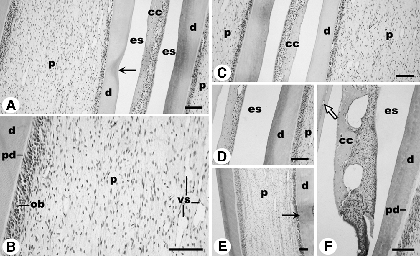
Light microscopic images from laser irradiated rabbit molar teeth.
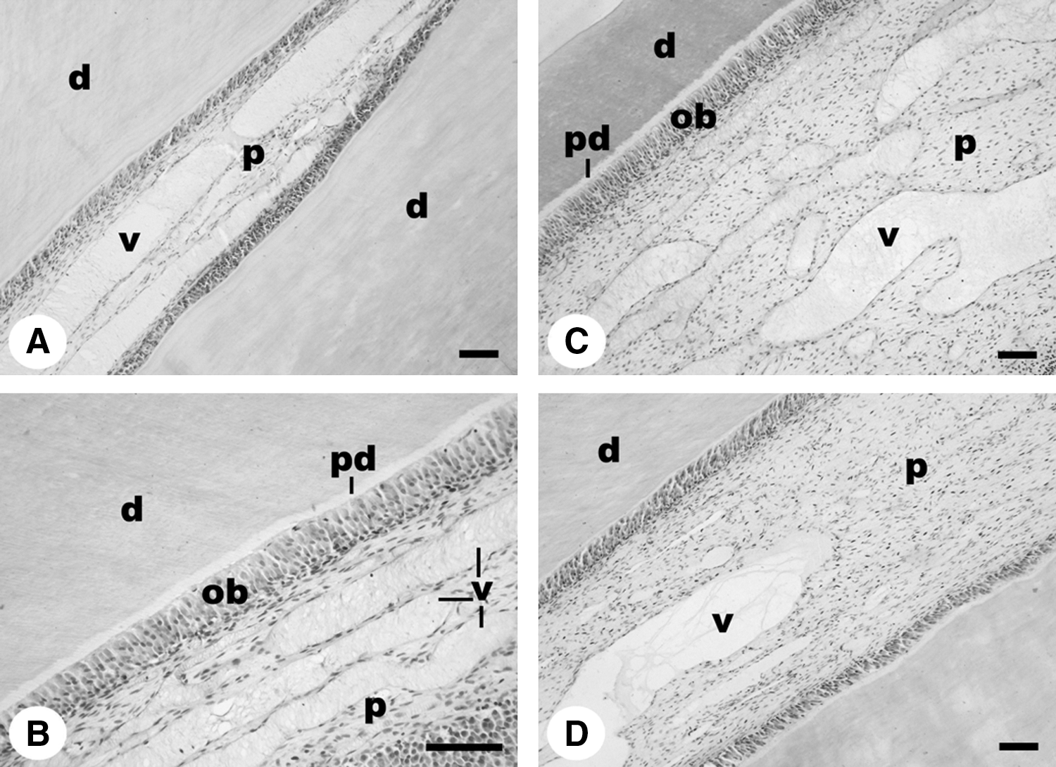
Light microscopic images from laser irradiated rabbit incisor teeth.
Discussion
In the present study, the histopathological effects on the pulp of rabbit teeth were evaluated following exposure to different power settings of a Nd:YAG laser.
Practitioners preparing cavities for restoration and scaling or root planing for periodontal health have traditionally relied on mechanical hard tissue surgery with low- and high-speed handpieces. However, these mechanical devices produce an irritating noise level, frequently with uncomfortable vibrations being transmitted throughout the patient's jaw and craniofacial tissues. Dentists have sought various alternative methods for dental procedures to find a more acceptable instrument lacking the negative aspects of traditional methods. Recently, the use of lasers for oral purposes has been the subject of numerous in vitro and in vivo studies. A number of studies 6,8,10,11,15,16 have reported the potential usefulness of pulsed Nd:YAG laser irradiation for different types of dental treatment, including alteration of various bacteria, treatment of perforation lesions during root canal preparation, induction of pulpal analgesia, treatment of acute and chronic dentin hypersensitivity, and removal of carious dentin. Since lasers were first introduced into dentistry, there have been investigations to establish the laser energy (wavelength, energy density, continuous or pulsed mode, time of exposure, focal spot) that would be the most useful and least harmful for the soft and hard tissues of the oral cavity. 8 In other words, laser treatment has been expected to serve as an alternative or adjunctive treatment to conventional mechanical therapy in dentistry. 17,18
The energy from a Nd:YAG laser is easily transmitted through dental hard tissue to the pulp, 19 and the resulting temperature increase caused by the laser can give rise to protein degeneration in the pulp. 20 The thermal effects of laser irradiation on the dental pulp have been studied, and researchers have measured the temperature increase in the pulp chamber both in the laboratory and in vivo as a means of investigating pulpal damage. 21 The increase in the pulp temperature has been shown to cause pathologic changes in the pulp tissue. 22 Continuous Nd:YAG laser irradiation of the enamel surface has been shown to injure the dental pulp. 23 These changes might occur because the dentinal tubules may act as waveguides that target the laser energy directly to the pulpal dentin surface. 20 Srimaneepong et al. 21 found that laser irradiation and use of a high-speed diamond bur created measurable pulpal space pressure and temperature changes. Wigdor et al. 24 prepared dentinal cavities in dog teeth using an Er:YAG and showed that the device produced minimal damage to pulp tissues compared with other types of lasers such as Nd:YAG and CO2, or the high-speed handpiece. In another study in dogs, the response of dental pulp to the application of an Er:YAG laser was not unlike that obtained with a high-speed handpiece. 25 The pulpal blood flow was also found to be strongly influenced immediately after Nd:YAG laser irradiation, showing an increase in rate. 26
Although rabbit teeth are not comparable to the narrow-apex teeth in human patients, for ethical reasons we used rabbit vital teeth in this study to readily evaluate the short-term pulpal reactions.
In conclusion, the present study suggests that Nd:YAG laser irradiation produced no irreversible tissue damage in the pulp of rabbit teeth with lasing for the purpose of different treatment methods at the power outputs of 1–4 W. The results support the premise that, for dental treatments, Nd:YAG lasers with suitable irradiation parameters would lead to better patient acceptance and fewer postoperative adverse events. However, the same effect may not be evident when the human tooth is irradiated for cavity preparation or for deep caries management; in these conditions, pulpal damage may be observed or may be increased. For these reasons, additional university-based controlled studies are needed to further corroborate these results. Nonetheless, the future use of lasers in dentistry has exciting potential, and research should continue on this promising new tool.