
Abstract
Introduction
Resident microorganisms found on different parts of the body generally do not cause disease. These microorganisms contribute to the development of the immune system and may make colonization by pathogenic microorganisms difficult. However, although they are not pathogenic in their usual locations, they may cause opportunistic disease or infection if they penetrate other sites. Some microorganisms may also favor pathogenic ones due to an imbalance of the resident environment. 5
Postsurgical infection is a potential complication of any surgical procedure and arises from multiple factors. Wound infection is the most common complication in healing wounds and causes both vascular and cellular responses in the tissue. 5,6 The repair of infected tissues occurs only after the resolution of the infectious process and removal of the necrotic tissue. Another problem is that infection causes increased lysis of collagen due to an increased amount of lysosomal enzymes from leukocytes. Additionally, fibroblasts compete locally with both other cells and microorganisms for oxygen and nutrients and cause reduction in their metabolism and consequently reduce collagen synthesis. The main etiologic factors of skin and nasopharynx infections are Staphylococcus aureus and the Streptococcus pyogenes, respectively. 6
Several therapeutic approaches have been proposed as being efficacious in improving wound healing, including the use of different light sources such as the laser. Previous studies have shown that the use of adequate protocols may improve tissue responses to different traumatic agents or to either systemic or local conditions. 2,7,8 Some wavelengths present positive photobiological effects on the healing process. Besides quickening the process, laser light affects the tissue at cellular levels, including a photo-activated increase in cell metabolism. In addition, it also causes analgesia and increases the rate of both inflammatory response and wound healing. 2,9 –11
The aim of this study was to histologically assess the effect of different wavelengths on the healing of wounds infected by Staphylococcus aureus.
Material and Methods
Following approval by the Animal Experimentation Ethics Committee of the School of Dentistry of the Federal University of Bahia, 24 young adult male Wistar rats (200–250 g) were obtained from the Central Animal House of Federal University of Bahia and kept at the Animal Experimentation Laboratory of the School of Dentistry of the Federal University of Bahia. The animals were kept in individual plastic cages with wood chip bedding and maintained at 22°C in a day/night light cycle and fed with standard pelted laboratory diet (Labina®, Purina) and had water ad libidum.
After regular quarantine and under intraperitoneal general anesthesia [47.5 mg/kg of ketamine (Ketalar®, Pfizer, São Paulo, SP, Brazil) and 12 mg/kg of xylazin (Virbaxil®, Pfizer, São Paulo, SP, Brazil)], the animals had their dorsa shaved and cleaned (2% chlorohexidine). A 1 by 1 cm cutaneous wound was created with a scalpel on the dorsum of each animal and left untreated. Four hours after wounding the lesion was inoculated with Staphylococcus aureus ATCC 6538. 6 Forty-eight hours after contamination, 6,12 the animals were randomly divided into two sets of four subgroups, with three animals in each subgroup: control, red laser light, infrared laser light, and red + infrared laser light.
Laser phototherapy (LPT) was carried out with a diode laser [λ680 nm/790 nm, P = 30 mW/40 mW, continuous wave (CW), laser, Ø = 3 mm, PD = 424 mW/cm2 and 566 mW/cm2, time = 11.8/8.8 sec, E = 0.35 J; BioWave®, Kondortech, São Carlos, SP, Brazil], started immediately after surgery, and repeated every other day for 7 d. Laser light was applied at four points around the wounded area (5 J/cm2). All irradiated subjects were treated with 20 J/cm2 per session. The groups irradiated at both wavelengths received 2.5 J/cm2 of each at four points totaling 10 J/cm2 of each wavelength. The time of the application varied according to the equipment used and was automatically set. The choice of the wavelengths, as well as the use of 20 J/cm2, was due to the lack of previous studies using the chosen model. The choice of treatment parameters was also made with regard to different wavelengths having different absorption and penetration.
Nonsteroidal analgesics were available if an animal presented any evidence of pain, but they were not needed in any group. Following macroscopic examination, each animal was killed by an overdose of general anesthetic either 8 or 15 d after contamination. Specimens were taken and kept in 10% formalin for 24 h, then embedded in paraffin, sectioned, and stained with hematoxylin and eosin. Histological analysis carried out by an experienced pathologist in a double-blind manner. One slide was made from each specimen, and the whole fragment was analyzed. The criteria used for this analysis are presented in Table 1. Depending on the criterion, it was scored as present or absent; complete or incomplete; or discrete, moderate, or intense. The percentage of occurrence of each score was analyzed using Minitab15® software (Globaltech). The significance level was set at 5%.
Results
Light microscopy revealed that control animal wounds were partially covered by keratinized epithelium at day 7. Fibrinous exudate, newly formed blood vessels, hyperemia, and intense inflammatory lymphocitary exudate were also observed at this stage (Fig. 1). Sirius red stain showed a moderate amount of delicate and fragmented collagen fibers. The amount of collagen fibers at day 14 was intense and the fibers were well organized (Fig. 2).
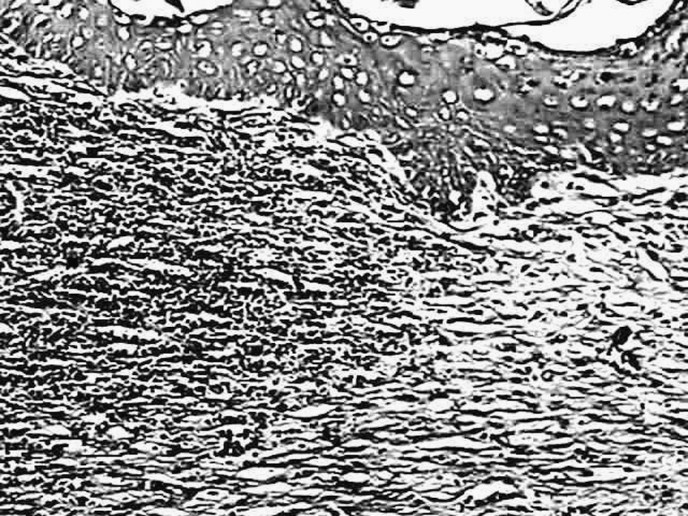
Photomicrography of control specimen at day 7 showing intense hyperemia of the granulation tissue rich in newly formed blood vessels. A moderate number of both fusiform and triangular fibroblasts were seen at this stage (hematoxylin and eosin [H/E] ×100).

Photomicrography of control specimen at day 14 showing intense and well-organized collagen matrix (Sirius red, ×100).
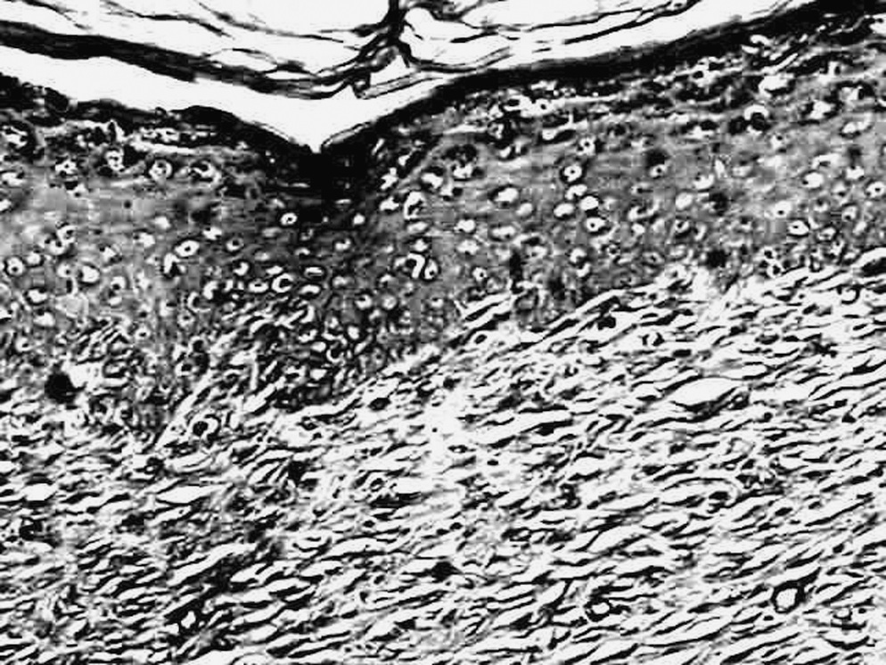
At day 7, specimens irradiated with λ680 nm showed some level of epithelial pavementing, granulation tissue formation, hyperemia, interstitial edema, and newly formed congested blood vessels. An intense chronic inflammatory infiltrate was also observed at this stage. Fibroblasts were also detected (Fig. 3). The collagen matrix was graded as moderate and was disorganized as seen with Sirius red staining (Fig. 4). At the end of the experimental period, epithelial pavementing was complete. On the dermis, a discrete lymphocitary inflammatory infiltrate could be seen. Moderate hyperemia and interstitial edema were also seen at this stage. Fibroblasts with varied shapes could also been seen. Sirius red staining showed a moderate amount of a well-organized collagen matrix.
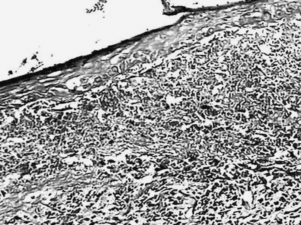
Photomicrography of specimen of the red laser light group at day 7 showing some degree of epithelial pavementing, granulation tissue, hyperemia, interstitial edema, and congested newly formed blood vessels. Intense lymphocitary infiltrate was also observed at this time (H/E, ×100).

Photomicrography of specimen of the red laser light group at day 7 showing a moderate amount of a disorganized and immature collagen matrix (Sirius red, ×100).
Specimens irradiated with λ790 nm showed some level of epithelial pavementing at day 7. Granulation tissue was rich in newly formed blood vessels, and hyperemia, interstitial edema, and lymphocitary inflammatory reaction were intense. Fibroblasts were also seen at this stage. Collagen matrix was scored as discrete at this time. At the end of the experimental time, epithelial pavementing was complete. Granulation tissue presented a chronic aspect and was associated with a moderate lymphocitary inflammatory infiltrate and discrete hyperemia (Fig. 5). Fibroblasts were also seen, and the amount of collagen matrix was intense at this stage (Fig. 6).
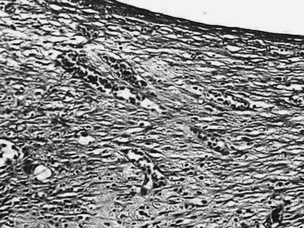
Photomicrography of specimen of the infrared red laser light group at day 14 showing complete epithelial pavementing, a moderate amount of granulation tissue, discrete hyperemia, and chronic inflammation (H/E, ×100).
Photomicrography of specimen of the infrared red laser light group at day 14 showing intense deposition of collagen matrix (Sirius red, ×100)
Subjects irradiated with both wavelengths (λ680 nm + λ790 nm) showed complete epithelial pavementing at day 7. Underneath, granulation tissue was rich in newly formed blood vessels and showed moderate hyperemia associated with intense lymphocitary inflammatory reaction (Fig. 7). Fibroblasts were also seen at this stage and the collagen matrix was scored as intense and was composed of mature and well-organized collagen fibers (Fig. 8). At the end of the experimental time, epithelial pavementing was complete. Granulation tissue showed large amounts of newly formed blood vessels and discrete hyperemia associated with a discrete chronic inflammatory reaction. Fibroblasts were seen and the collagen matrix was scored as intense and the fibers were mature and well organized. A summary of the results is shown in Table 2.
Photomicrography of specimen of the infrared red and red laser light group at day 7 showing complete epithelial pavementing covering the granulation tissue rich in newly formed blood vessels, moderate hyperemia, and intense lymphocitary inflammatory infiltrate (H/E, ×100).

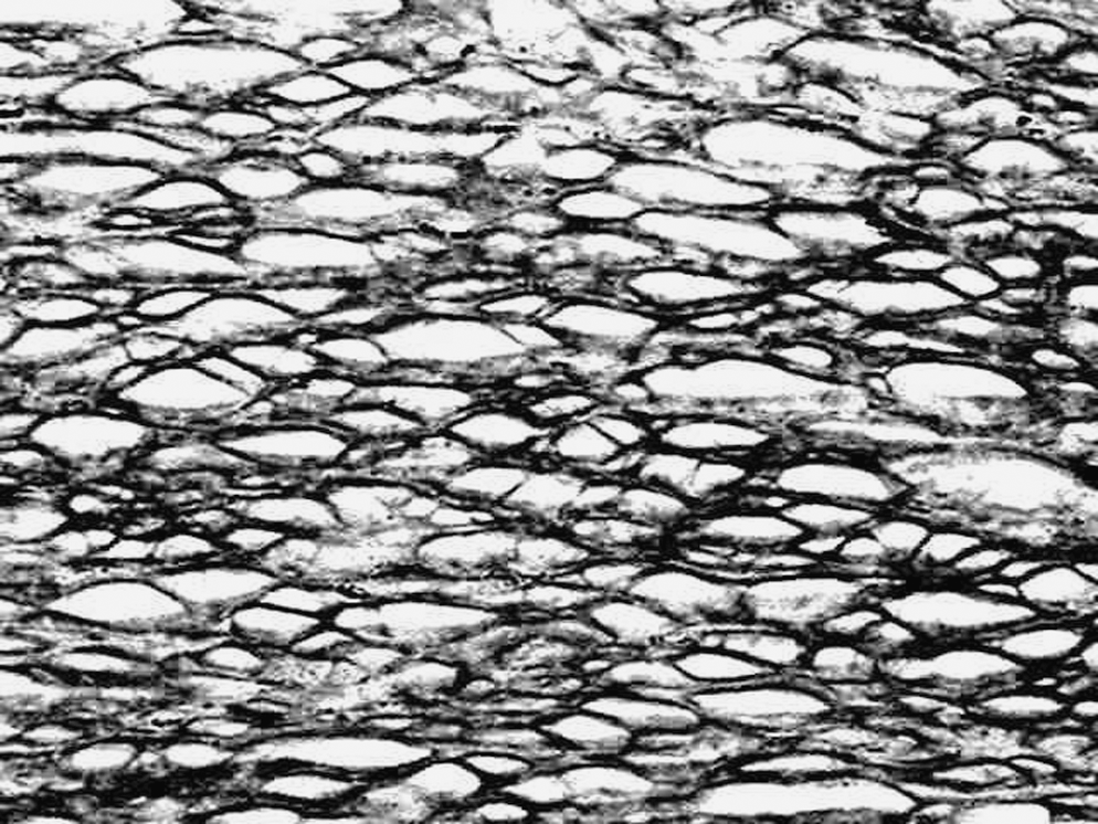
Photomicrography of specimen of the infrared red and red laser light group at day 7 showing an intense and well-organized mature collagen matrix (Sirius red, ×100).
0 = absent; 1 = discrete; 2 = moderate; 3 = intense.
0 = absent; 1 = present.
Discussion
Wound infection accounts for 50% of the complications observed in surgical wounds and it greatly affects wound healing. Infection impairs collagen metabolism, reducing its production and increasing the lysis. Bacterial infection also impairs local nutrition of the tissues. 1 In the present study we used a model of local infection using Staphylococcus aureus ATCC 6538, a model widely used worldwide.
Many authors have indicated that phototherapies positively affect wound healing by increasing cellular metabolism and proliferation as well as reducing pain and inflammation. 11,13 We aimed to verify the effect of laser photobiomodulation on infected wounds by using two wavelengths because previous reports had shown that the use of different wavelengths may cause different tissue responses. 2,14
Our results showed that control subjects presented a discrete number of blood vessels when compared to irradiated subjects as previously reported on the literature. 10,11,15 We also found that inflammation was at a more advanced stage of resolution in irradiated subjects when compared with controls. In normal tissue repair, the chronic inflammatory reaction starts around day 7 after wounding. In the present study, we found an earlier appearance of this chronic inflammatory reaction in irradiated subjects, which agreed with a previous report. 5
Several positive effects of LPT have been reported in the repair of cutaneous wounds. Both experimental and human studies have shown increased collagen synthesis and quicker inflammatory reaction, with these being attributed to increased ATP production, increased cell metabolism, and fibroblastic proliferation. 2,4,6,7,9,13
Previous studies have shown that laser light may positively affect both fibroblast proliferation 6,16,17 and collagen synthesis. 7,15,18,19 Rocha Júnior et al. 13 also found that LPT resulted in more advanced wound repair when compared with nonirradiated subjects. The same was observed in the present study. Laser light also influenced both the amount and quality of the collagen matrix. Irradiated subjects showed more intense expression of the collagen matrix and collagen fibers were mostly mature and well organized in these subjects at the end of the experimental time when both wavelengths were used, as seen previously. 20
A complete understanding of the effects of light on repair is still far away, and the mechanism of stimulation of specific cells, such as fibroblasts, remains unknown. This dearth of knowledge explains why it is so difficult to set effective protocols for the treatment of different wound types as well as wounds in systemically compromised subjects.
Another important parameter in our study was the energy density. Despite not being the focus of this report, we used an energy density similar to one previously reported. 4,7,19 The effect of energy density on this model will be reported in the near future.
Wavelength is another important parameter in phototherapy because it interacts with specific molecules. 21,22 It is accepted that visible light acts directly on the mitochondria, infrared light initially interacts with the cell membrane, and each type of light offers specific therapeutic actions. This indicates that the use of associated wavelengths may further improve the outcome of the treatment. 21,23,24 The positive results with the association of both wavelengths may also be attributed to different levels of absorption because both superficial and deep tissues receive the treatment. 11,13,21,22 These aspects may explain the finding of intense collagen expression and maturation at day 14 in subjects treated with paired wavelengths. 21
The results of the present study indicate that LPT has a positive effect on the healing of infected wounds and this is more evident with paired wavelengths of 680 and 790 nm.
Footnotes
Acknowledgment
We thank the Coordination for the Improvement of Higher Education Personnel (CAPES) for the scholarship that allowed this work.
Author Disclosure Statement
No competing financial interests exist.