
Abstract
Introduction
Cisplatin [cis-diammine-dichloroplatinum (II)] is one of the most potent chemotherapeutic anticancer drugs. A major factor limiting treatment using cisplatin is its nephrotoxicity, which is associated with mitochondrial injury. 6,7 Cisplatin is often combined with other chemotherapeutic agents in clinical practice to take advantage of the synergy effect between the agents, which occurs through various pathways, including different pharmacokinetic interactions. 8 The present study was designed to evaluate the anticancer effect of PDT using low-dose cisplatin combined with a hematoporphyrin-derivative sensitizer, Photogem®, in vitro and in vivo.
Materials and methods
Chemicals
Cisplatin (0.5 mg/ml) was purchased from Dong-A Pharm (Seoul, Korea) and Photogem® from Lomonsov Institute of Fine Chemical (Moscow, Russia). Dulbecco's modified eagle's medium (DMEM), fetal bovine serum (FBS), antibiotics, Dulbecco's Phosphate-Buffered Saline (DPBS) and trypsin were purchased from Hyclone (South Logan, UT). Dimethyl sulfoxide (DMSO) and 3-[4,5-dimethylthiazol-2-yl]-2,5-diphenyl-tetrazolium bromide (MTT) were supplied by Sigma-Aldrich (St. Louis, MO).
Cell cultures
Mouse colon cancer cells (CT-26) were purchased from Korean Cell Line Bank (Cancer Research Institute, Seoul, Korea) and were cultured in DMEM with phenol red, penicillin (100 units/ml), streptomycin (100 μg/ml) and 10% FBS at 37°C.
Animals
The hair on the backs of 6- to 8-week-old female BALB/c mice (SLC, Inc., Shizuoka, Japan) was removed 2 days before the CT-26 cells were inoculated.
Absorption Spectrometry
The absorption curve of cisplatin was measured using a visible/UV spectrophotometer (Biochrom Ltd., Cambridge, UK) which records spectra within the wavelength range of 200–900 nm.
Photosensitizer and photoactivation
Photogem® was dissolved in DPBS at a concentration of 10 mg/ml and was stored at −20°C. A 632 nm diode laser (BioLITEC, Jena, Germany) was used as the light source and a triangular prism box, base dimensions 15 × 15 cm, covered with aluminum foil, was utilized to provide an even distribution of energy.
Photodynamic activity in CT-26 cells
CT-26 cells (1 × 10⁵ cells/ml) were seeded into 96-well plates. After overnight incubation, in the cisplatin-only group, 100 μl of cisplatin (50 μg/ml) was added to the first row and the two-fold dilution method was used until the 10th row. The media were changed 24 h later and cell viability was determined through MTT assay after another 24 h. For the PDT group, cells were exposed to a 632-nm diode laser at 3.2 J/cm2 after 6 h incubation in different Photogem® concentrations (from 0 to 37.5 μg/ml). The MTT assay was performed 24 h after PDT. In the combination group, cisplatin concentrations of 0.1, 1, and 6 μg/ml were administered 24 h before Photogem® and the subsequent protocols were carried out congruently with the PDT-only group. For all MTT assays performed, 50 μl of MTT solution (2 mg/ml) was administered to each well and incubated for 2 h. The solution was then replaced by 150 μl of DMSO and the absorbance was measured. The percentage of cell viability was calculated by dividing the mean absorbance in each treatment group by the mean absorbance in the control group. The MTT assay was repeated three times for consistency.
Photodynamic activity in mice bearing CT-26 cells
The CT-26 cells (107 cells/ml) were inoculated into the backs of the mice to make xenograft models and treatments began when the tumor volume measured approximately 0.5 cm in diameter. The animals were divided into laser, cisplatin, PDT, and combination groups. Group 1 (n = 7) was irradiated with a 632 nm diode laser, group 2 (n = 7) was treated with cisplatin (3 mg/kg, intraperitoneal (IP)), and group 3 (n = 14) with laser 1 h after Photogem® administration (5 mg/kg, intravenous (IV)). Group 4 (n = 11) was treated with PDT as in group 3, and after 24 h, was treated with cisplatin as in group 2. In every group in which the subjects were irradiated, the energy density projected was 600 J/cm2. The tumor size was measured twice a week, and the volume was calculated by the following formula:
Statistical analysis
Data were expressed as mean ± SD. Data were analyzed by one-way ANOVA using the Statistical Package for Social Science (SPSS, Chicago, IL). Differences were considered to be significant when p < 0.05.
Results
1. The absorption spectrum of cisplatin
In order to exclude the possibility that cisplatin might act as a photosensitizer, the absorption spectrum of cisplatin was measured with the spectrophotometer. The maximum absorption recorded was approximately 220 nm (Fig. 1).
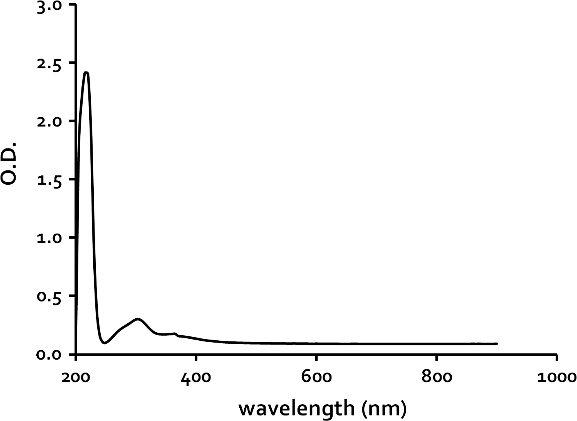
The absorption characteristics of cisplatin. The maximum absorption of cisplatin was detected at 220 nm.
2. The anticancer effects of combination therapy on CT-26 cells
Cell viability was 93.70 ± 0.39, 81.98 ± 1.14, and 30.92 ±2.59% for 0.1, 1.56, and 6.25 μg/ml of cisplatin, respectively (Fig. 2).There was almost no change in cell viability in cells treated with PDT when Photogem® concentrations were less than 1.17 μg/ml, but 2.34–18.75 μg/ml did induce remarkable cell death (Fig. 3). Whereas 0.1 μg/ml of cisplatin proved not to be cytotoxic and produced no additive effects with PDT (Fig. 4A), 6 μg/ml resulted in viability for only 24.36 ± 5.52% of the cells (Fig. 4C). A cisplatin concentration of 1 μg/ml was the most ideal concentration for application in combination treatment.

Cytotoxicity of cisplatin on CT-26 cells in vitro. Cell viability was measured by MTT assay 24 h after being treated with various concentrations of cisplatin.

Photocytotoxicity of Photogem®-mediated PDT on CT-26 cells in vitro. Cell viability was assessed by MTT assay at 0, 3, 6, 12, and 24 h after Photogem®-mediated PDT at various concentration of Photogem®.

The cytotoxicity of combined Photogem®-mediated PDT and various doses of cisplatin (0.1 μg/ml in
When Photogem® concentrations of 1.17, 2.34, and 4.69 μg/ml were combined with the 1 μg/ml of cisplatin, the percentage of cell viability dropped to 67.35 ± 0.88, 49.84 ±0.44, and 19.88 ± 0.41, respectively. Cell viability for PDT at a Photogem® concentration of 4.69 μg/ml was 47.13 ± 2.78 and that of 1 μg/ml of cisplatin was 80.31 ± 3.09, showing that the combination of the two concentrations resulted in an approximately 9% increase in effect compared with treatments with these two agents individually (Fig. 4B). The cell viability produced from 1 μg/ml of cisplatin and/or PDT was organized in a time-dependent manner and the additive effect of combination therapy was not observed until 12 h after PDT (p < 0.05, Fig. 5).
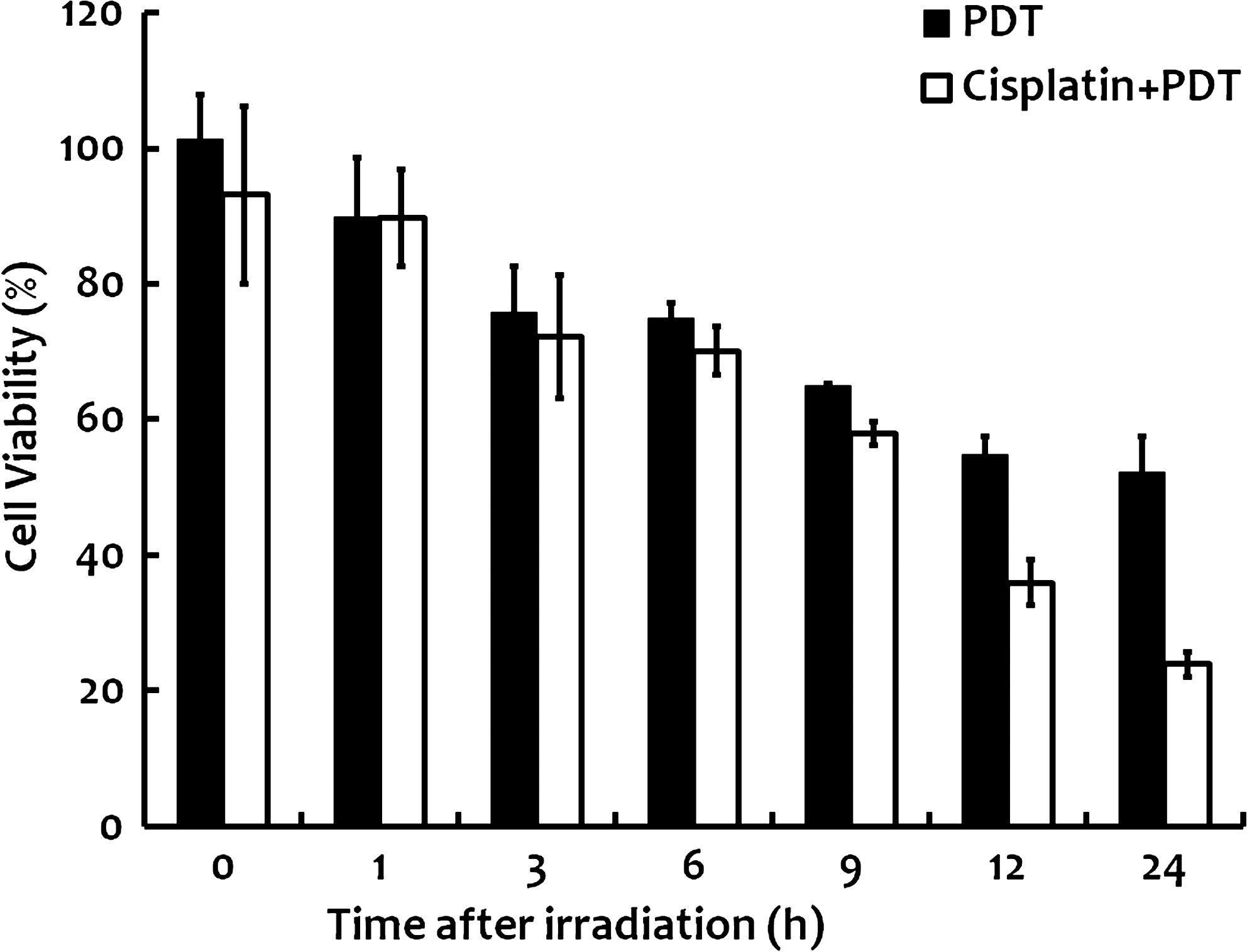
The cytotoxicity of combination therapy with 1 μg/ml cisplatin and 4.69 μg/ml of Photogem® mediated PDT on CT-26 cells. Significantly enhanced cytotoxicity of cisplatin and PDT was observed at 12 and 24 h after irradiation (p < 0.05).
3. The effect of combination therapy on mice bearing CT-26 cells
Three days after treatment, tumors of the subjects in the PDT and combination groups disappeared completely, while the tumors of those in laser-only and cisplatin-only groups grew continuously (Fig. 6). On the 10th day, 6 of the 14 mice in the PDT group exhibited recurrence rates of 42.86%, and on the 14th day, 78.57% recurrence rates were observed in 11 of the 14 mice. The combination group displayed rates of 0% and 27.27% on the 10th and 14th days, respectively (p < 0.05; Fig. 7).
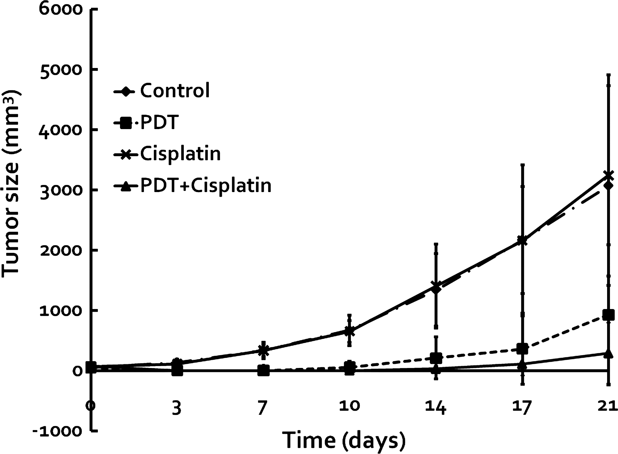
The effects of Photogem®-mediated PDT, cisplatin only, or combination treatment on tumor volume in nude mice bearing CT-26 cells. Tumor sizes were decreased in the PDT and combination groups.
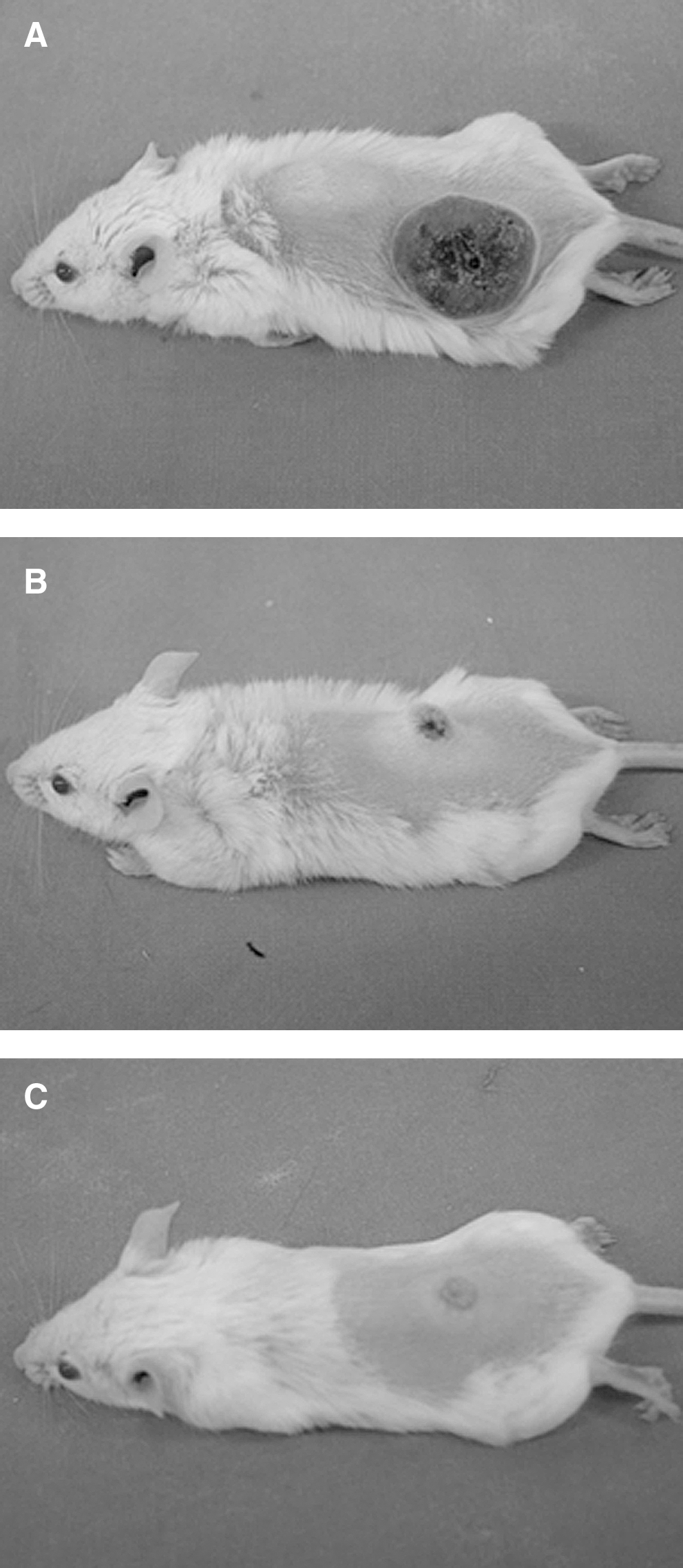
Gross tumor photographs at three weeks after the treatments.
Discussion
The combination of PDT and cisplatin presented in this study aims to reduce administration dosages, decreasing the risk of side effects. Cell viability was observed to be downregulated in accordance with the dosage of cisplatin. For Photogem®, there were almost no PDT effects at concentrations lower than 1.17 μg/ml (Fig. 2, Fig. 3). In combination therapy, 1 μg/ml of cisplatin was observed to have additive effects with those of PDT (Fig. 4). This is in agreement with other experiments, in which cytotoxic drugs such as cisplatin, mitomycin, and 5-fluorouracil were found to act synergistically with PDT in vitro. 9 –11 Some reports have suggested that cisplatin and other chemotherapeutic agents can also improve the anticancer effect of PDT in vivo, 12 –14 and in this study, the combined-therapy group prolonged the disease-free period, compared with the PDT-only group (Fig. 6).
Cisplatin can cause cell arrest in the S-G2 phase, 10,15 and PDT may also cause cell cycle arrests in the G2/M phase. 10,16 This disjointed phase-related effect could account for the favorable effect of the combined treatment; 10 cisplatin causes apoptosis by enhancing the expression of Fas/Fas-ligand on the surfaces of malignant cells 17 and PDT results in rapid cytochrome c release, which initiates the apoptotic cascade. 18 Cisplatin could also attack the mitochondria to impair cellular respiration in a caspase-independent system. 19
Uehara et al. reported that low-dose cisplatin, 5 mg/kg, alone had no effect on tumors but displayed an efficient antitumor effect when combined with PDT; 13 it has been suggested that low-dose cisplatin could make the tumor photosensitive and inhibit DNA repair processes 20 –22 by inactivating DNA polymerase. 23 In the present study, 3 mg/kg of cisplatin was tested in the subjects, and the combination group was found to display a significant decrease in the recurrence rate compared with the PDT-only group or cisplatin-only group. This raises the possibility of using low-dose cisplatin to enhance the antitumor effect of PDT while lessening the cytotoxic impacts of the drug. All experimental conditions concerning PDT were based on a previously done study by Ahn et al. 24
Because DNA damage induced by a sublethal dosage of PDT is suggested to be repaired within 15 min, 25 some time is required before the administration of PDT for cisplatin distribution to the cells to inhibit DNA repair. Studies that administered cisplatin 1 h 9 before PDT or immediately after 25 showed no enhanced antitumor effect in vivo. When cisplatin was administered 24 h before PDT, no antitumor effect was observed, but when the timing sequence was adjusted, even when cisplatin was administered 24 h after PDT, the tumor recurrence rate decreased in the combination group.
The photosensitizer has been observed to be localized in both the vasculature and in the tumor 1 h after injection. 26 The photosensitizer taken up by tumor cells may cause damage directly as singlet oxygens, 27 or may produce an anticancer effect indirectly by damaging the capillary vessels of the tumor tissue, resulting in vascular shutdown and inhibition of nutritional supply. 28 In order to utilize both approaches, a 1 h time interval was chosen. The in vivo mechanisms of PDT remain controversial, however, and because the disease processes in cancer are complex, only a well-designed combination therapy may have a great chance of success. A further study should be carried out, researching the relative effects of the sequence of administration of chemotherapeutic agents and the length of the time intervals in between administration.
Conclusion
The antitumor effect was enhanced when CT-26 cells were treated with combined cisplatin and PDT, both in vitro and in vivo. The combined regimen could decrease the required administration dosage of Photogem® and cisplatin, lowering toxic side effects on normal tissues. The mechanisms involved in this combined modality deserve further study, however, for better understanding and improved utilization.
Footnotes
Acknowledgments
Sincere appreciation and thanks to the Medical Laser and Device Research Center of Dankook University for their financial support and much more.
Author Disclosure Statement
No conflicting financial interests exist.