
Abstract
Introduction
The production of growth factors is a biological process that starts immediately after injury, aiming to stimulate the cells involved in the repair process to proliferate. Transforming growth factor β (TGF-β) is a multifunctional cytokine secreted by platelets, T lymphocytes, macrophages, endothelial cells, fibroblasts, and other tissues. It has a central action and presents anti-inflammatory and proliferative effects during tissue repair. Its effects include chemotaxis of leukocytes, fibroblasts, and smooth muscle cells. Besides it influences both formation and remodeling of the extracellular matrix; stimulates keratinocyte migration, angiogenesis, and fibroblastic differentiation; inhibits the proliferation of keratinocytes; regulates the expression of integrin and other cytokines; and also possess an auto-induction property. 7 –9
Previous studies found elsewhere in the literature have shown that red laser phototherapy (LPT) quickens both the inflammatory reaction and the healing process because of the effects of the light on the gene regulation of inflammatory cytokines, interleukin, and growth factors. 10,11 Other studies have also shown that red LPT stimulated TGF-β secretion on rodent model 12 and on cardiac cells in vitro. 13 LPT also stimulated TGF-β1 secretion on melanoma cell line in vitro, 14 on a rodent model, 15 and in a human oral tooth extraction wound healing model. 16 Another study showed that the expression of TGF-β2 was increased on human osteoblasts in vitro. 17
It seems possible that LEDPT presents beneficial effects similar to the ones observed when LPT is used, as several reports on the benefits of the use of LEDs operating at several wavelengths, both in vitro and in vivo, in both normal and pathologic conditions, have been published elsewhere. 2,3,6,18 –25 It is also possible that the mechanism involved is similar.
The aim of the present study was to assess the effect of LEDPT (λ700 nm, 5 J/cm2) on the immunohistochemical expression of TGF-β on cutaneous wounds in a rodent model.
Materials and Methods
The Animal Experimentation Ethics Committee of the School of Dentistry of the Federal University of Bahia approved this work (Process number 029/06). Twenty-four adult male Wistar rats weighing 200–250 g were obtained from the Central Animal House of the School of Veterinary Medicine of the Federal University of Bahia and kept in individual plastic cages bedded with wood chips, maintained at 22°C in day/night light cycle, fed with standard pelted laboratory diet (Labina®, Agribrands-Purina Ltda., Paulínia, São Paulo, Brazil), and had water ad libitum, at the Animal Experimentation Laboratory of the School of Dentistry of the Federal University of Bahia.
After regular quarantine, under intraperitoneal general anesthesia [60 mg/kg of ketamine chloridrate (Vetaset®, Fort Dodge Animal Health, Campinas, São Paulo, Brazil) and 10 mg/kg of xylazine (Coopazine®, Intervet Schering-Plough, São Paulo, Brazil)] the animals had their dorsum shaved and cleaned with chlorhexidine gluconate 10 mg/ml solution (Merthiolate®, Hypermarcas S.A., Barueri, São Paulo, Brazil). One excisional cutaneous wound (1 × 1 cm) was created with a scalpel on the dorsum of each animal and left without suturing or dressing. The animals were then randomly distributed into two groups with 12 animals in each: G0 (Control) and G1 (LED λ700 ± 20 nm, 16 mW, Ø = 16 mm*, Illuminated area = 2 cm2). Each group was subdivided into three subgroups according to the animal death timing (2, 4, and 6 days). LEDPT was performed using a prototype device, started immediately after surgery, and was repeated every other day during the experimental timing. LED light was applied over the wounded area, as a prototype device, the spot area was 2 cm2 and the illuminated area was 1 cm2 (Fig. 1). The device automatically set the time of the application. We performed a pilot study to determine the most suitable amount of energy to be used, as there are no definite comparative studies between laser and LEDs regarding equivalence of the dose X effect.

Diagram of the illumination protocol used in the study.
The spatial average energy fluence (SAEF), or the energy density in relation to the illuminated area (1cm2) per session was 5 J/cm2 (Table 1). If the animal presented any evidence of pain, non-steroid analgesic was used. This was not the case in any group.
According to manufacturer data.
Immunohistochemistry
Following macroscopic examination, each animal was killed by an overdose of general anesthetic [300 mg/kg of ketamine chloridrate (Vetaset®, Fort Dodge Animal Health, Campinas, São Paulo, Brazil)] and 50 mg/kg of xylazine (Coopazine®, Intervet Schering-Plough, São Paulo, Brazil) according to the experimental timing, after surgery. Specimens were taken and kept in 10% formalin for 24 h, and were then routinely cut and processed to wax. The specimens were immunomarked with polyclonal anti-TGF-β (1:4) associated to a background reductor (DAKO antibody diluent with background reducing components, Denmark, S3022) using the EnVision® system (DAKO, Glostrup, Denmark, K4061), DAB substrate chromogen system (DAKO, Glostrup, Denmark, K3466) and Meyer haematoxylin. Granulation tissue acted as positive controls, and negative control used PBS buffer as a replacement for the primary antibody.
Immunomarked sections underwent histological analysis performed by an experienced pathologist in a blind manner. Light microscope Motic B5® Professional Series with an assembled camera Moticam 2000® interconnected to Motic Image Advance 3.0® (Motic Instruments Inc., Richmond, British Columbia, Canada) software were used to capture images of three consecutive fields in each slide. The area of expression of TGF-β was measured with the Motic Image® software. The mean area of each specimen was calculated using ExcelMac®, Microsoft, Redmond, WA. The mean area of each subgroup was similarly calculated. The data were analyzed using Minitab15® software (Globaltech, Belo Horizonte, Minas Gerais, Brazil). For the statistical analysis, the ANOVA and Tukey's test were used to compare the mean area among groups. The significance level was 5%.
Results
We found small variations in the expression of TGF-β in all groups during the experimental time. It was found that in controls, the expression decreased with time. However, in irradiated subjects there was an initial reduction on the expression up to the 4th day and an increase on the 6th day. A more intense and well-located immunomarking was seen on controls. On irradiated subjects, the immunomarking was less intense and diffuse. The statistical analysis showed that the area of the expression of TGF-β on LED-irradiated animals was significant smaller than on the controls at day 2 (Table. 2). No significant differences were found at later times (Figs. 2 –8).
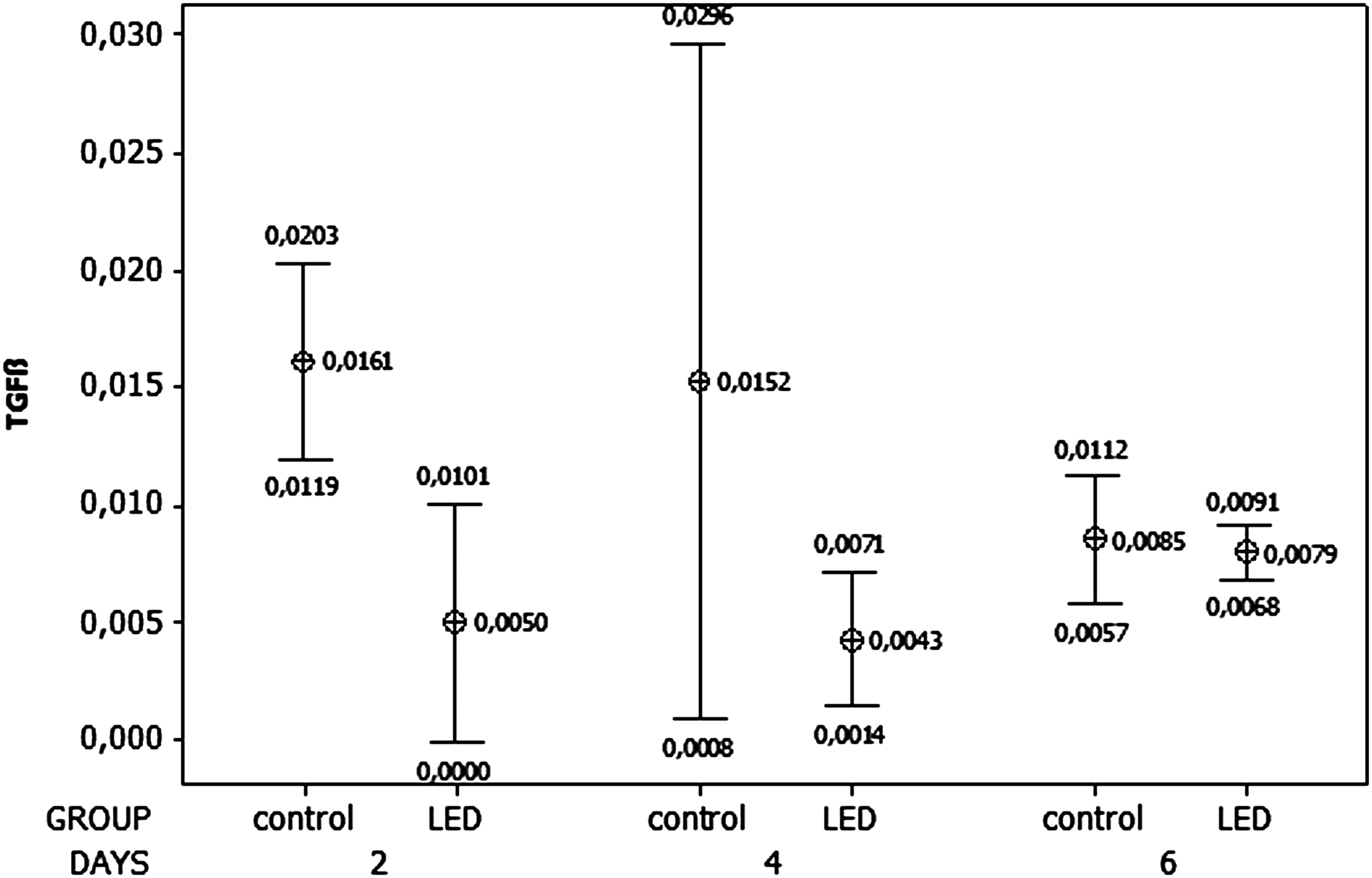
Expression of TGF-β on LED and controls at each experimental time.

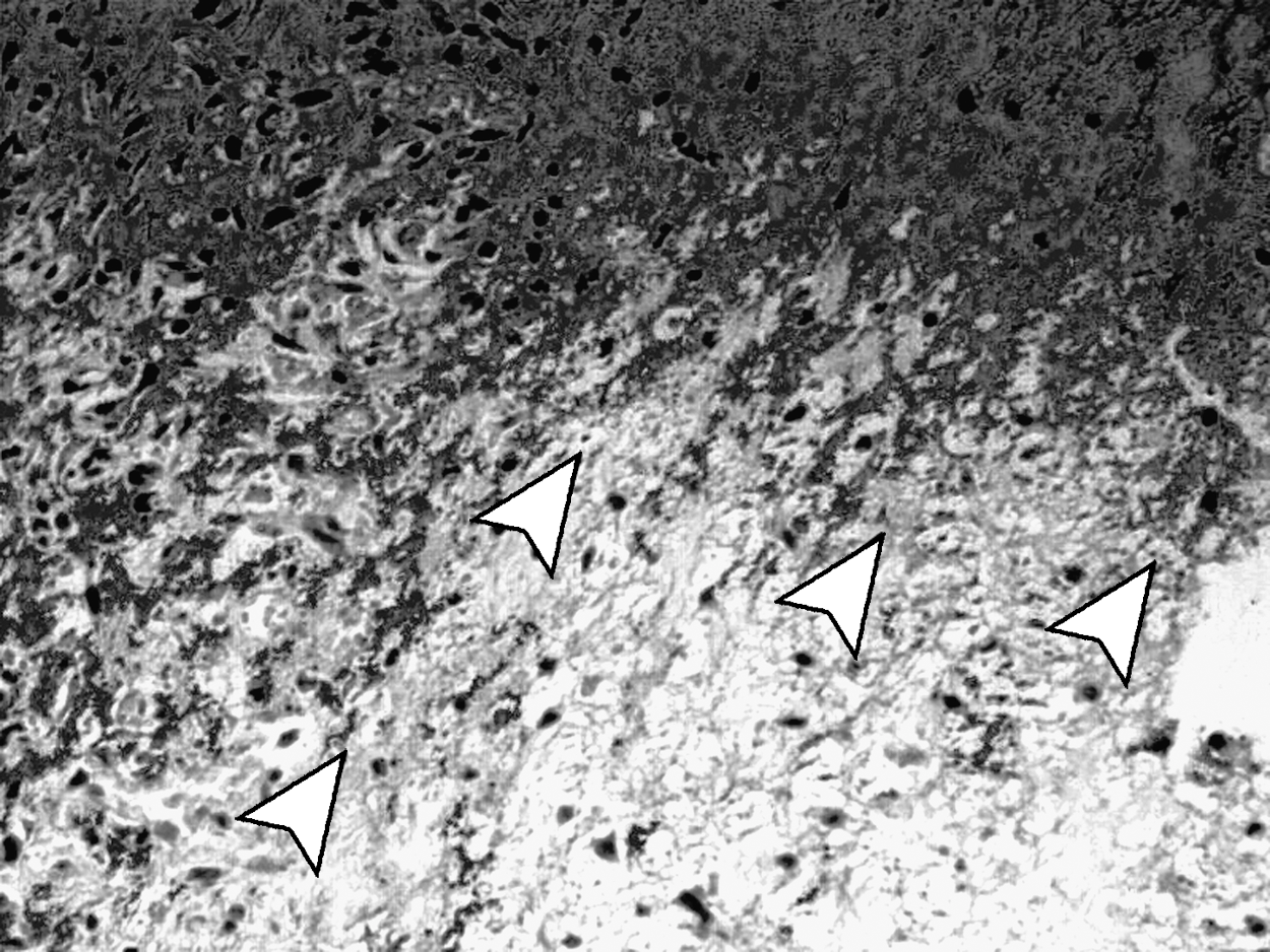
Photomicrography of a specimen from the control group at day 2 showing intense presence of immunomarked TGF-β concentrated in a determined area (arrowheads).

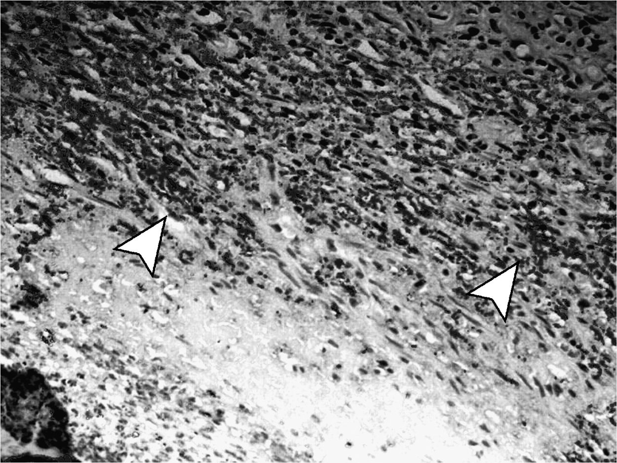
Photomicrography from a specimen from the LED group at day 2 indicating discrete and uniformly distributed immunomarked TGF-β (arrowheads).
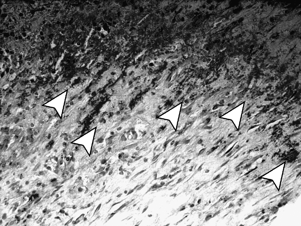
Photomicrography from a specimen from the control group at day 4 showing intense presence of immunomarked TGF-β differently distributed in the field observed (arrowheads).
Photomicrography from a specimen from the LED group at day 4 showing discrete presence of immunomarked TGF-β (arrowheads).

Photomicrography from a specimen from the control group at day 6 showing moderated distribution of immunomarked TGF-β (arrowheads).
Photomicrography from a specimen of LED group at day 6 showing discrete presence and limited distribution of immunomarked TGF-β (arrowhead).
Difference statistically significant between LED and control groups at day 2 (p = 0.013).
NS, not significant.
Discussion
It seems that the basic mechanism of interaction between LED light λ630–1000 nm and the tissues involves the mitochondria, and that the cytochrome C oxidase present at the cell membrane is responsible for the absorption of the light. 26 –29
Wong-Riley et al. 28,29 showed that LED light (λ670 nm, bandwidth 25–30 nm) was one of the most effective in the upregulation of cytochrome C oxidase among the wavelengths tested. On the other hand, LED light (λ728 nm, bandwidth 25–30 nm) was the least effective one. However, the λ700 nm (bandwidth 20 nm) was not tested in those studies. Because of the bandwidth, it may be reasonable to consider the possibility of some effect on the cytochrome C oxidase of the λ700 nn used in the present study.
A previous study has suggested that macrophages are the first target of non-coherent λ660 nm light. 30 Other studies also suggested the involvement of genomic expression, which may regulate, either positively or negatively, the response, as well as the ordination of the cascade of signaling pathway of the cells. 4,31,32
Unfortunately, the number of animals was small because of constraints imposed by the ethics committee. However, even with the small sample, statistical analysis was possible.
Our results showed a significant inhibition of the TGF-β up to 48 h after wounding, on LEDPT-treated animals. It is possible that this may reflect a similar inhibitory effect seen when using laser light with SAEF > 4 J/cm2. Despite not being significantly different from controls, this tendency was observed during throughout the experimental time. If we consider only this effect on the healing wound, a more severe inflammatory reaction would be expected to occur on LED-irradiated subjects, especially at early stages. This finding disagrees with the idea that LED light could always cause similar responses on tissues such as the ones observed with the use laser light. There are reports on the literature that show that red laser light causes positive effects on growth factors 10,11 and specifically on TGF-β1 and β2. 12 –17 This was not seen in the present study. Our results are also conflicting with reported anti-inflammatory effects of LEDPT on inflammatory conditions such as oral mucosites. 6,33,34
Secondary effects of LEDPT (λ630–1000 nm) might include the regulation of the expression of cytokines and growth factors influencing the regulation of the inflammatory reaction and cellular events observed during healing. However, few reports showed the effect of LEDPT on the expression of growth factors. Whelan et al. 5 reported increased expression of TGF-β following the use of λ880 nm LEDPT (4 J/cm2, 50 mW/cm2) on ischemic wounds. However, no difference was observed regarding the expression of VEGF.
The use of LEDs on living tissue is a real possibility, as it probably causes similar responses to the ones observed following the use of other light sources such as laser light, at lower costs. Our results showed that λ700 nm LEDPT, using the parameters and experimental protocol of this study, caused an inhibition of the expression of TGF-β at an early stage of the healing process. Many aspects of using phototherapies may lead to inhibition, including the wavelength. It is possible that either stimulation or inhibition demands a combination of factors such as wavelength, power density, and irradiation time. The total energy density may have been the reason for inhibition in the present study. It is concluded that the use of LED light, at these specific parameters, caused an inhibition of the expression of TGF-β at an early stage of the healing process.
Footnotes
Author Disclosure Statement
No competing financial interests exist.