
Abstract
Introduction
There are two key factors involving adhesion of resin cements to silica-based dental ceramics: (a) micromechanical interlocks formed as a result of the erosive effect of acid etching when applied to porcelain surfaces, and (b) chemical bonding achieved through a silane coupling agent. 5 However, luting aluminum-oxide ceramic represents a challenge because the high crystalline content and limited vitreous phase of alumina make it resistant to acid-etching or silanization. 6 Furthermore, glass infiltration enhances alumina density and makes it somewhat resistant to sandblasting, 7 which has just been recommended as an effective cleaning method prior to bonding aluminum-oxide ceramics. 8
Alternative surface treatments such as silica-coating (Co-Jet, Rocatec) 9,10 thermal silica-coating (Silicoater MD system), 11 flame-treatment (PyrosilPen), 12 and erosive techniques (irradiation with neodymium:yttrium-aluminium-garnet; Nd:YAG–laser) 13 have been reported to improve surface roughness and, probably, bond strength between alumina and resin cements.
Despite minor differences in the particles' size and composition, silica coating procedures consisted of air-abrading the ceramic surface with a layer of alumina particles coated with silica. 9,10 The blasting pressure determines the embedding of those silica particles in the substrate, rendering the ceramic chemically more reactive to the resin after silanization. 14,15
Another promising ceramic treatment is laser irradiation. 16 Pulsed laser irradiations cause micro-explosions that generate micro-retentions on the surfaces due to energy discharges. Alumina requires higher laser parameters than do feldespathic ceramics because of the lower silica content, 16 even though Nd:YAG laser has been reported to optimize bond strengths of Panavia F to alumina 13 and zirconia. 17 However, no previous research has been conducted on the bond strength of Nd:YAG laser-irradiated alumina when bonded by self-adhesive resin cements, such as RelyX Unicem cement that does not require previous adhesive application.
Ceramic frameworks can be fixed with conventional cements. However, higher clinical survival rates have been reported for ceramics restorations bonded with resin-based luting agents. 18 The resin cements can be classified as: (a) etch-and-rinse resin cements (used after application of adhesives requiring a separate acid etching), (b) self-etch resin cements (used after application of a self-etch adhesive) (e.g., Panavia F, Clearfil Esthetic Cement), and (c) self-adhesive resin cements (self-adhering cements used without application of any adhesive system) (e.g., RelyX Unicem). 19 Satisfactory in vitro results with CAD/CAM ceramics were reported using Panavia F or Clearfil Esthetic Cement (self-etching [14] M. Sarr, A. Mine, J. De Munck, M.V. Cardoso, A.W. Kane and J. Vreven et al., Immediate bonding effectiveness of contemporary composite cements to dentin, Clin Oral Invest (2009) [Epub ahead of print].resin cements) 20,21 or RelyX Unicem (self-adhesive resin cement) 22 . However, little data are available concerning their bonding stability. 4
The presence of chemical agents in the oral cavity (e.g., acids and proteolytic residues from oral bacteria and salivary esterase) is considered a potential factor responsible for bonds' degradation. 8,23 A short-term immersion in aqueous solution of sodium hypochlorite (NaOClaq) has been proposed as a suitable and less time-consuming ageing technique 24,25 to simulate the hydrolytic effect of salivary enzymes over time. 26,27 Such storage medium has been considered to have similar effectiveness and lower variability than water aging or thermocycling to assess bonds' stability. 25
Specimens were stored in NaOClaq solution to evaluate resistance to degradation of resin cements when bonded to conditioned alumina. The tested study question was whether alumina surface treatment or resin cement type did influence the bond strength and stability of cement/ceramic interfaces.
Material and Methods
Experimental design
Sixty-four In-Ceram Alumina CAD/CAM blocks (10 × 10 × 6 mm) with the following composition by weight: Al2O3 (82%), La2O3 (12%), SiO2 (4.5%), CaO (0.8%), and oxides (0.7%), were sintered and glass-infiltrated according to the manufacturer's instructions.
Ceramic blocks (batch no 7803, Al Cubes for Cerec, Vita Zahnfabrik; Bad Säckingen, Germany) were embedded in self-curing acrylic resin (Special Tray, Dentsply–DeTrey; York, PA). The exposed surface of each alumina block was polished with SiC water-proof sand paper Hermes of 500, 800, 1000, and 1200 grit mounted in a circular grinder (EXAKT-Apparatebau, Otto Herrman; Nortedst, Germany). Afterwards, sandblasting (BIO-ART; Sâo Carlos, SP, Brazil) with 110 μm aluminum-oxide (Al2O3) powder was applied perpendicularly to those ceramic surfaces for 20 s at a working distance of 10 mm, under a pressure of 2.8 bars.
Alumina blocks were randomly assigned to four experimental surface treatments (n = 16). (a) No further surface treatment was applied [NT]. (b) Rocatec system [Roc]: surfaces were treated by means of tribochemical silica coating (30 μm silica particles) that was applied perpendicularly for 20 s, at a working distance of 10 mm and a pressure of 2.8 bars; silanization was performed before bonding with Scothbond ceramic primer, as described in Table 1 (batch no 2721, 3M/Espe; Saint Paul, MN). (c) Nd:YAG laser irradiation [L]: a graphite powder stain (DEKA M.E.L.A.; Calenzano, Italy) was applied onto the ceramic surface and laser was applied at 1 mm of working distance, 100 mJ, 20 Hz, 2 W, and 141.54 J/cm2. The laser optical fiber (diameter: 300 μm) scanned the whole ceramic area. 13 (d) Nd:YAG laser followed by Rocatec system [LRoc]: both procedures were developed as previously described.
10-MDP: 10-methacryloxydecyl dihydrogen phosphate; bHEMA: 2-hydroxyethylmethacrylate; cCQ: camphoroquinone; d3-MPS: trimethoxysilylpropylmethacrylate.
Sixty-four composite specimens (height: 4 mm) were made by layering 2 mm-thick increments of a microhybrid composite (batch no 6BX, Filtek Z250, 3M/Espe) using a square-shaped silicon mould. Each composite film was condensed with a clean plastic filling instrument to avoid contamination, and light-cured for 40 s with a halogen light-curing unit (Translux EC, Kulzer GmbH, Bereich Dental; Wehrheim, Germany, output: 500 mmW/cm2).
The last increment of composite was covered with a glass microscope slide for obtaining a flat surface. After removing the specimens from the mold, an extra 40 s irradiation was performed on the portions that were previously in contact with the silicone pattern.
Luting procedure
Composite specimens were bonded to the ceramic conditioned surfaces using different dual-cure resin cements: a self-etching resin cement: Panavia F 2.0 (PF) (batch no 00023D & 00244F, Kuraray Medical; Okayama, Japan) and a self-adhesive resin cement: RelyX Unicem- (RXU) (batch no 275331, 3M/Espe; Seefeld, Germany).
All materials were handled following the manufacturer's instructions, at room temperature (23.0°C ± 1.0°C) and relative humidity (50% ± 5%). PF groups received application of an oxygen-inhibitor agent (Oxyguard II, Kuraray Medical) covering the cement layer for 10 min to ensure the resin cement polymerization. Application mode and chemical composition of the tested materials are detailed in Table 1.
Ceramic-to-composite luting procedures were carried out using a customized metallic tool to standardize a cementation pressure of 1 kg (1,249 MPa), leaving the resin cement to set in the self-curing modality during 5 min. Finally, the specimens were photoactivated for 40 s from each side of the blocks (Translux EC; Output: 500 mmW/cm2) in order to guarantee polymerization. Bonded specimens were stored for 24 h in a laboratory oven at 37°C and 100% relative humidity.
Microtensile bond strength test
After 24 h, all samples were vertically sectioned across the bonded interface into 1.0 ± 0.1 mm2 sticks, using a low-speed diamond disk n°-11-246 (Buehler; Lake Bluff, IL) at 3100 rpm under water cooling (Isomet 4000, Buehler), according to the nontrimming method of the microtensile test.
Half of the specimens from each subgroup were tested immediately for microtensile bond strength. The other half were stored in a 10% sodium hypochlorite aqueous solution (NaOClaq) for 12 h. 21,27 Those sticks were then retrieved from the challenging medium, rinsed with water for an hour, and tested in microtension.
Twenty microtensile sticks were obtained per subgroup. Each stick was attached with cyanoacrilate adhesive (Zapit, Dental Ventures of America; Corona, CA) to the flat grip of a Bencor Multi-T testing assembly (Danville Engineering; San Ramon, CA) and loaded in tension in an universal testing machine (Instron Model 4411; Canton, MA) at a cross-head speed of 0.5 mm/min until failure.
The detached area of the ceramic beams was measured with a pair of digital callipers (Sylvae Ultra-Cal). Bond-strength values were calculated in MPa. Failure modes were evaluated by a single operator under an optical microscope (BH-2 Olympus; Tokyo, Japan) at 100 × magnification and classified as adhesive (at ceramic/cement or cement/composite interfaces), cohesive (within the cement or the ceramic), or mixed (adhesive and cohesive fractures occurred simultaneously).
Statistical analysis
ANOVA was performed to study the influence of: (a) ceramic surface treatment, (b) cement type, and (c) degradation challenge on the bond strength of resin cement/ceramic interfaces. Post-hoc multiple comparisons were analyzed by the Student–Newman–Keuls test. Significance was set in advance at p < 0.05.
SEM analysis
Four representative sticks from each subgroup were rinsed with 96% ethanol, mounted on metallic stubs, gold-sputtered at 10 mA for 1 min (Unit E500; Polaron Equipment Ltd., Watford, UK) and evaluated under scanning electron microscope (SEM 1430 VP, LEO Electron Microscopy Ltd.; Cambridge, UK) at different magnifications in order to analyze the fracture pattern and the morphology of the debonded surfaces.
For qualitative analysis of the treated ceramic surface topography, two additional In-Ceram Alumina blocks (edge: 5 mm) for each subgroup of conditioning method were pretreated, gently ultrasonicated in 96% ethanol for 2 min, and air-dried before being processed for SEM evaluation at 1452x magnification.
Results
Ceramic surface pretreatment and NaOClaq immersion significantly affected bond strengths to alumina (p < 0.0001). Resin cement type did not influence bond strengths (p = 0.331). Mean microtensile bond-strength values (MPa) and results of post-hoc comparisons are presented in Table 2.
Different lower case letters in rows and upper case letters in columns indicate significant differences (p < 0.05).
After 24 h, L-treated sticks achieved the highest MTBS regardless of the cement type, whereas NT-samples recorded the lowest MTBS.
After NaOClaq challenge, PF luted to NT-specimens attained the lowest bond strength values. RXU recorded the highest MTBS when luted to L-samples. No significant differences exist between bond values of Roc-, LRoc-, and NT-sticks. All experimental subgroups significantly decreased bond strengths except RXU luted to NT-alumina. Significant differences between PF and RXU were neither recorded at 24 h, nor after chemical challenging in any subgroup of ceramic treatment.
Table 3 summarizes the failure mode distribution. After 24 h, the highest percentage of mixed failures was detected when RXU was applied to Roc or LRoc-treated ceramics. Few pure cohesive failures were registered when RXU was luted to L-samples, and when PF was luted to Roc- and LRoc-treated surfaces. Few samples failed adhesively, belonging to L-sticks luted with PF and NT subgroups.
Adhesive (A): between the ceramic and the resin cement; Cohesive (C): within the resin cement; Mixed (M): adhesive and cohesive.
After NaOClaq challenge, mixed fractures continued to be the most frequent in all groups, but the percentage slightly decreased. Cohesive failures within the resin cement augmented. Roc-sticks luted with PF and NT samples occasionally exhibited a complete detachment from the porcelain surfaces (Table 3).
SEM analysis
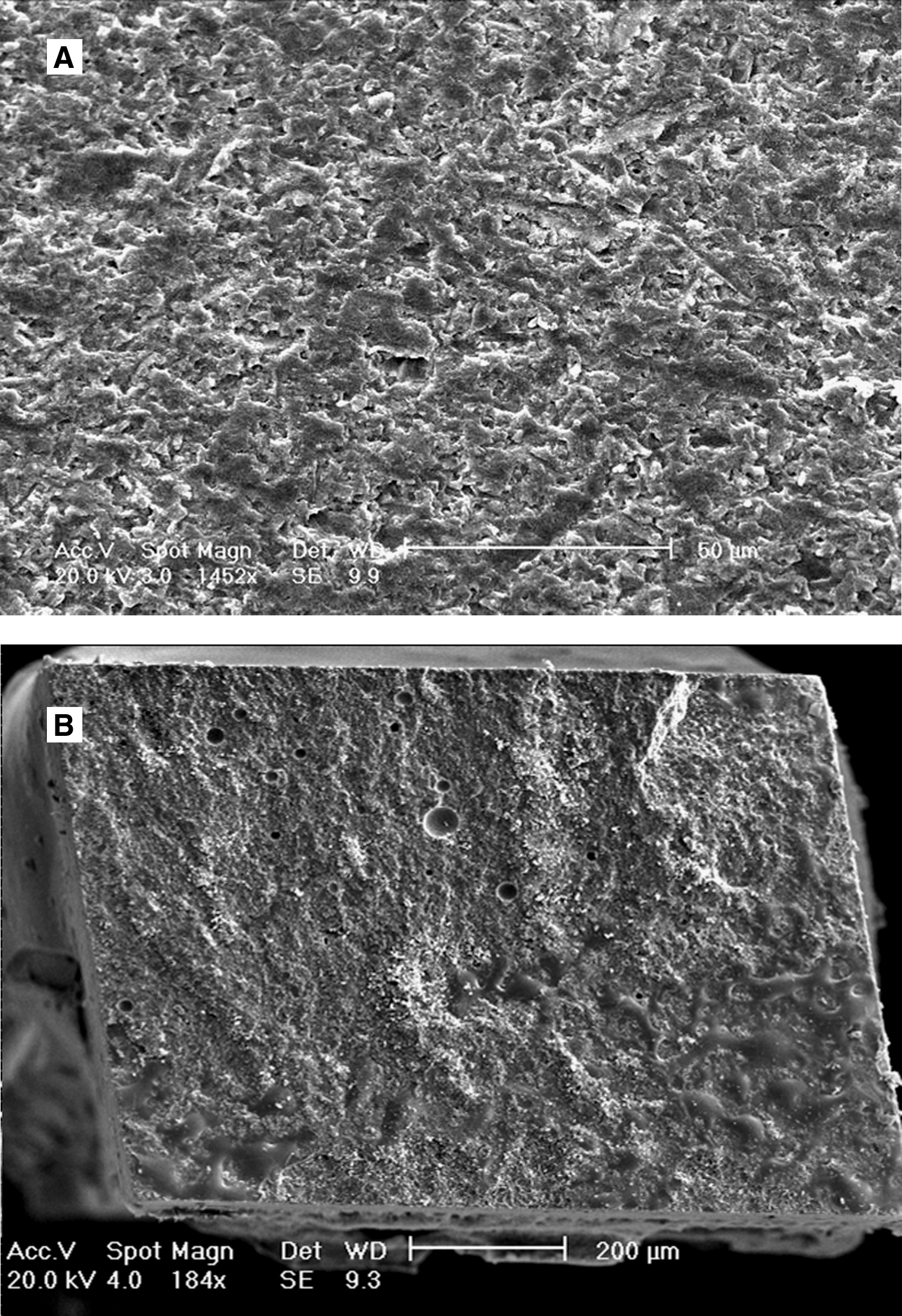
Both Sandblasting (Fig. 1A) and Rocatec (Fig. 1B) resulted in a rippled superficial structure with homogeneously distributed micro-irregularities and shallow pits.
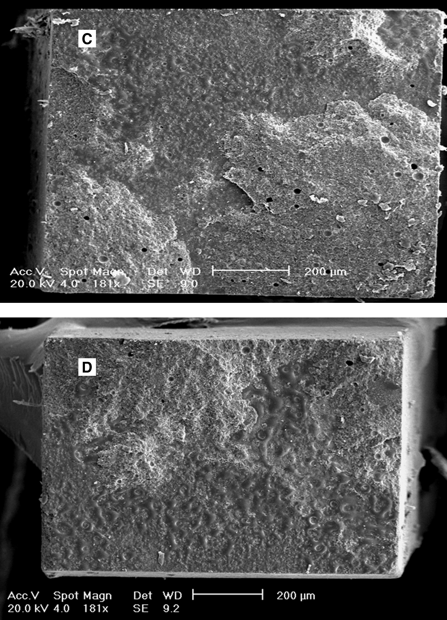
SEM images of the alumina ceramic surfaces after conditioning treatments (1452 ×; 20 kV; bar 50 μm) (SEM 1430 VP). Both
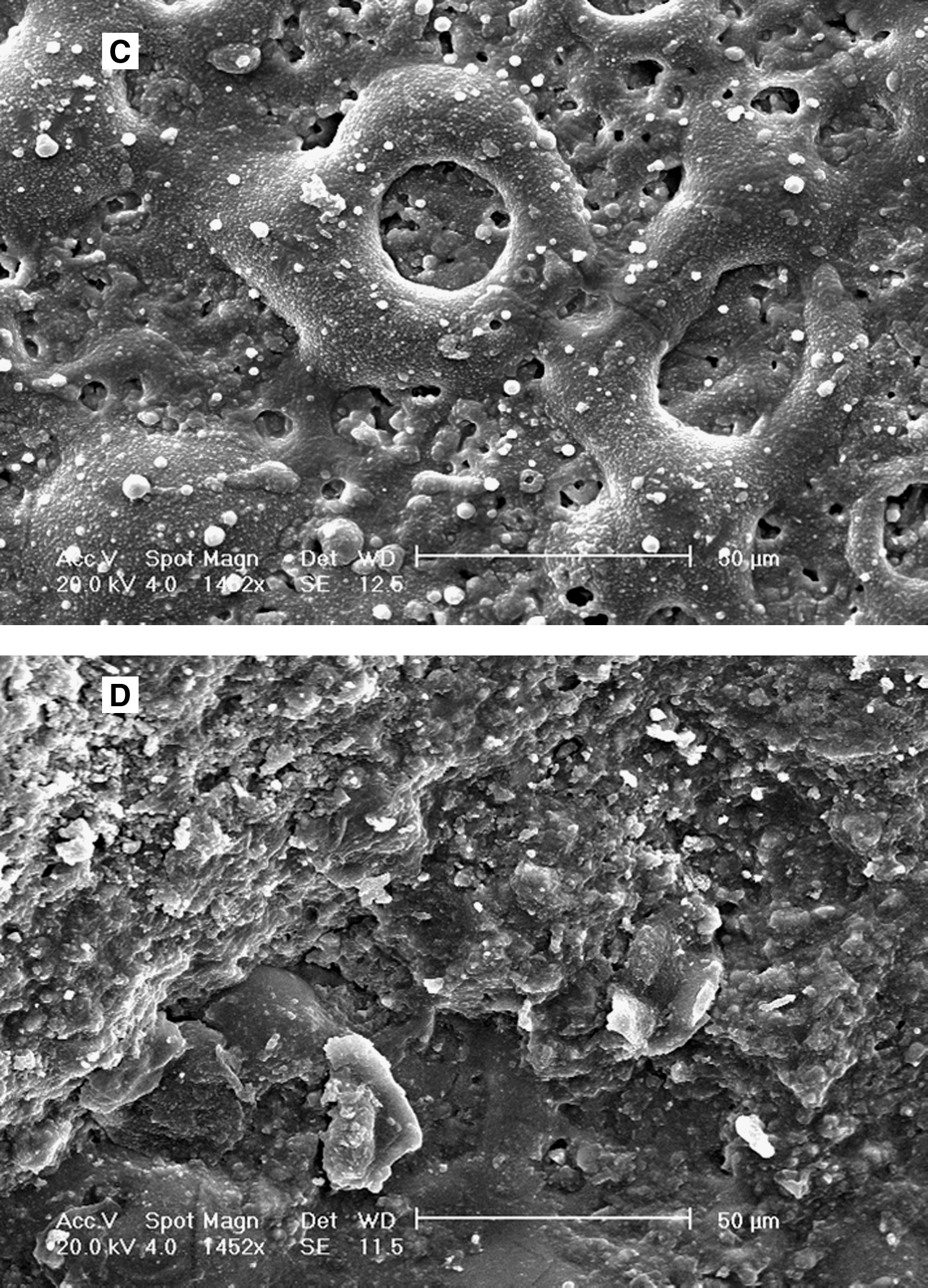
Nd:YAG laser determined the major modification of the porcelain substrate with holes exhibiting a melted-appearance (Fig. 1C). Nd:YAG laser plus Rocatec (Fig. 1D) caused wedge-shaped micro-retentions with partial coverage of the holes.
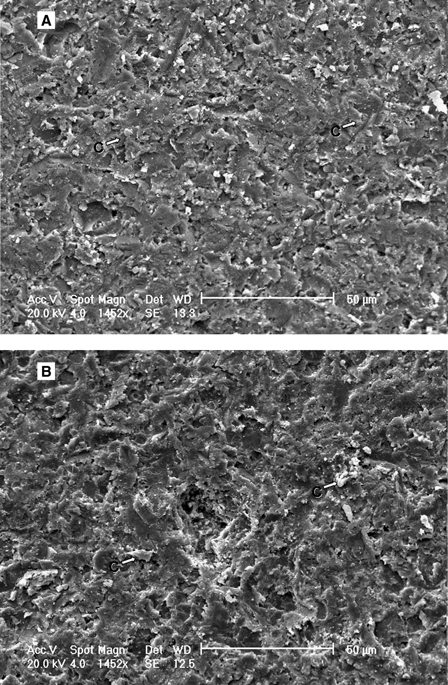
After 24 h, cement residuals of PF and RXU mainly luted to LRoc and Roc treated ceramics were frequently observed (Figs. 2A and C, respectively). Pores in remaining cement layers were detected (Figs. 2B–D).
SEM micrographs of alumina debonded surfaces after 24 h (SEM 1430 VP).

After NaOClaq immersion, cement layers of both PF (Figs. 3A and C) and RXU (Figs. 3B and D) persisted above the ceramic substrates, exhibiting patterns of micro-morphological alterations and porosities (Figs. 3C and D). In PF debonded samples, structural alteration of the resin cement may be observed if compared to those samples that were tested at 24 h, independently of the received surface treatment (Figs. 2A/3A–Figs. 2B/3C). In RXU debonded sticks, cement dissolution was evident after NaOClaq immersion (Fig. 3B).
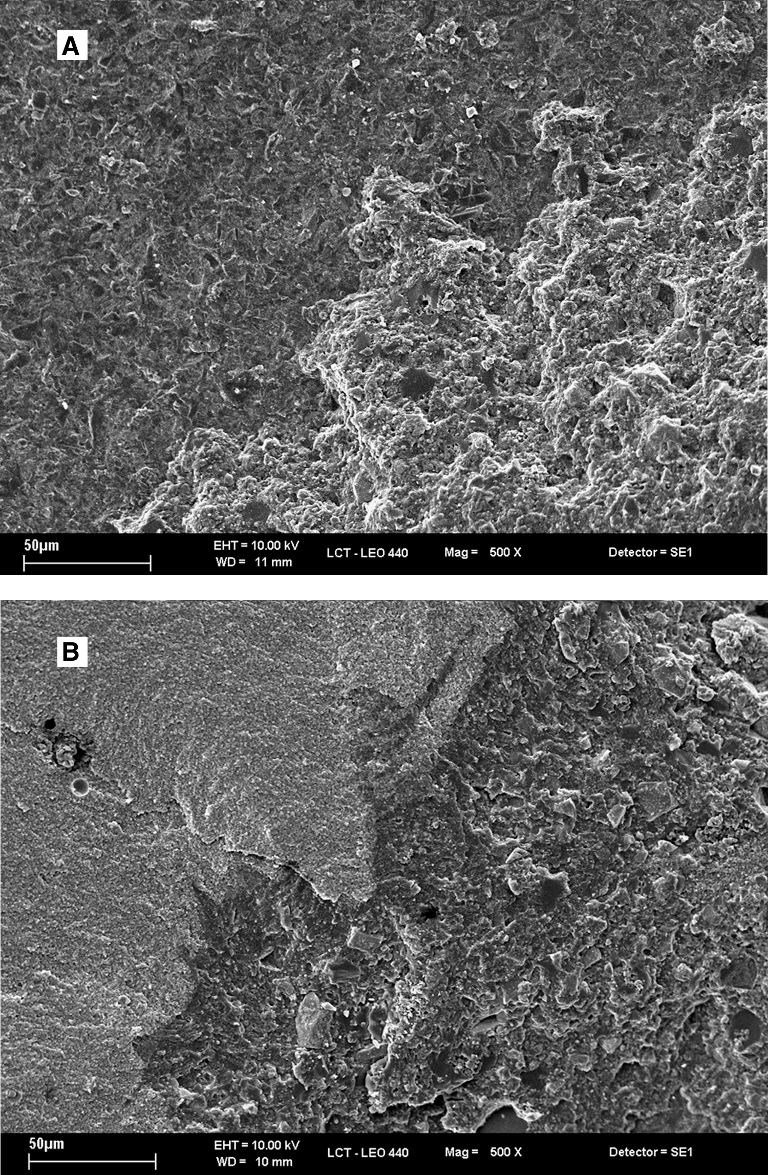
SEM images of alumina debonded surfaces after chemical challenging (SEM 1430 VP).
Discussion
The goal of the current research is to evaluate the effect of alumina surface treatment or resin cement type on the bond strength and stability of cement/ceramic interfaces.
Findings of the present study require partial rejection of the null hypothesis. After 24 h, L-sticks achieved the highest MTBS regardless of the cement type, whereas sandblasted samples recorded the lowest. L-irradiated beams showed the most evident micro-morphological changes in surface texture. Micro-retentive grooves on the ceramic surface with melted appearance holes were noticeable, probably as a result of local increases of the substrate temperature that generated an intense erosive effect 28 (Fig. 1C). Laser can also eliminate residual layers left by sandblasting alumina particles, which might interfere with proper adhesion. 29 Attained results are determined by the selected parameters when using the Nd:YAG laser on to the alumina surfaces. 13 Panavia F was previously shown to lute efficiently to Nd:YAG laser-treated alumina ceramic 13 and zirconia 17 by using self-etching cements under comparable experimental conditions, but no previous studies were performed using self-adhesive cements as RelyX Unicem.
NT-alumina registered the lowest MTBS. Glass-reinforced alumina ceramic is a high-toughness polycrystalline solid, 7 and the micro-hardness of the sandblasting-particles is similar to that of alumina crystals. 6,30 Interpretation of the published data indicates that chemical treatments of alumina provide higher bond strength and durability than do the simple air-abrading techniques. 9,10,31 Thus, sandblasting of CAD/CAM ceramic surfaces is recommended just as a cleaning method prior to any surface treatment. 8
Roc and LRoc-beams attained higher bond strength values than NT-sticks, regardless of the employed luting agent. The impact of Rocatec particles is supposed to produce both erosion and surface ceramication by means of a silica layer. Rocatec (30 μm particle-size) and sandblasting (110 μm) have been shown to produce similar alumina surface roughness with the tested parameters. 32 In the current study, LRoc surfaces showed micro-retentions, but after coating, melted-appearance areas seemed to be covered (Fig. 1D). The attained higher MTBS may be the result of the ceramicising of the surface promoted by Rocatec system and the performed silanization of the ceramic surfaces before bonding, which may account for chemical bonding. 9,10,31
Several in vitro methods have been used to test stability of resin bonds, trying to mimic those clinical conditions that may cause adhesive interfaces to fail. 24 Immersion in 10% NaOClaq has been previously employed, simulating the action of potential biodegraders, such as salivary proteases on resin-based cements. 8,23,33
NaOClaq immersion resulted in a detrimental effect in bond strengths for both cements. Structural alteration of PF layers can be observed by comparison of SEM images taken with and without chemical challenging (Figs. 2A/3A and Figs. 2B/3C). Cement dissolution with rounded-shaped margins was also noticed in RXU debonded sticks (Fig. 3B).
NaOClaq immersion may have affected the mechanical properties of both luting agents through degradation of the resin matrix of these polymeric materials 23,33 explaining the increase of cohesive failures within the resin cement, after challenging. Nonetheless, the main failure type (over 80%) continued to be mixed even after NaOClaq immersion (Table 3).
Silane-bond degradation may have contributed to the bond-strength decrease. Those groups without silane application were the less affected by NaOClaq immersion (NT and L-treated specimens luted with RXU). The trimethoxysilylpropylmethacrylate 3-MPS (present in PF) and the pre-hydrolyzed silane (employed in Roc and LRoc-treated beams luted with RXU) contain hydrophilic monomers that may expedite interfacial water sorption through hydrogen bonds and hydrolytic bond disruption. 34,35 It seems that silanization may increase the wetability of glass-infiltrated alumina and the immediate bond strength, 36,37 but there is conflicting evidence in the literature about the long-term efficacy of silanization when luting alumina ceramic with 10-MDP (PF) containing resin cements. 5
Not only chemical bonding but also micromechanical interlocking are key factors for guaranteeing a suitable and durable adhesion between resin cements and ceramics. 5 Mechanical interlocking of the bonded interface of RXU has been compared to that of silicate and zinc-phosphate cements, so that inorganic fillers (glass silicate) may react with acidic phosphoric esters forming a silicate gel in which glass particles are entrapped, facilitating microretention at the luted interface. 38
Despite published data, the precise bond strength of resin cements to aluminous ceramic still remains unknown. Further controlled long-term clinical trials should be conducted to determine the best bond strategy for alumina-based all-ceramic restorations.
Conclusions
Within the limitations of the present study, it may be concluded that Nd:YAG Laser irradiation is an effective surface treatment to enhance bonding of resin cements to alumina ceramic, regardless of the luting cement (self-etching [PF] or self-adhesive [RXU]). Both resin cements and silanization procedures are prone to hydrolytic degradation.
Footnotes
Acknowledgments
This investigation was supported by CICYT/FEDER MAT2008–02347/MAT, JA–P07–CTS–2568, JA–P08–CTS–3944, UNGR–08–1E–030, and FAPESP (PROC. 2005/01918–5). The authors are grateful to Prof. Carlos de Paula for scientific advice and support.
Author Disclosure Statement
No competing financial interests exist.