
Abstract
Introduction
Adhesive removal procedures following debonding may remove up to 55.6 μm of surface enamel. 3 The process of debonding a bracket from the tooth can potentially result in iatrogenic damage to the enamel surface. Potential sites of failure can therefore be located between the bracket and the adhesive, within the adhesive itself, or between the tooth surface and the adhesive. The incidence of ceramic bracket fracture (when conventional debonding techniques as recommended by manufacturers are employed) ranges from 10% to 35%. 4
Bracket debonding is based on the principle of degrading the strength of adhesive resin between the tooth and the bracket. The search for an efficient and safe method of adhesive resin removal following debonding has resulted in the introduction of a wide range of instruments and procedures. 2 These include manual removal with hand scalers or band-removing pliers, tungsten carbide burs with low- or high-speed handpieces, Sof-Lex disks (3M ESPE, St. Paul, MN), and special composite finishing systems using zirconia paste or slurry pumice, as well as ultrasonic applications. 1,3 Novel approaches involving the abovementioned laser-application and air-powder abrasive systems have also shown promising outcomes. 5 All the techniques described—whether classical or laser-based—produce different degrees of polish. Some introduced abrasion anomalies accompanied by a significant loss of enamel, and some also adversely affect pulpal tissues if not dissipated with an appropriate coolant. 2,6
It has been mentioned that brackets can be removed by laser irradiation, which can penetrate the bracket and reach the adhesive resin, and influence its strength bond to enamel. 7 Laser energy can degrade adhesive resin in three ways: by thermal softening, thermal ablation, and photoablation. 7 Thermal softening occurs when the laser heats the bonding agent until it softens. Clinically, this results in the bracket succumbing to gravity and sliding off the tooth surface. Thermal ablation occurs when heating is rapid enough to raise the temperature of the resin up to its vaporization range before debonding occurs through thermal softening. This results in the bracket being blown off the tooth surface. Photoablation also causes the bracket to be blown off the tooth surface. This occurs when very high-energy laser light interacts with the adhesive material and the energy level of the bonds connecting atoms of adhesive resin rises rapidly above their dissociation energy levels, resulting in decomposition of the material. Thermal softening is a relatively slow process, and can lead to a large rise in both tooth and bracket temperatures. Thermal ablation and photoablation proceed rapidly, so very little heat diffusion occurs, and therefore both tooth and bracket stay close to physiological temperatures. 8 The previous studies have confirmed that lasers can be used effectively to thermally soften the adhesive resin for the removal of ceramic brackets. 9,10
The aim of this study was to prepare a simple and reliable method for ceramic bracket debonding, ensuring minimal changes in the enamel structure and an acceptable temperature rise in the pulp.
Materials and Methods
Laser irradiation source – diode-pumped Tm: YAP microchip
The Thulium:Ytterbium-Aluminium-Perovskite (Tm:YAP) laser, is one of the most efficient lasers emitting in the 1900–2000 nm spectral band. 11 It has the main properties of the Yttrium Aluminum Oxide YAlO3 (YAP, YAlO) matrix—this material has good thermal and mechanical properties and the natural birefringence. 12 Because of the local absorption peak of liquid water at 1940 nm, it is an interesting materials for use in medical applications, namely urology or ophthalmology. 12
After preliminary experiments with different lasers 10 , the longitudinally diode-pumped Tm:YAP microchip laser 12 (Solid State Laser Laboratory - university prototype - Czech Technical University in Prague, Faculty of Nuclear Sciences and Physical Engineering, Czech Republic) was used as the source of irradiation. 12 The active material of this laser had a diameter of 3 mm and a length of 3 mm (simultaneously representing the resonator length). The optical resonator was formed by dielectric layers depositing on the front and back facets of a Tm: YAP crystal. From the pumping side, the layers had a transmittance level of ∼99% for the pumping 790 nm irradiation, and high reflectivity for laser emission at 1998 nm. The reflectivity level of dielectric layers for optimal output was settled at 97% at 1998 nm. 13,14 During the experiment, the microchip laser was mounted in a water-cooled copper block and was wrapped in an indium foil for excess heat removal. For pumping a fiber-coupled diode LIMO HLU30F400-790 was used. The 400 μm fiber tip was imaged into the Tm:YAP crystal by means of two achromatic doublet lenses (Thorlabs, Inc., AC508-075-B, Nord Newton, NJ) with a focal length of f = 75mm, resulting in a pumping beam mean diameter of ∼260 μm within the active medium. The Tm:YAP laser was operated in a continuous-wave regime, with a maximum output power of 5.5 W. The dependence of the Tm:YAP laser output power on the diode pumping power is shown in Fig. 1. The generated wavelength was 1998 nm, and the output beam profile was close to TEM00 mode (Fig. 2). In microchip construction Tm:YAP laser is simple, rugged, and almost maintenance-free, with good output parameter stability.
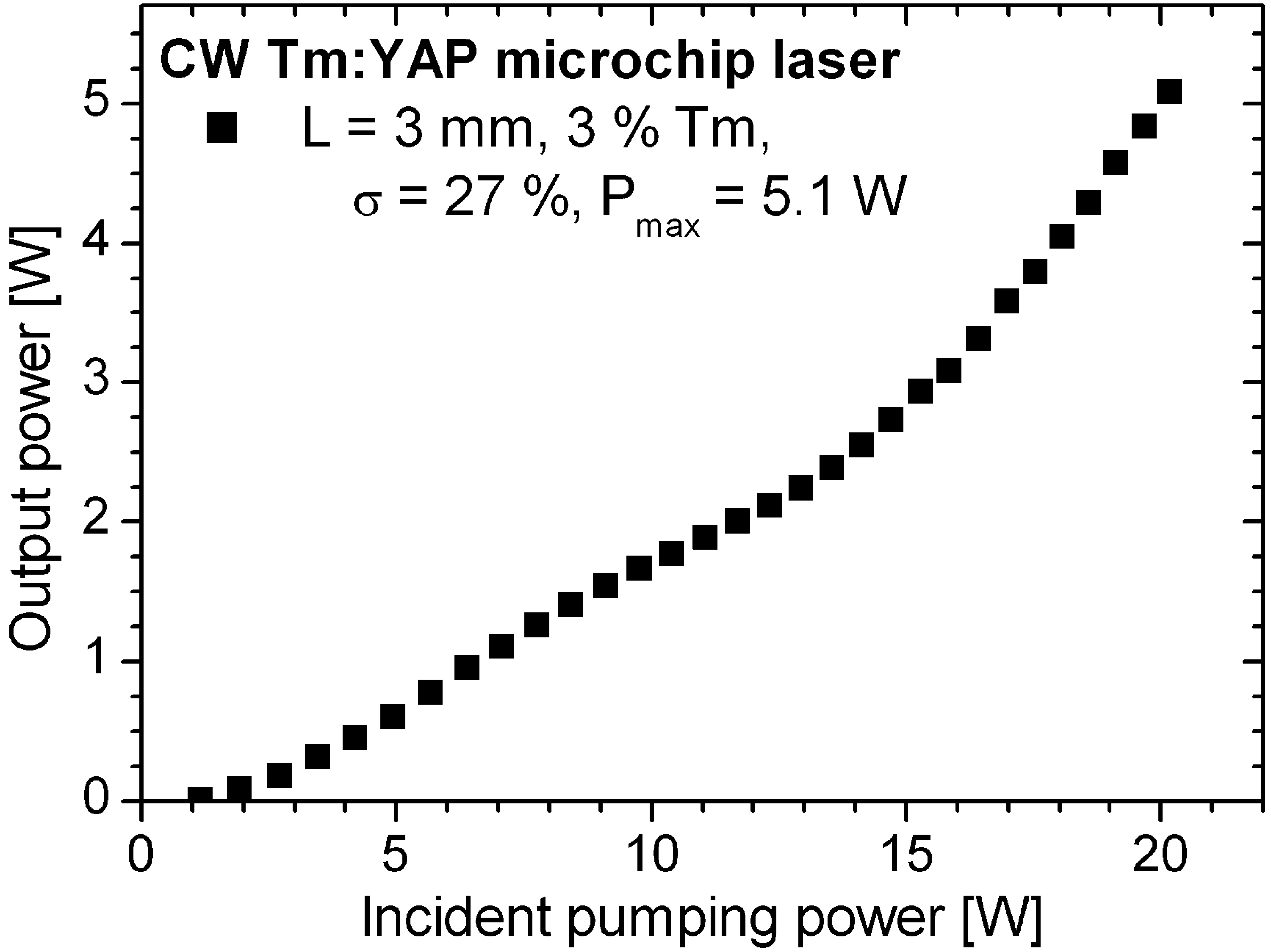
Dependence of Tm:YAP laser output power on diode-pumping power.
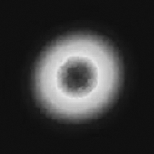
Tm:YAP laser irradiation output beam profile.
For the debonding experiment, output irradiation divergence was modified by the CaF2 lens with the focus length 100 mm placed at a distance of 100 mm from the output microchip laser face. The beam diameter behind the lens was 3 mm and, because of the small beam divergence, it can be supposed that the ceramic bracket had a similar spot diameter, namely 3 mm. The irradiation condition was: power – 1 or 2 W, time – 60 sec; which means the fluence was 849 or 1698 J/cm2 (the irradiance was 14 or 28 W/cm2). The water flow was 2ml/min. The whole experimental system is illustrated in Fig. 3.
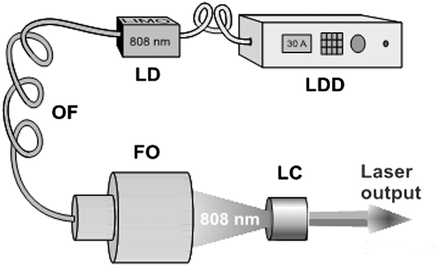
Tm:YAP laser system.
Tissue material
Thirty premolars and third molars of adolescent patients (age 11–18), extracted for orthodontic reasons, were used. Fascination 2 (Dentaurum, Pforzheim, Germany) ceramic brackets were bonded on ten teeth, using ConTec LC adhesive (Dentaurum, Germany) BIS-GMA, urethan dimetacrylates, tetraethyleneglycol dimethacrylate (TEGDMA), hydroxyethy-metacralate, butylated hydroxytoluene (BHT), and amines. The second group of 10 teeth was connected with Charity SL APC (3M Unitek Orthodontic Products) adhesive, precoated ceramic brackets using Transbond plus (3M Unitek Orthodontic Products) (Bis-GMA/ TEGDMA (triethylene glycol dimethacrylate–based SEP adhesive system) self-etching primer.
Six groups (10 samples per group) were analyzed (Table 1): Ceramic brackets Fascination 2 (no irradiation; Tm: YAP irradiation at power 1W and duration 60-sec; Tm: YAP irradiation at power 2W and duration 60-sec); and Charity SL APC (no irradiation; Tm: YAP irradiation at power 1W and of duration 60-sec; Tm: YAP irradiation at power 2W and duration of 60-sec).
The enamel surface was cleaned using non-fluoride polishing paste, rinsed in a stream of water, and dried in a stream of oil-free air. An etching agent (ConTec Etch - Dentaurum, Germany) was then applied to the labial enamel surface for 15 sec and rinsed in a stream of water for 10 sec. After air-drying, the enamel showed a chalky-white hue. ConTec Primer was applied to the prepared etched surfaces, slightly dispersed with air, and polymerized for 20 sec, using an Elipar Free-Light 2 (3M ESPE, Seefeld, Germany) halogen lamp. The ceramic brackets were coated with a sufficient amount of ConTec LC adhesive, and each ceramic bracket was polymerized for 20 sec in a halogen-lamp frontal light beam.
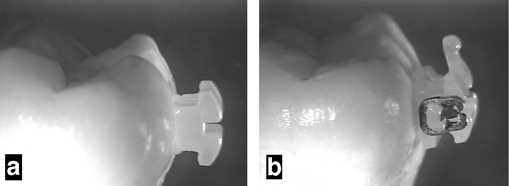
For the Charity SL APC groups, every labial tooth surface was cleaned with a prophy rotary brush and burst of air, following which the self-etching primer was applied for 3–5 sec, after which the ceramic brackets (with pre-coated adhesive) were stuck on the tooth surface and polymerized for 20 sec. Both types of ceramic brackets connected to the teeth can be seen in Fig. 4a and b. To check the temperature rise by thermocouple, a hole was prepared opposite the ceramic bracket, inside the tooth. All teeth with bonded ceramic brackets were placed in a physiological solution and stored at room temperature.
Methods of analysis and measuring instruments
To characterize the laser irradiation, the mean power, spectrum, time development, and spatial beam structure were measured. The following instruments were used for this purpose: Molectron energy/power meter EMP2000 with PowerMax probe PM3 or PM10 (Molectron-Coherent, Portland, OR); Oriel monochromator model 77250 (50 μm wide slit) (Newport Corporation, Irvine, CA); IR-sensitive Pyrocam III pyroelectric camera (Ophir-Spiricon Inc., North Logan, UT); and a TDS3052B Tektronix oscilloscope (500 MHz, 5 GS/s) (Tektronix, Portland, OR) with an InAs/InAsSbP photodiode (model PD36-05, IBSG Co, Ltd., spectral range 0.8–3.8μm, rise time 150 ns) (Lambda Photometrics, Ltd., Hertfordshire, UK).
All teeth with ceramic brackets were photographed (before and after treatment) using a Nikon SMZ-2T (Osaka, Japan) stereomicroscope connected to a Mintron color video camera (MTV-73X11P-R, Mintron Enterprise, Fremont, CA) and a computer; the temperature changes inside the tooth during irirradiation of the ceramic bracket were recorded using a GMH 3210 digital thermometer; the spatial distribution (progress and attenuation) of tooth surface temperature was monitored using an Optilas – Electrophysics PV320L2E thermal imaging infrared camera; the surface of the enamel was analyzed after removal of the ceramic bracket using a JSM 5510 LV Jeol (Jeol, Tokyo, Japan) electron microscope. The teeth were processed in a “low vacuum” (10 Pa) regime without desiccation. Back-scattered electron images were recorded using this technique.
Enamel structures were checked below the ceramic bracket and on the enamel/bracket border. The SEM record and photograph of the tooth were classified into grades according to enamel damage. Grade 0, no damage; Grade 1, minimum damage – enamel removal <55.6 μm3; Grade 2, medium damage – enamel removal up to 55.6 μm; Grade 3, high damage – enamel removal >55.6 μm; Grade 4, maximum damage – enamel carbonization. Temperature increase was measured within the pulp cavity.
Experimental procedure
Tm:YAP laser output irradiation was directed into the particular ceramic bracket, with cooling water being simultaneously directed onto the tooth, thereby helping to maintain the pulp at the accepted temperature. According to our preliminary experiment, 10 two power levels (1–2W) were used within a period of 60 sec. Laser power was measured using the Molectron EPM 2000e power meter with a PM3 probe. Temperature changes inside the tooth were recorded using a NiCr-Ni thermocouple and a GMH 3210 digital thermometer. Spatial distribution of temperature on the tooth surface was simultaneously monitored using a PV320L2E thermal imager (Electrophysics, Sofradir EC, Inc., Fairfield, NJ). Irradiation was discontinued after a period of 60 sec and the ceramic bracket was removed from the tooth surface mechanically, with 3M Unitek band-removing pliers (Unitek, Monrovia, CA). This ceramic bracket debonding instrument was placed in the center of the self-ligating ceramic bracket, perpendicular to the arch-wire slot. The ledges of the instrument were symmetrically positioned against the labial surfaces of the ceramic bracket. The instrument handles were then squeezed (pressed together) until the ceramic bracket collapsed or deformed. To completely separate the ceramic bracket from the enamel, the ceramic bracket was gently rocked in the mesial or distal direction.
Both these parts – the tooth and removed ceramic bracket – were photographed using Nikon a SMZ-2T stereomicroscope and subsequently analyzed by scanning using a JSM 5510 LV Jeol electron microscope (Jeol, Tokyo, Japan) to investigate any possible damage to surface enamel.
Similar procedure (but without laser irradiation) was used for the control group. Ceramic brackets Fascination 2 and Charity SL APC were mechanically removed and both parts were photographed and scanned for assessment of possible damage to enamel.
The adhesive remnant index score was calculated. The overall and affected area (with the remnants) was estimated using image processing software ImageTool, ver. 3.00 UTCSCSA, (University of Texas Health Science Center, San Antonio, TX). Statistical analysis was performed using Statistical Package Excel 2007 (Microsoft). The average value and standard deviation were derived.
Results
Debonding procedure and temperature measurement
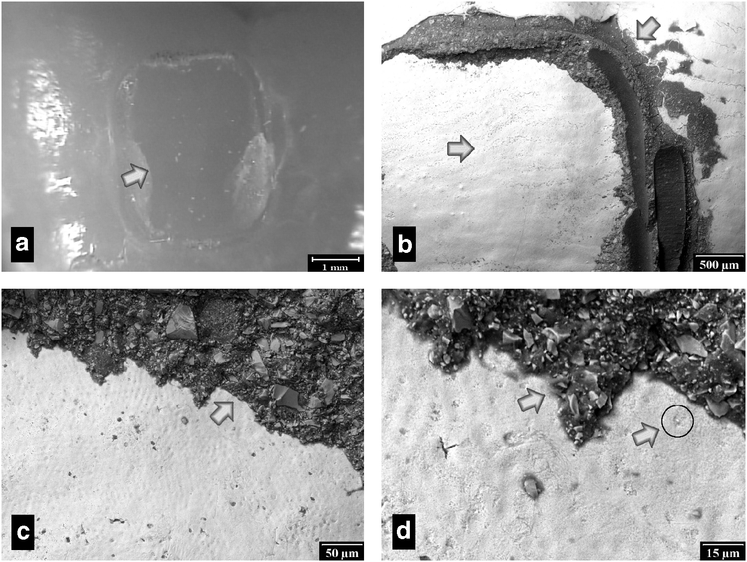
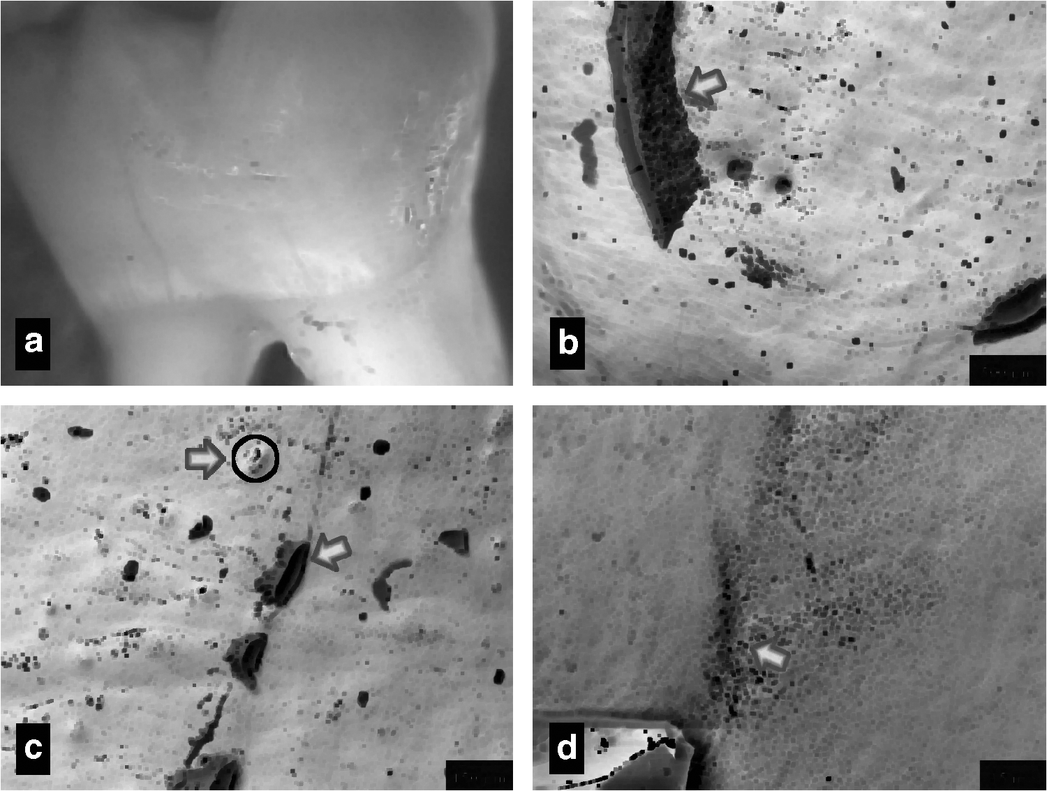
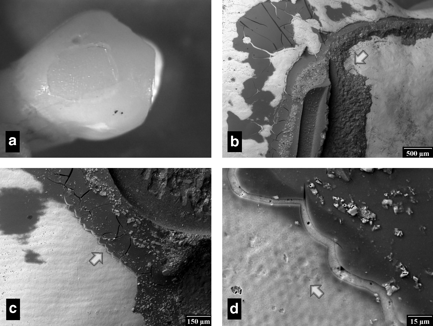
When Tm:YAP irradiation was used during the debonding procedure, heat from the laser was concentrated inside the ceramic bracket and adhesive resin, and after 60 sec the ceramic bracket could be removed. 10 The temperature rise was different due to the metal component in the Fascination 2 ceramic bracket Charity SL APC (Figs. 5 and 6). In both cases, when Tm:YAP irradiation of the ceramic brackets was used, the temperature increase was safe and it was concentrated within the ceramic bracket alone (Figs. 7 and 8). In Fascination 2 and Charity SL APC, temperature rises of 3°C and 3.8°C, respectively, were recorded. Following laser irradiation, the ceramic bracket can be easily removed without cracks. The result from the control group, which was not affected by laser irradiation, is shown in Fig. 9a and the SEM record is shown in Fig. 9b–d. It can be seen that the adhesive remained on the surface of the tooth (Fig. 9b) and that the enamel microstructure was relatively intact (Fig. 9c and d). Enamel loss was visible below the ceramic bracket, leaving behind Tomes' processes, pits, fine scratches, and facets (Fig. 9c) – Grade 2, medium damage – enamel removal up to 55.6 μm.
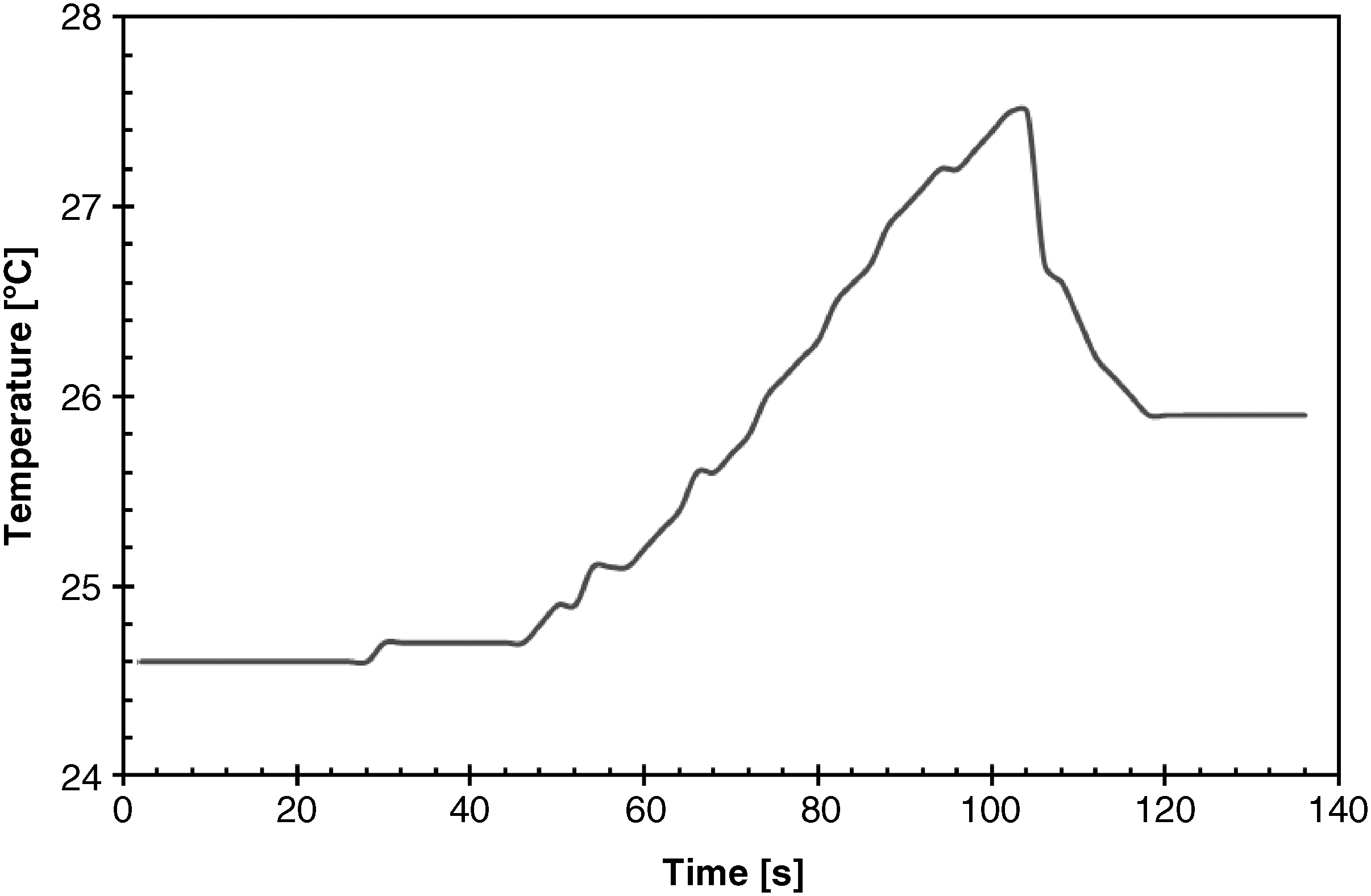
Temperature increase during irradiation of Fascination 2 ceramic bracket using Tm:YAP laser (1 W, 60 sec).
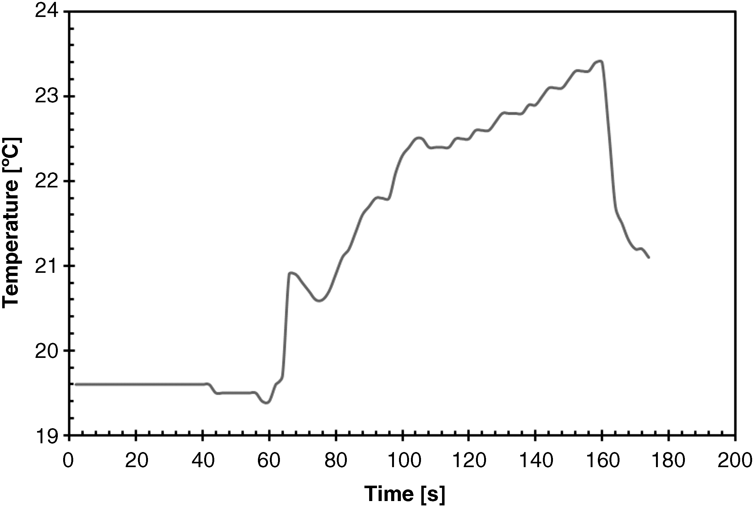
Temperature increase during irirradiation of Charity ceramic brackets using Tm:YAP laser (1 W, 60 sec).
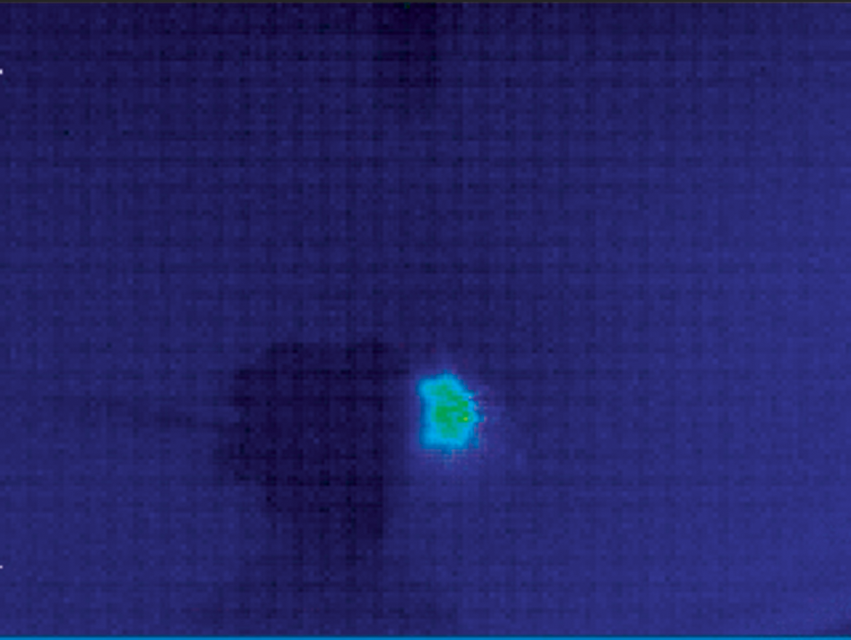
Thermocamera image: temperature increase during irirradiation of Fascination 2 ceramic bracket using Tm:YAP laser (1 W, 60 sec).

Thermocamera image: temperature increase during irirradiation of Charity ceramic bracket using Tm:YAP laser (1 W, 60 sec).
Application of mechanical pliers for debonding only.
In the group that was irradiated with the Tm:YAP laser, the enamel microstructure was identical to the intact, normal enamel surface, which exhibited some parallel perikymata, typical shallow arcades, irregular Tomes' processes, and enamel cap. Enamel damage and scratches were not detected, and almost no adhesive remained (Fig. 10a–d) – Grade 1, minimum damage – enamel removal <55.6 μm. As relatively greater force was required to remove the metallic or ceramic brackets without laser irirradiation, it is not surprising to find that the enamel surface in both group one and group two manifested many microscopic scratches and various degrees of gouges. Rougher surfaces could potentially contribute to plaque accumulation, stain, odor, and demineralization through microbial activity. The rest of the adhesive resin is visible on the enamel surface (Fig. 9). It is evident that after the ceramic bracket had been removed, some changes occurred in the SEM record on the enamel surface (Fig. 9b). Minimum damage to enamel was observed when 1 W power was applied for a duration of 60 sec using Tm:YAP irradiation. The honeycomb enamel structure can be seen in detail in Fig. 9d. Grade 1, minimum damage – enamel removal <55.6 μm – was observed. By increasing irradiation power (up to 2 W), the temperature changes of the enamel are visible (Fig. 11a–d), and even enamel melting can be seen (Fig. 11c and d) – Grade 3, enamel removal >55.6 μm.
Tooth structure following application of Tm:YAP laser irradiation (1 W, 60 sec) for debonding.
Tooth structure following application of Tm:YAP laser irradiation (2W, 60s) for debonding.
Figure 12 shows the differences between the control and laser-treated groups. The irradiated fully ceramic bracket Fascination 2 was removed from the enamel surface with a majority of the adhesive (82% of adhesive remnant remains on bracket; SD 16%). When the classical debonding was used, only 27% of adhesive remained on the ceramic bracket (SD 8%). It was observed that the metal part of ceramic bracket (Charity SL APC) blocked the laser radiation (32% of adhesive remnants remained on bracket; SD 15%). It was confirmed that the metal part of the ceramic bracket affects the amount of the adhesive after debonding (14%; SD 9%). The difference was significant.

Enamel area without adhesive remnant with standard deviation. Significant difference between Tm:YAP laser (1 W, 60 sec) and classical laser debonding.
Discussion and Conclusion
Many orthodontists are familiar with the term “laser”. However, there is a lack of information regarding its application in orthodontic practice 13 , where lasers can be used for gingivoplasty, gingivectomy, removal of gingival overgrowths, and frenectomies. 4,15,16 Various laser techniques have recently also been investigated for application in ceramic bracket debonding. One removal technique, already in use, is the application of heat generated by laser irradiation to ceramic brackets. For this method, the CO2, krypton fluoride (KrF), xenon chloride (XeCl), Nd:YAG or Tm:YAP lasers were used.5-8,10,15,16
The Er:YAG laser can also be used to remove composite remnants following orthodontic debonding. 17,18 It is also known that the Er:YAG laser system offers optimal wavelength and the ability to be absorbed in the most efficient manner for successful dental hard-tissue ablation. 19 The effectiveness and safety of a laser is also directly related to the adequate setting of working patterns, chiefly water flow. 20 To protect the enamel surface, we tried to find a new laser technique that would overheat the local bracket without destroying the surface. Tm:YAP laser irradiation has a different absorption level in the ceramic bracket, resin, and enamel than do other lasers investigated so far. It can even operate continuously; therefore, irradiation steadily adds to the quantity of energy/heat in the ceramic bracket and resin. In this study, we show that only 1 W of power is needed for ceramic bracket removal. It was evident from the photographs and videos taken by thermocamera that enamel was not directly affected by microchip Tm:YAP irradiation (1 W, 60 sec). The measurements were confirmed through electron microscope scans. Tm:YAP laser irradiation aids in softening the adhesive layer prior to removal of the ceramic bracket from the tooth surface with band-removing pliers.
It is known that during the thermal debonding procedure, a temperature gradient exists between the tooth surface and the pulp. The temperature gradient rise from the tooth surface through the enamel and dentin does not follow a linear relationship during transient heating. 16,21,22 Six-second lasing by the scanning method using an Er:YAG laser was found to be the most effective and safest way of removing ceramic brackets without causing damage to enamel and pulpal tissues. The temperature was increasing proportionally to extended duration of the lasing process. 23 In our previous experiment it was shown that for successful ceramic bracket debonding, a continuously running Tm:YAP laser can also be used. 10 Our detailed investigation using a thermal imager found that 1998 nm irradiation is concentrated mainly in the ceramic bracket and bonding agent material and it does not influence the internal part of the tooth tissue and does not remove enamel. 9 Zach's measurements 7 confirmed that temperature increase during our laser irradiation debonding was saved. In our experiment it was shown that a radiant power of 1 W (14 W/cm2), administered over a 60-sec period, together with moderate cooling, resulted in a temperature rise from 3 to ∼ 4°C in the approximate root location (freshly extracted, non-vital teeth), which is acceptable to keep the vital tooth alive. This irradiation is also suitable from the point of view of damage to below-the-ceramic bracket enamel during debracketing. Only slight damage to the enamel was registered by SEM compared to conventional bracket removal.
Use of a Tm:YAP laser (wavelength 1998 nm, power 1 W, irradiance 14 W/cm2, time 60 sec) which is at the same time compact and small enough to be used in the dental practice, together with moderate cooling, could be an efficient tool for debracketing.
Footnotes
Acknowledgment
This research has been supported by a grant of the Czech Ministry of Education, No.MSM6840770022 “Laser systems, irradiation, and modern optical applications”; and by an IGA MZCR 9991-4 grant.
Author Disclosure Statement
No conflicting financial interests exist