
Abstract
Introduction
The “contact mask” process is an industrial technique that utilizes a metallic mask situated directly over a substrate during treatment by a laser. In this way, the laser creates an imprint of the mask pattern onto the substrate. In the present study, we present a novel technique for treatment of dentin, using a hard-tissue laser and a metallic meshwork placed directly over the dentin surface during laser treatment.
Materials and Methods
Four non-carious human third molars were embedded and ground using a universal grinding machine (Ecomet, Buehler, IL) to 600-grit.
Group 1
Two specimens were treated with Er,Cr:YSGG laser treatment (without masking). The Er,Cr:YSGG laser (Millenium, Biolase, San Clemente, CA) was set to 3.5W power, with 100% water and 80% air settings. Laser treatment was done by hand at a distance of 1 mm above the sample. The angle of laser tip to tooth surface was set at ∼15 ∘ to prevent damage of the laser tip by reflecting laser energies. The samples were treated for 60 sec inside a 5 × 5 mm square surface delimited by masking tape (Fig. 1A).
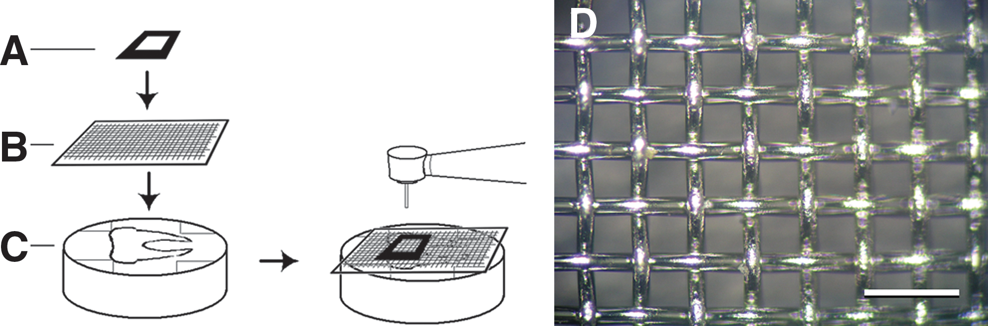
Schematic diagram of experimental procedures.
Group 2
Two specimens were treated using Er,Cr:YSGG laser treatment with a metallic meshwork situated over the dentin specimen (Fig. 1B). The metallic meshwork used was a plain weave 100 μm aperture stainless steel mesh cloth with 50μm thickness stainless steel wires (Ying Ho Wire Netting Co., Taipei, Taiwan) (Fig. 1D). This meshwork was adapted to the specimen using double-sided tape situated at the periphery of the tooth-resin specimen (Fig. 1C). Laser treatment was the same as previously described.
Light-microscopic observations were conducted under an Olympus stereomicroscope. Before SEM observation (JSM-6700F, JEOL, Tokyo, Japan), all surfaces were desiccated and coated with platinum-silver (Pt-Ag) film.
Results
Under SEM, Group 1 exhibited a step-like appearance, with mostly open dentin tubules. Some debris and cracking of the dentin surface could be distinguished (Fig. 2A). Group 2 exhibited uniformly distributed craters ∼100μm wide. Depths of the craters were ∼150μm (Fig. 2B). Marked cracking could be observed inside some of the craters, and large debris could also be found. Surfaces inside the craters were found to exhibit open dentin tubules with a step-like dentin surface (Fig. 2C). The surface adjacent to the craters exhibited a surface covered with smear, with occluded dentin tubules.
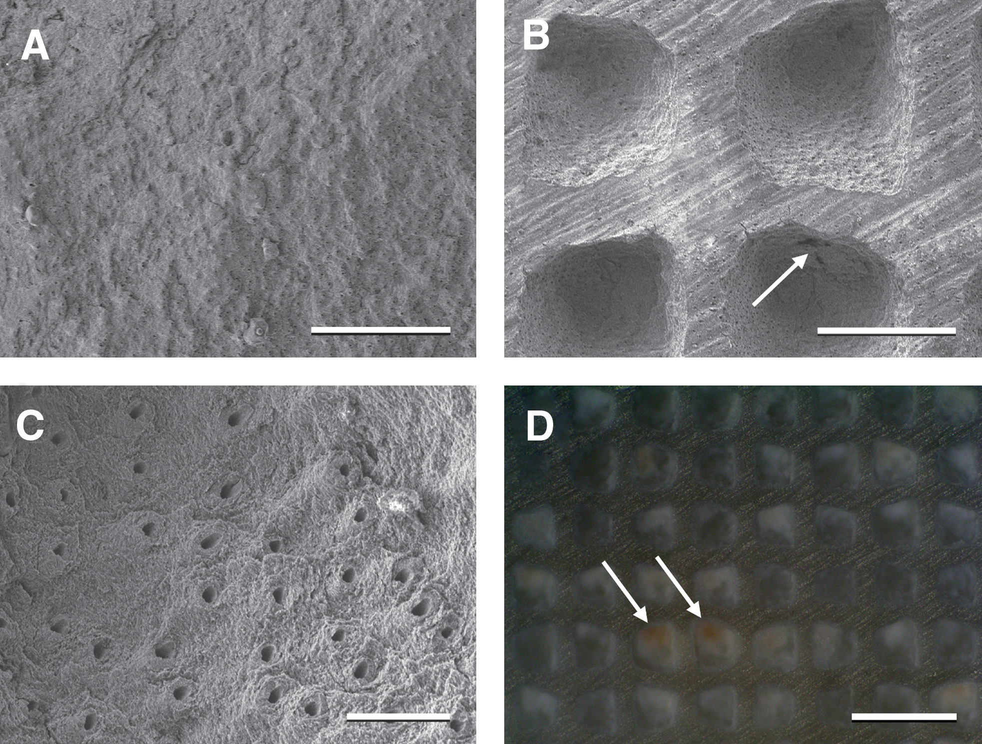
Representative SEM and light micrographs.
Under light microscopy, Group 2 also exhibited uniformly aligned craters. A brown/yellow hue could be seen in some of the craters, which suggests some degree of charring (Fig. 2D). We were unable to focus the light microscope to such a degree as to permit visualization in the Group 1 sample. However, we could observe no evidence of charring in the Group 1 sample through visual examination.
Discussion
The size of the craters seen in Group 2 corresponded to the aperture size of the stainless steel mesh. Within these craters, the microstructure exhibited a step-like appearance with open dentin tubules, devoid of smear. This appearance is indistinguishable from that of dentin surfaces treated by laser in this and previous studies. Cracks and debris could be found inside some of the craters. This may be because of the inability of the water spray to reach the deeper areas. Although we have deliberately set the water spray settings to the highest level available on this laser system, it did not prevent charring inside some of the craters. The surfaces surrounding the craters appeared to be covered with smear, suggesting that these areas were unaffected by the laser. As was observed in the original study by Hibst and Keller who utilized the Er:YAG laser, which has similar properties to the Er,Cr:YSGG laser, we observed a cloudy appearance under light microscopy for Group 1. 10 We were unable to focus the microscope to such an extent as to permit adequate visualization, so this observation was discarded. We have included the light microscopic image of Group 2, as it demonstrated evidence of charring.
In a previous study, we have not found Er,Cr:YSGG laser-treated dentin surfaces to exhibit greater shear bond strengths to composite resin compared to conventional phosphoric acid etching. 7 Other investigators have found conflicting results. 8,9 Ceballos et al. suggested that the rugged surface created after laser treatment was beneficial to bonding, but that damage to the collagen microstructure prevented adequate hybridization of the bonding agent. 11 In our present study, we found unaffected dentin covered with smear adjacent to craters in Group 2. It is likely that the smear-covered, unaffected dentin, when treated with an etch-and-rinse or self-etch bonding system, will produce superior hybridization compared to the laser-affected dentin inside the craters. Furthermore, the uniform cratering observed in Group 2 demonstrated an abundance of mechanically retentive areas, which may enhance mechanical retention.
Researchers have utilized power parameters ranging from 0.25–5W for dentin treatment. 12,13 The power setting of 3.5W was chosen in the present study because we have previously observed that a low power setting (i.e., 1 or 1.5W) was not particularly efficient and may not allow for penetration through the meshwork layer. A high power setting (i.e., 5 or 6W) would be prone to overheating the dentin surface, especially as the meshwork would block much of the water spray from contacting the dentin surface. We have also used higher water/air settings than previous studies, to prevent overheating. In light of our findings that some craters in Group 2 exhibited slight charring, additional cooling, such as concomitant water rinse using a three-way syringe, and/or using lower power settings, may be advised.
We have chosen the stainless steel mesh with the described dimensions for the following reasons: (1) It is more flexible than thicker meshes; (2) It is semi-malleable, and may be adapted to an irregular surface by burnishing; (3) It is a mass-produced product, and is economical; (4) It is durable, and does not easily break under laser irradiation; and (5) It enables craters to be generated in the 100 μm range, which is potentially an adjunct to retention by hybridization and resin tag formation (in the 1 μm range). However, using the stainless steel mesh as a mask does have a few drawbacks: (1) The operator must ensure that the meshwork mask does not shift during laser irradiation; (2) Craters in the 100 μm range may not be the ideal size for bonding; and (3) After adapting the meshwork to an irregular tooth surface through burnishing, there is a gap between the tooth surface and the mask, which may impede adequate formation of craters.
This study is a preliminary look at the feasibility of utilizing a masking technique for laser dentistry. The surface presented appears to offer the following advantages: a larger surface area, mechanical undercuts, and unaffected dentin between the craters. There are numerous questions that will need to be addressed before this technique may be utilized clinically, such as: What is the optimal material for masking? What is the optimal surface design of the dentin/enamel? What is the optimal laser? What is the optimal setting of the laser? How should the operator adapt the mask to the tooth?
Presently, it is beyond our ability to address all of these issues, but our findings suggest that the use of a mask during hard-tissue laser treatment of teeth may improve mechanical retention where it is traditionally lacking, such as for porcelain veneers bonded to dentin, or anterior composite resin restorations. Further studies should be conducted to test the bond strength of materials bonded to this surface, other mask designs, and laser settings.
Footnotes
Author Disclosure Statement
This study has been funded by the authors and Chung-Shan Medical University School of Dentistry. No conflicting financial interests exist.