
Abstract
Introduction
Some studies have shown that whereas TiF4 is more effective at preventing dental erosive demineralization than is sodium, stannous, or amine, 1,4 –6 it cannot completely protect against enamel erosion. 6 –10 However, the preventive effect of TiF4 against demineralization may be dependent upon the agent used, as it was recently shown in vitro that a TiF4 varnish exhibited a higher protective potential than a TiF4 solution on enamel erosion. 8
In order to improve the effect of different fluoride compounds on erosive and carious demineralization, laser irradiation has been discussed as an appropriate tool. 11 –15 Previous experiments showed favorable effects of neodymium-doped yttrium aluminum garnet (Nd:YAG) laser irradiation on the acid resistance of dental hard tissues. 16,17 The Nd:YAG laser can diminish the permeability of enamel because it induces enamel melting and resolidification. Melted enamel surface shows a crystal growth that can reduce the enamel interprismatic spaces and consequently, the diffusion of acids into the enamel during the acid challenges reducing the demineralization. 16,17 Moreover, laser irradiation favors the formation of pyrophosphate that can reduce the hydroxyapatite dissolution, induces water loss, and replaces the more soluble carbonate apatite with hydroxyapatite or fluorapatite (in the presence of fluoride). 18
Although the effect of Nd:YAG laser irradiation on sodium and amine fluoride has been analyzed 13,14,18,19 , there is no available information on the interaction between Nd:YAG laser irradiation and TiF4. It is assumed that Nd:YAG laser irradiation can increase the efficacy of TiF4 in preventing enamel erosion through the melting and recrystallinization of the glaze-like surface layer as is observed after the application of TiF4 or by creating an altered surface on which more fluoride and titanium could be applied upon the application of TiF4, as was expected in the case of CO2 laser. 15
Therefore, the aim of this in vitro study was to analyze the influence of Nd:YAG laser irradiation on the efficacy of TiF4 and NaF varnishes and solutions in protecting enamel against erosion. The null hypotheses formulated were: (a) there is no significant difference between F salts (TiF4 or NaF), whether the agent is varnish or solution, in prevention of dental erosion; (b) for each type of fluoride (NaF/TiF4), there is no significant difference between varnish and solution in the prevention of enamel erosion; (c) the laser does not increase the effect of F, regardless of the salt, agent, or protocol of application, in the prevention of enamel erosion.
Materials and Methods
Specimen preparation
Enamel specimens (4 × 4 × 3 mm, N = 202) were prepared from the labial surfaces of bovine incisors crowns. The specimens were cut using a ISOMET low speed saw cutting machine (Buehler Ltd., Lake Bluff, IL) with two diamond disks (Extec Corp., Enfield, CT), which were separated by a 4-mm thickness spacer. The specimens' surfaces were ground flat with water-cooled silicon carbide discs (320, 600, and 1200 grade papers; Buehler, Lake Bluff, IL), and polished with felt paper wet by diamond spray (1 μm; Buehler). The specimens were cleaned using an ultrasonic device for 2 min and checked regarding the presence of white spots and cracks using a microscope (x40). In addition, they were selected using microhardness values between 320 and 390 KHN (Knoop diamond, five indentations, 25 g/10 s, Shimadzu Corporation, Tokyo, Japan) and randomly allocated to groups. Prior to the experiment, two layers of nail varnish had been applied to half of the surface area of each specimen to maintain a reference surface for enamel loss determination after the experiment. Before the experiment, all the specimens were stored in 100% humidity.
Twelve enamel specimens were randomly allocated to each of 16 test groups: NaF varnish (Duraphat-Colgate, São Paulo, Brazil, 2.26% F, pH 4.5), experimental TiF4 varnish (FGM-Dentscare, Joinvile-SC, Brazil, 2.45% F, pH 1.2), NaF solution (2.26% F, pH 4.5), TiF4 solution (2.45% F, pH 1.2), placebo varnish (FGM-Dentscare, pH 5.0), Nd:YAG (84.9 J/cm2), Nd:YAG prior to or through NaF varnish, Nd:YAG prior to or through TiF4 varnish, Nd:YAG prior to or through NaF solution, Nd:YAG prior to or through TiF4 solution, and Nd:YAG prior to or through placebo varnish. Ten specimens remained untreated (control).
Nd:YAG laser treatment and fluoride application
Laser irradiation was performed with a commercially available Nd:YAG laser (1064 nm, PowerLase™ ST6, Lares Research, Chico, CA). Prior to the laser irradiation, the enamel specimens had been coated with a photoabsorber dye solution of coal paste, which was composed of coal triturated in a porcelain mortar for 10 min, resulting in particles of 10 μm in diameter, then diluted in equal parts of deionized water and 99% ethanol. The paste was applied using a microbrush (Carbo Activatus, Bionatus, São Paulo, Brazil) in a layer thickness of ∼100 μm 20 . The absorption of the laser by the enamel is highly dependent upon the presence of the coal paste. 20
The irradiation was performed perpendicularly to the sample with an optical fiber (320 μm in diameter) in contact mode, at 300 μs pulse duration, 60 mJ/10 Hz, resulting in an energy density of 84.9 J/cm2. The exposed surface of the specimens (free from nail varnish) was irradiated three times for 10 s each, with intervals of 10 s between the irradiations, by one calibrated dentist moving the laser probe tip continuously in contact with the enamel surface. The coal paste was reapplied before each laser scanning. After laser irradiation, specimens were rinsed with distilled water for 15 s. 21
To prepare the F solution, solid salts (Aldrich Chemical Company, Milwaukee, Wisconsin) were dissolved in deionized water. The pH of all solutions and varnishes was measured by electrode and indicator paper (±0.5 units), respectively. The 5% NaF (Sigma–Aldrich) solution was adjusted to pH 4.5 by adding 12.6 g 5 M H3PO4/100 ml, in order to present a composition similar to the NaF varnish. The pH of the 4% TiF4 (Sigma-Aldrich, St. Louis, MO) solution was native (pH 1.2). The fluoride solutions were applied using a micropipette (50 μL/sample), and were left on the surface for 1 min. 5,10,22 Excessive solution was removed from the surface by a cotton roll.
The varnishes were applied in a thin layer using a microbrush. After 6 h, the varnishes were carefully removed using acetone and a scalpel blade, taking care to avoid touching of the enamel surface. Complete removal of the layer was checked microscopically (40 ×). 8,22 The composition of the products is displayed in Table 1.
For groups for which the irradiation was performed before F application, the fluoride agent was applied immediately after the last laser irradiation. Regarding the laser application through fluoride, the fluoride agent was first applied for 50 sec, the coal paste was placed on the surface, and then the first laser irradiation applied for 10 s, completing 60 s. After that procedure, the specimens were washed in deionized water and the laser irradiation was repeated twice as described previously.
Erosive cycling
Ten specimens per group were submitted to a 5-day de- and remineralization cycling. Erosion was performed with fresh Sprite Zero (pH 2.6, 30 ml/sample, unstirred, 25°C, Coca-Cola Company, Porto Real-RJ, Brazil) four times daily for 90 s. After demineralization, the specimens were rinsed with tap water and transferred into artificial saliva (30 ml/sample, unstirred, 25°C) for 3 h. After the last daily erosive treatment, the specimens were stored in artificial saliva overnight. 15,22 The artificial saliva was renewed daily and consisted of 0.2 mM glucose, 9.9 mM NaCl, 1.5 mM CaCl2.2H2O, 3 mM NH4Cl, 17 mM KCl, 2 mM NaSCN, 2.4 mM K2HPO4, 3.3 mM urea, 2.4 mM NaH2PO4, and ascorbic acid (pH 6.8). 23 After the experiment, all the specimens were stored in 100% humidity.
Profilometric measurement
Enamel loss (μm) was quantitatively determined by contact profilometer (Hommel Tester T1000, VS, Schwenningen, Germany) at the end of the experiment. For profilometric measurement, the nail varnish was carefully removed using a scalpel and acetone solution (1:1 water) and the specimens were slightly dried. 8 The diamond stylus was moved from the reference to the exposed area (2 mm length). Three profile measurements were randomly performed in the center of each specimen and the differences in height between control and exposed area were averaged (μm) using the equipment's software. 8,15,22
Scanning electron microscope (SEM)
To show the interaction between the fluoride agents and laser with enamel, specimens (n = 2, 4 × 4 mm) were freshly treated (not exposed to erosive cycling) according to the groups described previously. Next, the specimens were carefully dried with paper, coated with gold/palladium using sputter coating, dried by vacuum, and examined with a scanning electron microscope (XL 30 FEG SEM; Philips, Eindhoven, The Netherlands, with field emission gun at 15KV).
Statistical analysis
Statistical analysis was performed with GraphPad InStat version 2.0 for Windows (GraphPad Software, La, Jolla, CA). The assumptions of equality of variances and normal distribution of data were checked for all the variables tested, using the Bartlett and Kolmogorov-Smirnov tests, respectively. Then, the data were analyzed by one-way ANOVA followed by Tukey's post-hoc tests, after the means were log-transformed. The log transformation was done because the values of standard deviation of the groups significantly differed. The level of significance was p ≤ 0.05.
Results
Enamel loss (mean ± standard deviation [μm]) after 5 days of de-remineralization cycles is presented in Table 2. One-way ANOVA revealed significant differences among the groups. Only TiF4 varnish, laser prior to TiF4 varnish, and laser prior to TiF4 solution significantly reduced enamel erosion compared to control. However, in specimens treated with the placebo varnish, enamel erosion was significantly increased compared to control.
Values with different superscript letters indicate statistical significance between the groups (p < 0.05).
Data were submitted to log transformation before analysis.
The SEM image of the enamel specimens treated with NaF varnish (Fig. 1a) showed a polished surface, as was observed for the placebo varnish (not shown). Enamel treated with TiF4 varnish presented a coating layer (Fig. 1b). NaF and TiF4 solutions produced a slight demineralization, as is shown in Fig. 1c and d, respectively. Figure 1c shows the delimitation of the prismatic areas. Additionally, Fig. 1d shows that the TiF4 solution induced demineralization of the enamel surface with the presence of microcracks.
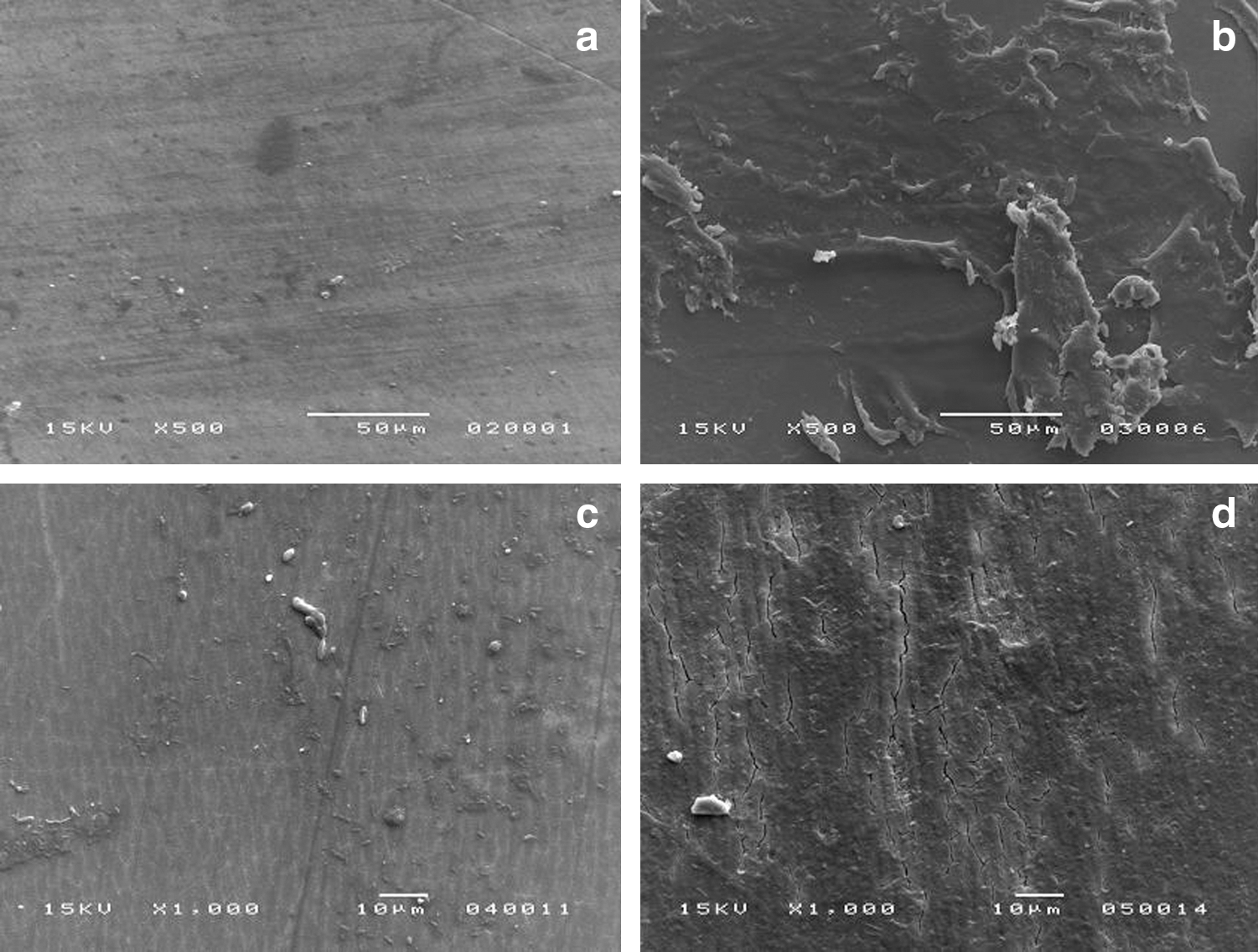
SEM image of NaF varnish treated enamel (
According to the SEM images of the enamel specimens treated with laser irradiation, the laser application alone was not homogeneous and induced surface microcracks and the appearance of pores (Fig. 2a and b). The application of Nd:YAG prior to NaF varnish/solution produced a superficial alteration (Fig. 3a and b) similar to that of enamel treated with laser alone. On the other hand, specimens treated with Nd:YAG prior to TiF4 varnish and solution presented a different surface than the other groups, with fewer microcracks and without pores, compared to specimens treated with laser alone (Fig. 3c and d).
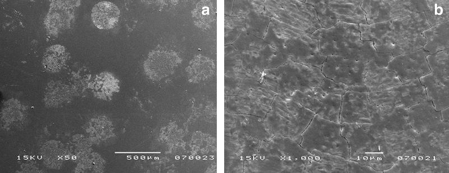
SEM images of laser treated enamel, showing the presence of microcracks and pores. Original magnifications: (

SEM image of enamel specimen treated with laser prior to NaF varnish, showing microcracks and pores as compared to laser alone (
The application of laser through NaF varnish did not induce surface alteration (Fig. 4a), whereas in specimens treated with laser through TiF4 varnish, it was possible to see several surface cracks (Fig. 4b). The application of laser through NaF or TiF4 solution caused damage and demineralization of enamel surface, as is shown in Fig. 4c and d, respectively.
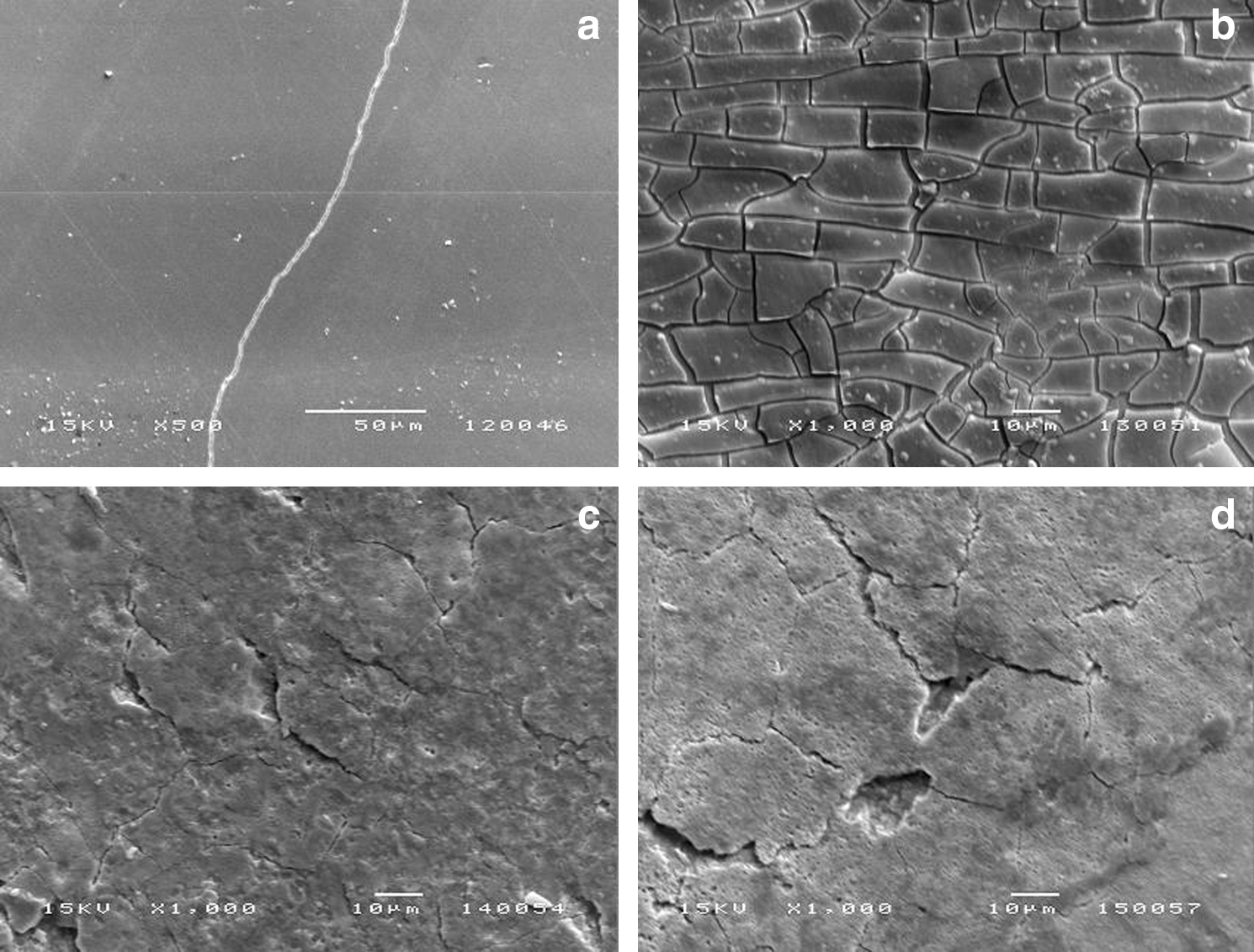
SEM image of enamel specimen treated laser through NaF varnish, showing non surface alteration (
Discussion
NaF varnish/solution and TiF4 solution did not prevent enamel erosion under the conditions of the present study, whereas TiF4 varnish reduced enamel loss independent of laser irradiation. Nd:YAG laser irradiation alone was not able to decrease erosive enamel loss, but enhanced the capability of TiF4 solution to reduce enamel erosion. Therefore, the first hypothesis of this study was rejected, and the second and third hypotheses were partially rejected.
The F products and the laser were applied only once in order to simulate the standard clinical procedure with a single professional application. 8,15,22 In contrast to Vieira et al., 24 –26 the varnishes were completely removed after 6 h in the present study, before the de-remineralization cycling. This was done to focus on the chemical effect of the varnishes rather than on the mechanical protection. Therefore, the varnishes were removed to simulate the clinical situation in which the varnishes might be removed post-application through regular toothbrushing and mastication.
However, it must be noted that the fluoride agents used in the present study show differences in the fluoride concentration and the pH. As a positive control, a commercial NaF-Duraphat varnish (Colgate, São Paulo-SP, Brazil, 2.26% F, pH 4.5), which is frequently used in clinics, was chosen. The fluoride concentration of the TiF4 varnishes (2.45% F, pH 1.2) was selected according to a previous study that showed favorable results for this TiF4 varnish in preventing enamel erosion. 8 The TiF4 agents show a naturally lower pH than does the NaF product. It was previously shown that the erosion-inhibiting effect of TiF4 is pH-dependent and that TiF4 is less effective at higher pH (when it is buffered). 27
Irradiation was performed with a Nd:YAG laser because a previous in vitro study showed promising results. 14 In the study by Rios et al., 14 the efficacy of a NaF varnish and APF gel in preventing enamel erosion could be increased by Nd:YAG laser irradiation (52.5 J/cm2), after 5 and 10 days of erosive challenges (20 and 40 min of erosion, respectively). However, in the present study, the energy density was used according to Zezell et al. 21 (84.9 J/cm2) with a photoabsorber dye solution, in order to allow a better absorption of the laser by enamel, and consequently more chemical (hydroxyapatite to fluorapatite) than morphological (melting and resolidification) surface modifications, with no pulp damage.
According to the SEM images, the laser caused some morphological changes to the enamel surface. When the laser was applied prior to the application of TiF4 solution, it probably enhanced the formation of an acid-resistant layer richer in titanium and fluoride, when compared to use of the TiF4 solution alone. 3,10,27 We are predicting that the application of laser prior to TiF4 solution could have created an altered surface, by removing carbonate apatite (chemical changes) or enhancing the temperature (morphological changes- melting and resolidification), on which more fluoride and titanium could be incorporated upon the application of TiF4 solution. This reaction could contribute to the formation of an acid-resistant layer on enamel. The mechanism of the layer formation following application of TiF4 is still not clear. It is likely that within this process a new compound might be formed (hydrated titanium phosphate). 28 This might help to explain the good results for this group. However, a better effect of the laser applied through TiF4 solution was not found, as we had imagined at the beginning of this study. This finding might be related to the increase of temperature during the contact of the solution with enamel, reducing the reaction between titanium and fluoride with apatite. However, no chemical alterations could be detected by the SEM equipment used in this study. Further studies should apply other methodologies, such as EDS and infrared, to answer this question.
In accordance to a previous studies, 8,22 the TiF4 solution alone was not effective in protecting against enamel loss, whereas TiF4 varnish showed the highest efficacy to prevent erosion. This result was justified in a previous study, which showed that TiF4 solution led to a demineralization of the surface, whereas TiF4 varnish led to the formation of a mechanical and chemical barrier. 8 This finding is in agreement with the SEM images of the present study. An interesting finding of this study was that the application of TiF4 solution with a pipette induced less enamel demineralization than the microbrush application used in a previous study. 8 This effect might be explained by the mechanical impact of the microbrush.
Laser application prior to TiF4 varnish or solution produced a different surface compared to laser alone or to TiF4 solution, which showed more similarities with the samples treated with TiF4 varnish alone. The SEM images are comparable to the profilometric results after 5 days of pH cycling. However, when the laser was applied through TiF4 varnish, the positive effect of this F product disappeared. It can be hypothesized that alterations in the TiF4 varnish induced during laser application resulted in several enamel microcracks, thereby facilitating the diffusion of the acid and decreasing the demineralization protection. It might also be assumed that the layer rich in titanium and fluoride could not be deposited by the application of TiF4 varnish under laser irradiation because of the high temperature and consequent laser-induced alterations in the varnish.
Within the limitations of the present study, it should be acknowledged that even though bovine enamel is widely used in erosion research, it presents different susceptibility to the acid challenges 29 and different behavior in contact with TiF4 than does human enamel. 7 This information should be taken into consideration when the data are extrapolated to the clinical situation. The same limitation should be considered in the case of laser application.
NaF varnish and solution were not able to reduce erosive enamel loss. It is known that NaF—especially at high concentration, low pH, and increasing duration of contact, induces the formation of CaF2 on enamel surface. 30 However, the CaF2 globules or layers could not be seen by SEM in the present study. Additionally, the NaF solution application induced a slight enamel demineralization, because of the low pH and high diffusion. If any F precipitates occurred on the enamel surface, it can be assumed that these precipitates were completely dissolved by the erosive challenges (30 min in total), because this group did not show any protective effect against erosion. In this case, the Nd:YAG laser irradiation did not have any influence on the effect of NaF, although it was expected to either increase the formation of a CaF2 layer on the enamel surface when applied prior to NaF, or cause melting/resolidification of the CaF2 rich layer when applied via NaF. It is worth noting that the application of NaF or placebo varnish before laser irradiation probably acted as a mechanical barrier for the irradiation, reducing the energy delivered to enamel, and the temperature as well, which in turn might have prevented any surface alteration. On the other hand, the application of NaF solution/varnish after irradiation did not reduce the undesired alterations produced by the laser in the present study.
Whereas several studies found a protective effect of Nd:YAG laser irradiation on demineralization development and progression, especially related to dental caries, 17 –21 Nd:YAG laser irradiation alone was not effective in reducing erosive enamel loss in 5 days of experiments, according to Rios et al. 14 In this sense, important aspects to take into account are the energy density, average power, repetition rate, time and mode of irradiation (contact or non contact), pulse width, and use of photoabsorber. These laser parameters define the degree of absorption or penetration as well as the temperature of the irradiation, which in turn has a decisive influence on the chemical (low temperatures) or morphological (high temperatures) tooth surface changes and side-effects to the pulp. In the present study, SEM images showed the laser-induced inhomogeneous surface microcracks and pores, which might be considered an undesirable effect.
Heating enamel produces chemical changes such as water and carbonate losses. When the surface temperature increases up to 650°C the major carbonate component in the phosphate position decreases and the acid phosphate ions condense to form pyrophosphate ions. 19 It is important to point out that carbonated apatite has a higher solubility than does hydroxyapatite and, that pyrophosphate concentrates can reduce the hydroxyapatite dissolution rate to zero. At 650–1,100°C, the main changes are thermal recrystallization and crystal size growth, and pyrophosphate reacts with apatite to form PO4 along with the formation of beta-tricalcium-phosphate (β-TCP). 31 The main change at temperatures >1,100°C is that the β-TCP is converted to α-TCP. 32 Fowler and Kuroda 33 hypothesized that heating to temperatures >1,200°C may increase the susceptibility of dental enamel to acid dissolution because the β-TCP and α-TCP phases are more soluble than hydroxyapatite and dental enamel. Besides, the high temperature could cause not only chemical, but also morphologic changes such as cracks and pores. In order to create a better understanding of the laser effects on enamel, further studies should test the impact of different Nd:YAG laser protocols on the prevention of enamel and dentin erosion by slight, mild, and high erosive and abrasive challenges in vitro and in situ.
Conclusions
Under the conditions of the present study, it can be concluded that Nd:YAG laser irradiation did not influence the efficacy of F, except in the case of TiF4 solution, whereas TiF4 varnish protected against enamel erosion, regardless of whether or not laser irradiation was performed.
Summary
The effect of Nd:YAG laser irradiation on sodium and amine fluoride was analyzed; however, no information about the interaction between Nd:YAG laser irradiation and treatment with TiF4 is available so far. It is assumed that Nd:YAG laser irradiation might increase the efficacy of TiF4 to prevent enamel erosion, for example by melting and recrystallinization of the glaze-like surface layer observed after application of TiF4 or by creating an altered surface on which more fluoride and titanium could be incorporated upon the application of TiF4. In the present in vitro study, bovine enamel samples were pre-treated with fluoride and laser, alone or combined. The samples were then submitted to erosive pH-cycling for 5 days. Only TiF4 varnish, laser prior to TiF4 varnish, and laser prior to TiF4 solution were able to significantly reduce enamel erosion compared to control. SEM images showed that specimens treated with TiF4 varnish presented a surface coating. Based upon the results, it can be concluded that Nd:YAG laser irradiation did not have an influence on the efficacy of F, except in the case of TiF4 solution, whereas TiF4 varnish protected against enamel erosion, regardless of whether or not laser irradiation was performed.
Footnotes
Acknowledgments
The authors thank LELO-USP (Special Laboratory of Lasers in Dentistry—University of São Paulo), São Paulo, Brazil (Nd:YAG laser, FAPESP 2007/55497-0), Edimauro de Andrade (SEM analysis), and PIBIC-CNPq (The National Council for Scientific and Technological Development) for granting a scholarship to A.C.R.
Author Disclosure Statement
No competing financial interests exist.