
Abstract
Introduction
The effect of laser light on cell proliferation is controversial, as its use caused either stimulation or inhibition of the proliferation of cell cultures. 3 Moreover, the action of this type of energy may be influenced by the irradiation protocol. 3 –8
There is evidence that low-level laser therapy (LLLT) similarly affects neoplasic and non-neoplasic cells and a proliferative effect will depend upon the parameters of the irradiation. Both red (λ685 nm) and infrared (IR) (λ830 nm) laser light are capable of stimulating cell proliferation under different parameters. It is known that the nutritional status of neoplasic cells in culture affects the outcome of the treatment. Cells with nutritional deficit seem to be more prone to be affected positively by the laser light. 8 The amount of energy delivery to the cells is very important as different responses including stimulation, inhibition, or no effect may occur. 9
Little is known about the interaction between laser light and malignant tissues in vivo. We therefore evaluated, histologically, the effect of LLLT (λ660 nm) on DMBA chemically induced cancer of the oral mucosa of golden Syrian hamsters.
Methods
The Animal Experimentation Ethical Committee of the School of Dentistry of the Federal University of Bahia approved the present study. Fifteen male 6- to 8-week-old golden Syrian hamsters (Mesocrietus auratus) were obtained from the Animal House of the School of Veterinary Medicine of the Federal University of Bahia. The animals were randomly selected, weighed, and divided into three groups of five animals. Group 1: killed after cancer induction of 8 weeks; Group 2: 8 weeks of cancer induction plus 4 weeks with no treatment, and death at the 12th week; and Group 3: 8 weeks of cancer induction plus 4 weeks of LLLT and death at the 12th week. All animals were induced to develop tumors by using 0.5% DMBA (Sigma-Aldrich Laboratory, St. Louis, MO) in mineral oil. The carcinogen was applied to the medial wall and bottom of the right cheek pouch of each animal with a brush three times a week for a period of 8 weeks. Before each application each animal was anesthetized (0.5 mg/kg intramuscular Zoletil® 50, Zolazepam, Lab Virbac do Brasil, São Paulo, SP, Brazil) making it cooperative with the procedure. At the end of 8 weeks of tumor induction, animals in Group 1 were killed and the presence of tumors confirmed histologically. At this time, the animals in Group 3 were anesthetized (0.5 mg/kg intramuscular Zoletil® 50, Zolazepam, Lab Virbac do Brasil, São Paulo, SP, Brazil) and irradiated using a diode laser every other day for 4 weeks. The cheek pouch was held manually. Laser parameters can be seen in Table 1. Animals in Group 2 were given no further treatment for a period of 4 weeks. At a further 4 weeks, the animals in Groups 3 and 2 were killed and samples taken and routinely processed to wax, cut, and stained with hematoxylin and eosin (HE) at the Laboratory of Surgical Pathology of the School of Dentistry of the Federal University of Bahia. An experienced pathologist performed histological analysis in a blinded manner using light microscopy (Axiolab®, Zeiss, Germany).
The criteria used in our study to grade the lesions were based upon the histological grading system of the World Health Organization (WHO). 10 Thus, the tumors were graded into: well-differentiated SCC (resembles closely normal squamous epithelium); moderately differentiated SCC (contains distinct nuclear pleomorphism and mitotic activity, including abnormal mitosis and there is usually less keratinization); and poorly differentiated SCC (immature cells predominate with numerous typical and atypical mitoses and minimal keratinization). The results were statistically analyzed using Minitab15® software (Minitab, Belo Horizonte, MG, Brazil) using Fisher exact test with significance level set at 5%.
Results
A summary of the number of animals per group and histological grade classification for SCC can be seen in Table 2.
Group 1
Animals in this group received only DMBA applications during 8 weeks, and 100% of specimens showed features of malignancy, 100% described as well-differentiated SCC (Fig. 1).

Photomicrograph of a well-differentiated squamous cell carcinoma displaying epithelial islands with keratin pearls throughout the lamina propria.
Group 2
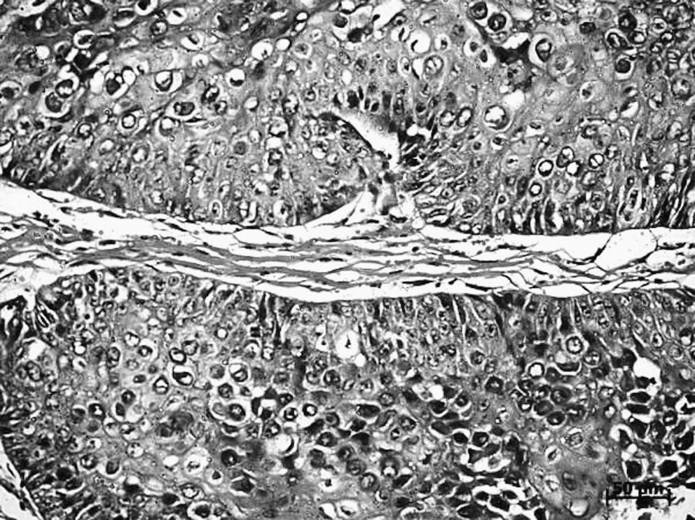
Animals in this group received DMBA applications during 8 weeks and another 4 weeks of no treatment. We found 20% of the tumors to be moderately differentiated SCC and the other 80% to be well-differentiated SCC (Fig. 2).

Photomicrograph of a moderately differentiated squamous cell carcinoma displaying anastomosing epithelial and papilliferous cords invading the lamina propria.
Group 3
Histological analysis showed 40% well-differentiated SCC, 40% poorly differentiated SCC, and 20% moderately differentiated SCC (Fig. 3).
Photomicrograph of a poorly differentiated squamous cell carcinoma displaying epithelial islands with hyperchromic nuclei, pleomorphic cells, and palisade-like peripheral cells.
Statistical analysis
Statistical analysis showed no significant differences in the amount of well-differentiated tumors between Groups 2 and 1. However, significant differences (p=0.02) were seen between Groups 1 and 3, with a larger number of lesions seen in Group 1. Significant difference was also seen between Groups 2 and 3 (p=0.04), with larger numbers found on Group 2. No significant difference was seen between groups with regard to the amount of moderately differentiated SCC. Significant difference was also seen among all groups with regard to the amount of poorly differentiated tumors (p=0.04). Poorly differentiated tumors were seen only in Group 3.
Discussion
In this study, the induction of chemical carcinogenesis was performed using DMBA, which has been successfully used for the induction of SCC on the oral mucosa of golden Syrian hamsters. 2,11 –20
A previous study showed that the use of 0.5% in mineral oil is the best concentration of DMBA for a rapid induction of epithelial tumors in the oral mucosa of hamsters. 19 This concentration produces the maximum response of the tumor with minimal latency period, without loss of animals caused by toxicity. 19 According to many studies this concentration should be chosen because the use of higher concentrations might cause precipitation of the drug. 7,8,14 –16,18,19 In this study, we used 0.5% DMBA in oil mineral three times a week for a period of 8 weeks for the induction of tumors in the oral mucosa of hamsters.
Laser radiation is noninvasive, well tolerated by tissues, and does not have mutagenic effects with the wavelengths clinically used. 21 However, the use of LLLT on cancerous conditions may cause both stimulatory and inhibitory responses on cell proliferation. Despite several studies showing the stimulation of malignant cells in culture following laser irradiation, it is important to state that studies using cell cultures do not reflect the reality of the complex biology of both the cancer and host responses. 3,22,23
The effects of LLLT on cell proliferation are influenced by the time, dosimetry, and wavelength. In a manner similar to that of our experimental protocol, previous studies have assessed cell proliferation using λ685- and λ830-nm laser (4 J/cm2). We used λ660-nm laser with much higher energy density, and we also found an increased progression of the tumors in irradiated animals when compared to controls. 7,8
Other studies, using cell culture, were conducted to verify the effect of λ635- and λ670-nm laser on the proliferation of HEp-2 cells. 3,24,25 The authors concluded that λ670-nm irradiation significantly increased cell proliferation. This finding is consistent with our results, in which the group treated with LLLT showed increased development of tumors.
Another study assessed the effects of LLLT using different wavelength (λ670 , λ780 , and λ830 nm) and energies (0.5, 1, 5, and 10 J/cm2) on the proliferation of normal bone cells (osteoblasts) and cells from osteosarcoma in vitro. It was found that each cell line responded differently to the wavelengths and combinations of energies, it was concluded that the use of λ830 nm resulted in a proliferative stimulus on osteoblasts, but caused no effect on cell proliferation on osteosarcoma. The use of λ780 nm reduced the proliferation of osteoblasts and increased it in osteosarcoma. The use of λ670 nm showed a moderate stimulatory effect on the malignant cell line. The results of this study demonstrated that, in fact, LLLT had an effect on cell proliferation. In addition, this effect might have been influenced by both wavelength and energy. 23
Studies using a cell line derived from oral human SCC showed decreased mitotic index with energy densities between 2–8 J/cm2 and wavelengths of λ805 , λ630 , and λ635 nm. 26,27 This finding is not aligned with our study.
Clinical phototherapy protocols of superficial lesions usually use visible red light. 28 It has been suggested that the biological effects of most types of light are similar on both prokaryotes and eukaryotes, being most of them observed on all bands of visible light. 29 Moreover one major effect of the absorption of visible laser wavelengths is the stimulation of the mitochondria. This result in an increase in the production of energy as well as in triggering the synthesis of nucleic acids. 21 In the present study, we used the visible red laser (λ660 nm) because of its somewhat superficial effect. 21 Red light can cause changes in the intermediate filaments that are part of the cytoskeleton and are related to cell division. 30 Previous studies using cells in culture have suggested the occurrence of depolarization and disorganization of the cytoskeleton 48 h following irradiation with red laser. However, non-irradiated cells in culture did not show these changes. 30 –32
A previous study investigated the effects of LLLT on the mitochondria, nucleus, and cytoskeleton of CHO K-1 cells by using a semiconductor laser (
Our study showed 100% effectiveness of the chemical induction of malignancies after 8 weeks. This finding was previously reported 34 and used similar methodology to ours. Group 2 (that remained without any treatment for an additional 4 weeks) continued to show 100% of malignancy at the end of the experimental time. This may be indicative of the irreversibility of the chemical carcinogenesis, even in the absence of further stimulation. 35,36 The animals that spent the same period of 4 weeks with laser treatment, showed an increase in the progression of the lesions, including 40% of poorly differentiated tumors. LLLT may have hastened the progress of the lesions toward becoming poorly differentiated carcinomas with a worse prognosis. 10
Although many studies have focused extensively on the role of LLLT on different tissues and cell lines, there are still many questions about its proliferative effects on malignant tissues. However, the effect of LLLT on cell proliferation observed in the present investigation may have been influenced by the wavelength, time, and treatment parameters as reported in previous studies elsewhere on the literature. 23 –25
Finally, one may question the statistical analysis used in the present study, because of its small sample. The data were statistically analyzed using Fisher exact test, which is a statistical significance test, used in the analysis of a small sample. 37,38
LLLT, within the parameters used in the present study, caused a significant progression of the severity of SCC of the oral cavity on hamsters.
Footnotes
Acknowledgments
We thank the Conselho Nacional de Desenvolvimento Científico e Tecnológico– CNPq for providing financial support for this project.
Author Disclosure Statement
No conflicting financial interests exist.