
Abstract
Introduction
Based on the clinical success of PDT in the treatment of CL 17,24 and the unique characteristics of MB in PDT, we performed a series of in vitro studies and evaluated a clinical case to determine the efficacy of PDT and MB in the treatment of CL. We hypothesized that the use of PDT with MB and a non-coherent light source such as infrared would be an effective treatment for CL.
Methods
Chemicals
Anhydrous MB was purchased from Sigma-Aldrich Corporation (Milwaukee, WI). The concentration of the stock solution was determined using absorbance measurements from spectrophotometric analyses and standardized absorbance versus concentration curves. 25
Parasites
Leishmania (Leishmania) amazonensis (MHO/BR/73/M2269 strain) was used in the in vitro study. The parasite was cultured at 26°C without CO2 in medium 199 supplemented with 20% heat-inactivated bovine fetal serum, 60 ng/mL penicillin G sodium salt, 100 ng/mL kanamycin sulfate, 50 ng/mL flucytosine, 10 ng/mL chloramphenicol, 2 mM L-glutamine, 40 nM HEPES, 0.1 mM adenine (in 50 mM HEPES), 5 mg/mL hemin (in 50% triethanolamine) and 1 mg/mL biotin (M199-C). Infective-stage metacyclic promastigotes were isolated from 5-day-old stationary cultures using a uniform procedure based on a modified density gradient purification.
MTT assay
The number of viable Leishmania was determined enzymatically based on the conversion of a tetrazolium salt, 3-(4,5-dimethylthiazol-2-yl)-2,5-diphenyltetrazolium bromide (MTT), into a colored, insoluble formazan product. The amount of product depends on the number of viable parasites present. 24,26,27
PDT
After the parasite samples were exposed to MB for 1 h, the samples were centrifuged at 2465×g for 30 min at 4°C, and the medium containing MB was removed. The parasites were washed with PBS and fresh PBS was added. The parasites were then transferred to 96-well ELISA plates and exposed to a 9 J/cm2 light using a home-built LED light source delivering 5 mW/cm2 (30 min) at 7 cm from the source (wavelength of maximum emission at 650 nm). Following the irradiation of the cells, 1 mL of fresh growth medium was added. After incubation at 26°C for 18 h, an MTT assay was performed to determine cell survival. As a control, we used a promastigote that was cultured for 3 h at 26°C without light. It was not possible to use the RL50® in the in vitro cell studies because the irradiation with this light source increased the temperature of the medium (there is some infrared emitted from this source), which complicated the analysis of the cell death curves. 28
Statistical analyses
The mean±the standard deviation were obtained from six independent experiments. The Student's t-test was used to analyze statistical significance (p<0.05).
Clinical treatment protocol
One patient was enrolled in this study and was given a low-dose of SbV (5 mg/kg/d) for 20 days. 29 –32 A single lesion was chosen to test the MB/PDT protocol. The patient received an intralesional injection of 0.5 ml of a sterile aqueous solution composed of 0.5% MB with 1% lidocaine. After an intralesional injection of the MB/lidocaine solution, RL50 was applied for 10 min to deliver 20 J/cm2 per session. RL50 is a non-coherent polychromatic light source with spectral irradiance from 570 to 750 nm and an irradiation diameter of 50 mm, delivering 35 mW/cm2 at a distance of 7 cm from the source. The irradiation protocol, including the wavelength, power of the source, and irradiation time, has been optimized considering the absorption of MB and the previous clinical data obtained by the group. 18, 21 It has been shown to be more effective in the in vivo studies than LED-only irradiation 28 A total of four weekly treatments were performed. RL50 and the MB/lidocaine solution protocol has been approved to study the effectiveness of PDT in melanomas. 21 Lesion evolution was monitored on a weekly basis to document changes in the CL lesion. ImageJ was used to calculate open wound (unhealed) and ulcer (lesion) surface areas from the serial images. The side effect that may be observed during the treatment is the pain caused by local skin photosensitization. In the protocol described here the patient reported only slight pain in the illumination area.
Results
In vitro study
The dose-dependent response of L. amazonensis to MB with and without light stimulation clearly shows that cell toxicity increases with the increase in MB concentration (5, 10, 20, 30, 40, 50, 70, 80, and 100 μM) and with light stimulation (Fig. 1). The IC50 calculated in the absence of illumination was 100 μM. This effect is highly amplified by light absorption (IC50 20 μM), causing an increase in toxicity in the parasites. It is important to note that 50 and 100 μM of MB and illumination lead to a partial killing of the parasites (Fig. 1). Based on the effect of MB/PDT in Leishmania promastigotes, we decided to observe the effect of MB/PDT in vivo using a human clinical case.
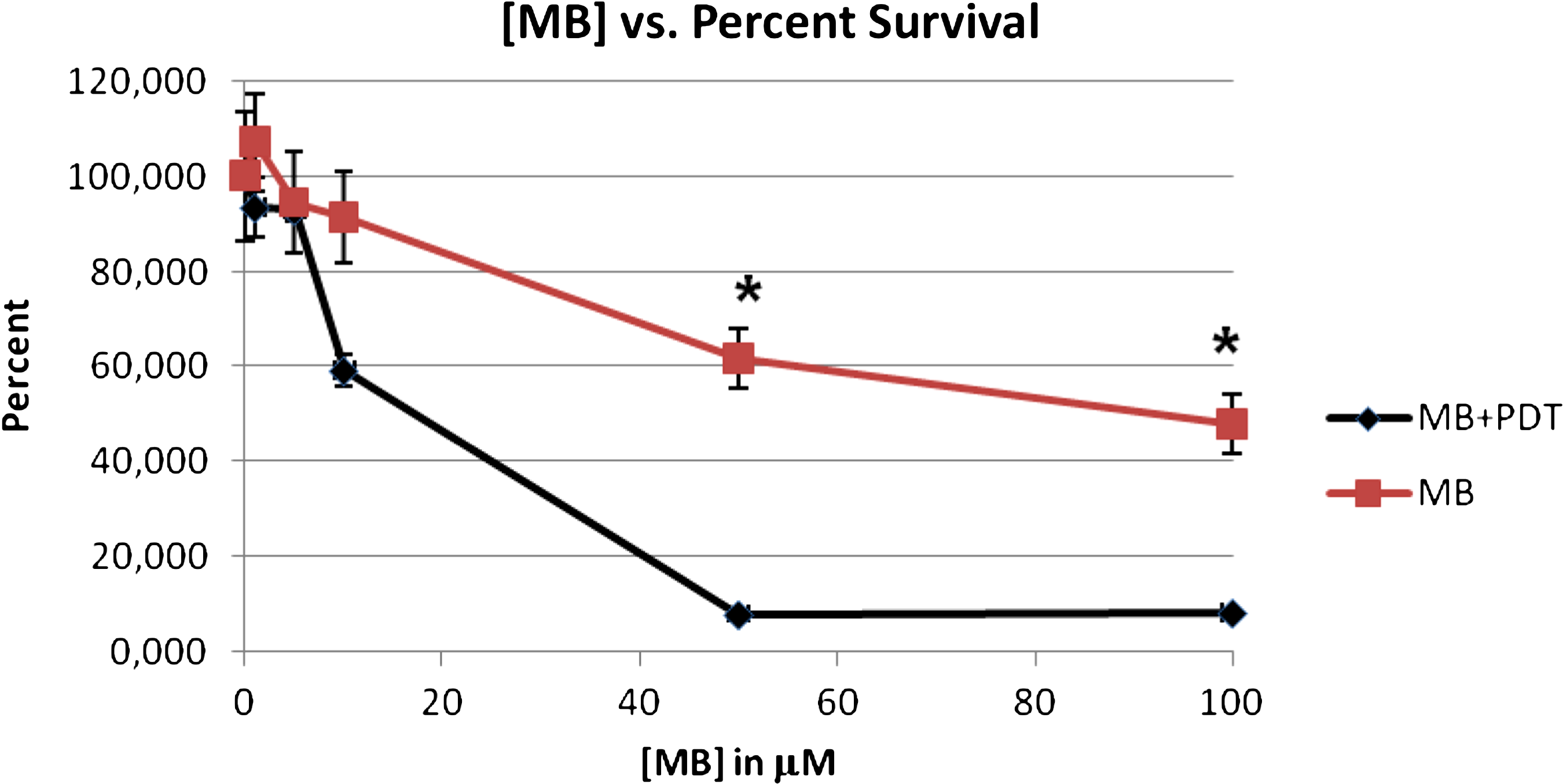
Dose response curve for L. amazonensis exposed to methylene blue (MB) alone and MB+light (PDT). The Student's t-test (p<0.05) indicates a significant decrease in parasite survival with the addition of PDT to 50 or 100 μM of MB. N=6 for each data point.
Clinical case
A male patient had three CL lesions localized in the left posterior flank, one lesion in the left ear and another in the left cheek, confirmed by Giemsa stain. The efficiency of PDT with MB was evaluated by comparing the results of two large lesions. The experimental lesion was treated with PDT and by low dose SbV, and the control lesion was treated with PA alone. The evolution of the lesions was followed by taking pictures and measuring the percentage of area occupied by open, unhealed wounds of 227 mm2 (base) as well as the total ulcer surface area of 227 mm2. As illustrated in Fig. 2, the size of the open wound was considerably reduced for the lesion treated with PDT. The diameters of the open wound regions treated with PA and PA +MB/PDT are plotted in Fig. 3 as a function of the number of days after starting treatment. One week after the second PDT dose (14 days), the lesion treated with PDT no longer contained an open wound. In the case of the lesion treated only with PA, the open wound persisted until 28 days after the initiation of treatment (Fig. 3).
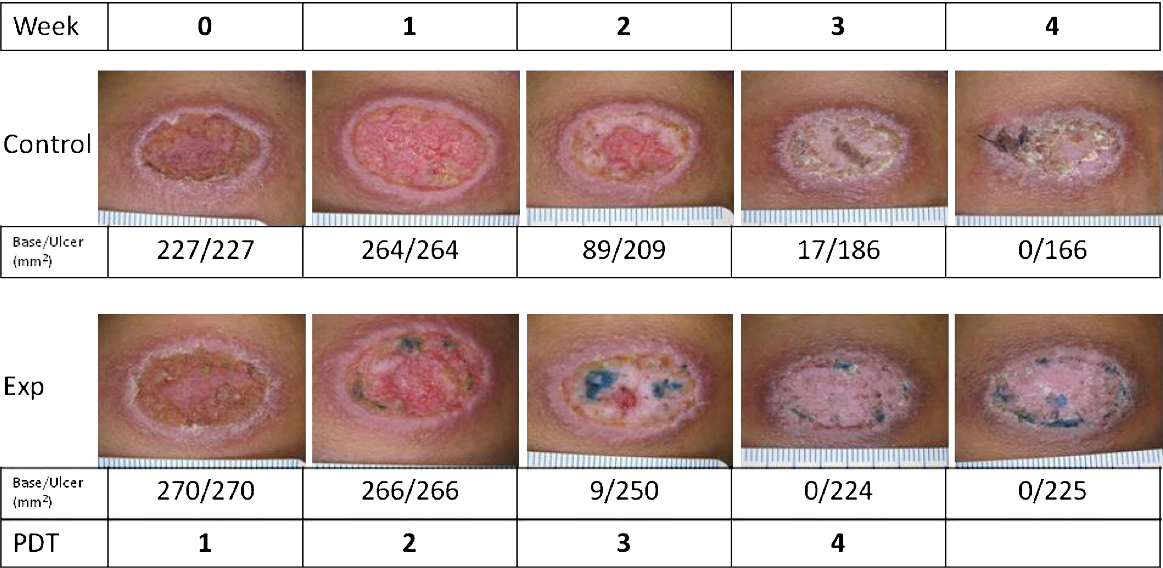
Pictures of the leishmaniasis lesions for the control (low dose SbV only) and experimental (low dose SbV+MB/PDT) lesions. Photographs were taken during weeks 0–4, during which PDT treatments 1–4 were given. Below each image is the area (mm2) of the open wound (base) versus the area of the lesion (ulcer).
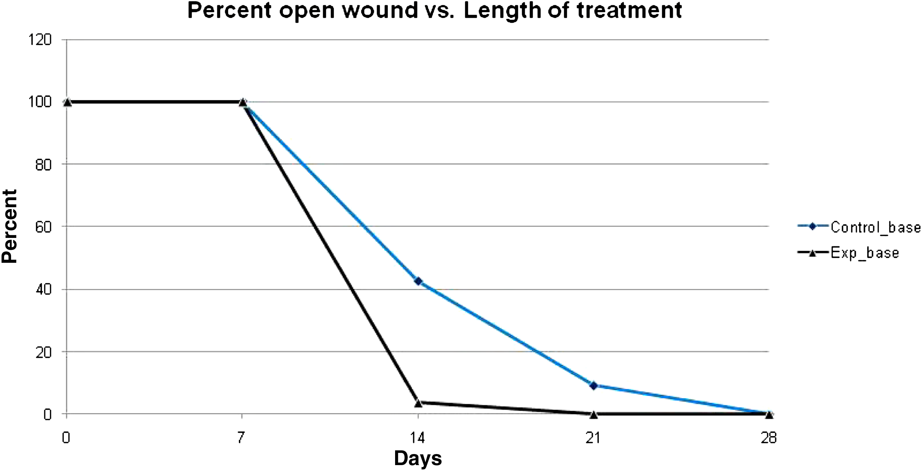
Percent of open lesions versus days of treatment. The graph indicates that the time to wound closure decreases when the lesion is treated with PDT/MB versus treatment with MB alone.
Discussion
We began our work with several in vitro experiments to test the parasiticidal effect of MB against L. amazonensis promastigotes with and without light. As anticipated, MB exhibited an increased parasiticidal effect that was proportional to its concentration, not only in the absence of light excitation, but also with light exposure.
One major difference between this study and prior PDT studies for leishmaniasis has been the addition of SbV to the experimental regimen. Prior studies, which were performed in the Middle East, dealt with forms of CLs that are at a lower risk for converting to the mucocutaneous disease than the leishmaniasis found in Brazil. As a result, the clinical cure of cutaneous lesions was the standard end point. However, in Brazil, because of the probability of the cutaneous disease converting into a mucocutaneous disease (2–8%), decades after healing the cutaneous lesions 5 systemic treatment is still required. Therefore, because of ethical and clinical concerns, a low dose of SbV therapy was added to the experimental treatment regimen. Although this makes it more difficult to clearly determine the efficacy of PDT as a treatment for CL, a comparison with low-dose SbV therapy alone will allow us to distinguish any added effect of the combined therapy. In addition, by combining both a systemic and a local therapy, we can minimize the side effects and costs associated with using SbV to treat CL. Concurrently with our in vitro studies, an initial clinical trial was performed on an individual affected by CL. By using the patient as his own control, we were able to compare the effect of the addition of MB/PDT to the healing process of his cutaneous lesions. Our analysis of the healing process revealed that although a low-dose of SbV did result in cutaneous lesion closure, the addition of MB/PDT accelerated the process, resulting in faster wound closure. It is important to mention that this protocol is inexpensive, allowing it to be used in the public health systems of underserved areas.
Future work should be aimed at understanding if the infected host cell has an effect on parasite susceptibility to the treatment and localization of MB. Because the Leishmania amastigote is more metabolically active, it is unclear if this form or if an infected host cell environment will confer increased resistance to treatment. It will also be important to elucidate if parasite viability and host cell viability are mutually exclusive in MB/PDT. Prior studies using ALA in Leishmania have shown that because of the biochemical properties of ALA/PpIX, parasite death occurs through host cell death. 26 However, MB does not require an enzymatic conversion to become active, and therefore we anticipate that the cell viability will depend upon the type of cell, host or parasite.
Conclusions
Our data show that MB causes damage, decreasing parasite viability fivefold compared with the dark control and decreasing the time necessary to heal a leishmaniasis lesion. These results show that the low-cost PDT protocol using MB and illumination with a non-coherent light source has the potential to be useful in the treatment of leishmaniasis and can be a practical solution for treating this disease in underserved populations. Further studies are needed to test the efficiency of this protocol in a statistically significant population of patients.
Footnotes
Author Disclosure Statement
No conflicting financial interests exist.