




Abstract
Introduction
Aminolevulinic acid hydrochloride (5-ALA), a secondary generation photodynamic reagent, is an endogenous precursor in the biosynthesis of human hemoglobulin, which transforms to produce protoporphyrin IX under the catalytic action of enzymes. Protoporphyrin IX can produce fluorescence under a certain wavelength of light, which can then be used in diagnosis and treatment. The clinical application of 5-ALA has been approved in the literature to be safe. 6 –8 Topical application is effective for photodynamic therapy of skin malignancies. 6 Fluorescence guidance during surgery for the treatment of malignant gliomas enables neurosurgeons to differentiate tumor tissue from normal brain, 7 and intravesicular injection has proven to enhance the visual detection of bladder malignancy. 8 However, the application of 5-ALA for the identification of the parathyroid glands has only been reported rarely in the literature. In this study, we investigated the feasibility of 5-ALA-induced fluorescence in the detection of the parathyroid glands in an animal model. The results may have potential clinical applications and provide new methods for the intraoperative identification of parathyroid glands, in an attempt to decrease the rate of the dangerous hypoparathyroidism after thyroidectomy.
Materials and Methods
Reagents
5-ALA hydrochloride (molecular weight, 167.59 Da) was obtained (Shanghai Fudan Zhangjiang Bio-Pharmaceutical, Shanghai, China) in white powder form. The powder was suspended in 0.9% sodium chloride at a concentration of 3% immediately before injection.
Animal models
Ten Sprague-Dawley (S-D) rats with an average weight of 200 g were obtained from the laboratory animal center of Sun Yat-sen University, China. The rats were housed and experimented on in accordance with the regulations of the experimental animal committee of the university (SYXK [YUE] 2007-0081).
The pentobarbital sodium (Shanghai Solarbio Bioscience & Technology Co. Ltd., Shanghai, China) was injected intraperitoneally at a dosage of 30 mg/kg to anesthetize the rats. After anesthesia was administered, the 5-ALA solution was injected intraperitoneally at the dosage of 300 mg/kg for this experiment. One hour later the rats were euthanized by cervical dislocation. Surgical dissection was then initiated. A longitudinal anterior collar incision was performed to expose the trachea and bilateral thyroid glands under conventional ambient light. The parathyroid glands were identified first under the same conditions. Then, blue light with a wavelength of 405±3 nm produced from a semiconductor laser producer (Fuda Minguang Electrical Technology Co., Fu Zhou, China) was used to illuminate the surgical field. Re-detection of the parathyroid glands was attempted with the use of the naked eye under the blue light.
Imaging and histology
The fluorescence of parathyroid glands was detected and recorded by photography (Panasonic automatic camera FX01). The tissue with fluorescence was removed surgically and sent for paraffin embedding. Routine hematoxylin-eosin staining was performed to confirm the presence of parathyroid glands under light microscopy.
Results
5-ALA-induced fluorescence in rat parathyroid glands
A single parathyroid gland was identified on each side of the trachea in the rat. The parathyroid gland was ovoid, tiny, and could not be visualized by the naked eye under daylight (Fig. 1). One hour after the intraperitoneal injection of 5-ALA solution at a dosage of 300 mg/kg, the rat was dissected. Blue light with a wavelength of 405±3 nm was used to light the surgical field. Red fluorescence was observed clearly with the naked eye bilaterally in the posterior thyroid glands (Fig. 2). The red fluorescence was present in all 10 rats.
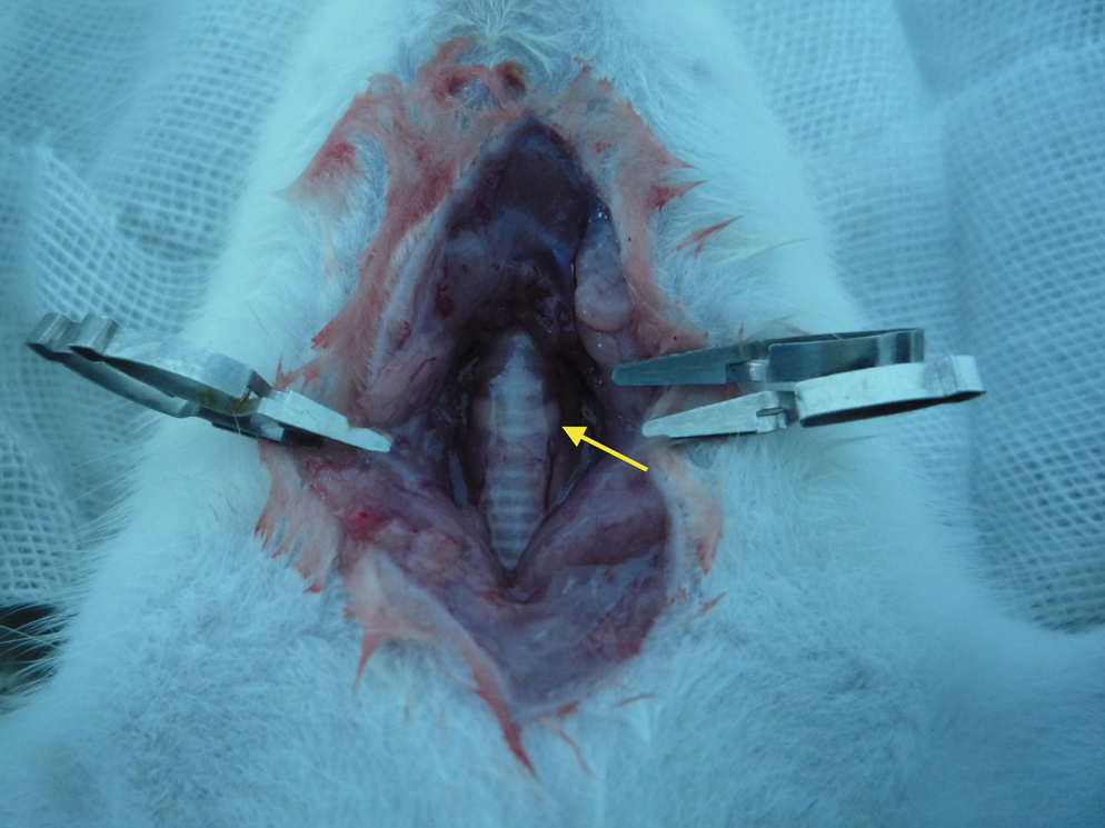
Surgical dissection revealed the trachea and bilateral thyroid glands. The parathyroid glands could not be identified under ambient light alone. The arrow shows the normal tissue of the thyroid gland lateral to the trachea.

After photosensitization with 5-ALA with fluorescence induced by blue light with a wavelength of 405±3 nm, bilateral red fluorescence could be seen from the posterolateral thyroid glands. The arrow shows the fluorescent parathyroid gland.
Histological confirmation
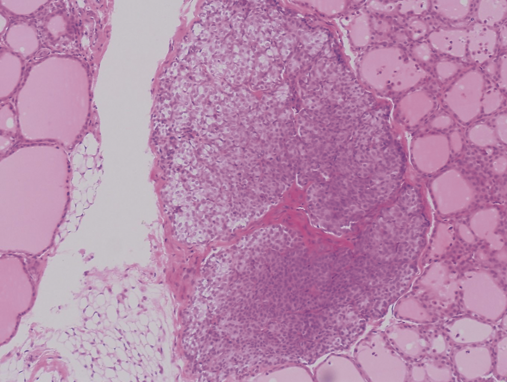
The tissue with red fluorescence was harvested to perform histology analysis. All tissues from the 10 rats were confirmed under light microscopy to be the parathyroid glands (Fig. 3 and 4). Under low magnification, the parathyroid gland was seen to be located outside of the thyroid capsule, surrounded by thyroid follicles and a thin layer of connective tissue. Under high magnification, clear parathyroid gland cells were seen to be arranged into columns and nests with a rich network of capillary vessels and sparse connective tissue.

Under low magnification, histological examination of the tissue with red fluorescence confirmed the identification of the parathyroid gland. The arrow shows the parathyroid gland under light microscopy.
High magnification (original × 20) showed parathyroid gland.
Discussion
This experiment demonstrated that exogenous application of 5-ALA could induce parathyroid gland fluorescence in an animal model, which could then enable the identification of the parathyroid glands under direct vision. The results suggest that this method might improve the detection of parathyroid gland tissue intraoperatively.
The production of fluorescence in parathyroid gland is a complicated process including accumulation of 5-ALA in the target organ and transformation to protoporphyrin IX under catalytic effect of enzymes. The detailed molecular biological mechanisms have not been known yet. Further basic research should be done to study the mechanisms of induced fluorescence by 5-ALA in parathyroid glands.
In the literature, only two groups have reported the application of 5-ALA to identify parathyroid gland fluorescence. Gahlen et al. and Asher et al. demonstrated that the intraperitoneal injection of 5-ALA could induce red fluorescence in the parathyroid gland in rats 9 –12 . They also investigated the optimal dosage of 5-ALA, which may be affected by different commercial sources of 5-ALA. They also used different ways of tissue sampling to confirm the microscopic diagnosis of the parathyroid gland. In the study by Gahlen et al., randomized sampling was applied; however, Asher et al. used whole thyroid specimens to perform the pathological identification of the parathyroids. 9,12 In comparison, we only sampled the thyroid tissue with red fluorescence to confirm the identification. This method was used to ensure that only the tissue with red fluorescence was the either the parathyroid gland or not. Furthermore, in our experiment we used a semiconductor laser producer to stimulate fluorescence, which could produce blue light with a wavelength of 405±3 nm. This was a portable device and could be used easily during the operation. In addition, observation of the fluorescence with the naked eye was used in this study because it had practical value in a clinical setting. As a result, the kinetics of 5-ALA fluorescence including the intensity and wavelength of the spectra were not measured in the current study.
Although the experiments showed that 5-ALA could induce visible red fluorescence in the parathyroid glands, the application of this method has been used only rarely in humans to date, and the published literature is comprised of case reports only. 13 –15 However, 5-ALA has been used clinically and approved to be safe in neurosurgery and urological surgery, and the optimal dosage and pharmacokinetics have been determined in humans. 7,8 As a result, we believe that this method could help surgeons to identify parathyroid glands during thyroidectomy, and assist with the differentiation of parathyroid glands from the surrounding tissues. It also can help to confirm parathyroid tissue during the transplantation of these glands.
Conclusions
In summary, 5-ALA-induced fluorescence detection of parathyroid gland can clearly identify the glands from surrounding tissue under direct vision. This method can be used intraoperatively and has potential clinical value.
Footnotes
Author Disclosure Statement
No competing financial interests exist.