
Abstract
Introduction
Low-intensity laser therapy has become an accepted tool for several clinical applications and has been successfully established in regenerative medicine and dentistry because of its anti-inflammatory, analgesic, and regenerative effects. 4 The latter effect, also called photostimulation, produces non-destructive events as well as increased cellular activity on tissues at the cellular level, and it has been used in a variety of medical therapies. 5,6
In the last two decades photodynamic therapy (PDT) has been successfully employed for clinical treatment of cancer using different photosensitizer molecules. Photosensitizers are the active agents in the photodynamic process. PDT is based on the photoactivation of photosensitizers when irradiated by light in a specific wavelength window (generally the maximum absorption band of the photosensitizer compound). The light-excited photosensitizer generates reactive oxygen species (ROS) that induce reduction or destruction of tumors by multifactorial mechanisms. 7
The explanation for the photobiological effects of laser light is based on the light absorption by primary endogenous chromophores (mitochondrial enzymes, porphyrins, flavins, and cytochromes). 8 Many in vivo and in vitro studies have reported on the influence of laser irradiation on the cellular functional state. 9 –11
The exact mechanism of action of the laser irradiation in living cells is not yet understood, although the photostimulation and therapeutic effects of low-power visible irradiation of different wavelengths and light doses are well known. 1,4,9 –15 Experimental and clinical studies have suggested that low-intensity laser irradiation affects cellular metabolic processes, leading to enhanced regeneration of biological tissues. 16,17 The biological effects of irradiation depend upon wavelength, power, time of exposure, and irradiation dose or fluence. 5,18
Lubart et al. 19 have suggested that low-intensity laser irradiation might promote changes in the cellular redox state, playing a pivotal role in sustaining cellular activities, thus promoting photobiostimulative processes. Other studies, however, have emphasized that different biological responses can be achieved, depending on the applied dose, wavelength, irradiation time, and the conditions of the treated tissue. 20 –22 Laser light processes present some special features that allow them to selectively and precisely influence some targets, including biological ones. 11,12,18 Small doses induce stimuli, large doses depress physiological processes, while extreme ones cause destruction of biological structures (for example, lipoperoxidation or heating, which causes protein denaturation). 5,18,23 –26
Some in vitro effects of photostimulation on the extracellular matrix, collagen production, macrophage stimulation, and fibroblast proliferation have been described. 27,28
Some authors 20,29 have suggested that the biological mechanism behind the effects of light used in therapy is related to absorption of red and near-infrared light by chromophores contained in the protein components of the respiratory chain located in the mitochondria, particularly in cytochrome c oxidase. Moreover, stimulation of the latter enzyme in isolated extracts has been recently demonstrated. 8
Therefore, the purpose of this work is to associate the photosensitive drug aluminum phthalocyanine chloride and low intensity laser in order to study its efficiency in biocellular biostimulation and in vitro osteogenesis through the photodynamic process for a possible acceleration of the bone damage recovery. The main purpose of this study was to investigate the in vitro effects of visible light radiation at 670 nm, as obtained from an Eagle diode laser (Quantum Tech, São Carlos, SP, Brazil) at low doses varying between 0.5 and 3 J/cm2, combined with the previous application of an aluminum phthalocyanine chloride (AlClPc) dye as a photosensitizer, on the growth and differentiation of osteoblastic cells using a primary osteoblastic cell line isolated from the bone marrow of rats. New evidence that low-intensity laser irradiation may play an important role in promoting bone formation is presented on the basis of the MTT assay, collagen content, total protein content, alkaline phosphatase (ALP) activity, and bone-like nodule formation.
Methods
Materials
All the solutions were prepared using pure apyrogenic water (Millipore Direct-Q, Millipore, Barueri, SP, Brazil). All the reagents were of the highest commercially available purity. Epikuron, Poloxamer 188, Polomazer 170, and soybean phospholipids were acquired from Lucas Meyer, France. Miglyol 812 N Hulls (Witten, Germany) was purchased from Puteaux, France. Aluminum chloride phthalocyanine or (chlorine [29H, 31H-phthalocyaninato] aluminum) (AlClPc), 3[4,5-dimethylthiazol-2-yl]-2,5-diphenyl tetrazolium bromide (MTT), β-glycerophosphate, sodium lauryl sulfate (SDS), Lowry solution, phenol reagent of Folin-Ciacalteau, bovine albumin, alizarin red, and dexamethasone were obtained from Sigma-Aldrich Co., St. Louis, MO. Hank's buffer, α-MEM, trypsin, ascorbic acid, fetal bovine serum, gentamicin, and fungizone were supplied by Gibco – Invitrogen Technologies (Grand Island, NY). Acetic acid, chloramine T, and Ehrlich's reagent were provided by Acros, Pittsburgh, PA. Glutharaldehyde and sodium cacodylate were obtained from Electron Microscopy Sciences, Hatfield, PA. ALP activity was measured using a commercial kit (Diagnostic Labtest, Belo Horizonte, MG, Brazil).
In addition to these materials, different organic solvents and other inorganic salts commonly used in the laboratory were employed. All the solvents were analytical grade.
Preparation of nanoemulsions
The nanoemulsions (NE) were obtained by the spontaneous emulsification process described by Yu et al. 30 Briefly, the organic phase (acetone) was prepared with oil, soy phospholipid, and AlClPc, at 55°C. The photosensitizer had been previously dissolved in Miglyol 812N and was added to the phospholipid organic solution at a concentration of 0.05 mg/mL. The organic solution was slowly added to the aqueous phase containing Poloxamer 188, under magnetic stirring. After total addition of the organic solution and spontaneous emulsification, the organic solvent was removed by evaporation under reduced pressure, at 65°C. Finally, the formulations were concentrated to a final volume of 10 mL. Formulations without the drug were prepared under the same conditions, for use as reference compounds in the spectroscopic analyses. All formulations were characterized by their mean size, polydispersity index (PdI), and ζ potential, as described by Siqueira-Moura et al. 31 The mean size and PdI of the colloidal dispersions were determined at 25°C by laser light scattering at an angle of 173o, whereas the ζ potential was measured by electrophoretic mobility using a Zetasizer® (Nano ZS, Malvern PCS Instruments, Worcestershire, UK). Data are the mean (±SD) of three different independent preparations.
Rat bone marrow cell isolation and culture
Cells were obtained and cultured according to Maniatopoulos et al., 32 with modifications standardized by Simão et al. 33 Bone marrow was obtained from young adult male rats of the Wistar strain weighing 110–120 g. The femora were aseptically excised, cleaned of soft tissues, and washed three times (15 min each) in culture medium containing 10 times the usual concentration of antibiotics.
The femoral epiphyses were cut off, and the marrow was flushed out with 20 mL culture medium. Released bone marrow cells were collected in a 75 cm2 plastic culture flask containing 10 mL osteogenic culture medium, which allows for the osteoblastic differentiation and is composed by α-MEM supplemented with 15% fetal bovine serum, 50 μg/mL gentamicin, 0.3 μg/mL fungizone, 10−7 M dexamethasone, 5 μg/mL ascorbic acid, and 2.16 mg/mL β-glycerophosphate. 33 Cells were cultured until subconfluence enzymatically-released and first-passage cells were cultured in the same medium, at a concentration of 2×10⁴ cells per well, in 24-well microplates (Falcon, Franklin Lakes, NJ). Cells were cultured up to 21 days at 37°C in a humidified atmosphere of 5% CO2 and 95% air, and the medium was changed every 48 h.
Cytotoxicity assays
The methodology used to evaluate cell viability was the classical MTT assay. The tetrazolium salt (3-[4,5-dimethylthiazol-2-yl]-2,5-diphenyl tetrazolium bromide) produced the highly coloured formazan dye upon NADH reduction, which reflects a living cellular dehydrogenase. 34 To investigate the toxicity of the photosensitizer, a suspension of 2×10⁴ osteoblastic cells in 1000 μL medium was added into each well in the 24-well microplate. After culturing in CO2 incubator for 24 h, monolayer cultures of osteoblasts were treated with AlClPc/NE at a final concentration of 0.5 μmol/L (for the darkness toxicity and photobiological toxicity assays). Three hours after incubation, the cells that were going to be used for the darkness toxicity experiment were washed twice, and the volume was completed with addition of 1000 μL α-MEM to each well, overnight.
The 1.0 mg/mL MTT solution (250 μL per well) was added to the cells placed in the 24-well microplates, followed by incubation for 4 h, at 37°C. The crystals formed as a result of interaction between the mitochondrial dehydrogenases and the MTT reagent were dissolved with 2-propanol. The samples were shaken until complete dissolution of the formed product. After the reaction was finished, the absorbance at 560 and 690 nm of each well was measured by means of the Safire II multiplate reader from TECAN (Tecan Trading AG, Männedorf, Switzerland), for determination of mitochondrial dehydrogenases and MTT, respectively. Cell viability is expressed as the percentage of cells incubated without AlClPc/NE.
Photobiological assays
For the photobiological tests, the cells were washed twice and the volume was completed by addition of 1000 μL phosphate buffer to each well of the microplate. Then, the wells were irradiated with the following light dosages: 0.5, 1.0, and 3.0 J/ cm2, using the Eagle diode laser (Quantum Tech, San Carlos, SP, Brazil). The light source was a continuous-wave laser operating at 670 nm, with maximum power of 50–300 mW in the end of the optical fibers. The Eagle diode laser model is a laser system that operates in a set wavelength of 670 nm of the visible electromagnetic spectra. The system is attached to an optical fiber system (200 μm) for light delivery. For the red laser, the active material is a diode emitter operating with a 50–300 mW potency through an optical fiber, continuous operation.
The laser equipment used presents all the internationally required specifications and a calibration point that guarantees that the programmed dosage be the same as that discharged by the outlet spot of the laser. The description of laser irradiation parameters used in this article are shown in Table 1.
After irradiation, the phosphate buffer was removed, the volume was completed by addition of 1000 μL osteogenic medium to each well, and the solution was incubated overnight.
The percentage of cell viability was determined using the MMT assay and is expressed as the percentage of cultures without either photosensitizer or laser irradiation.
Total protein content
Total protein content was calculated at 7 days using a modification of the Lowry method. 35 Culture medium was removed, and the wells were washed three times with PBS at 37°C and filled with 2 mL 0.1% sodium lauryl sulfate. After 30 min, 1 mL of this solution (from each well) was mixed with 1 mL Lowry solution and left for 20 min at room temperature. After this period, 0.5 mL of the solution of phenol reagent of Folin and Ciocalteau was added, and the solution was left to stand for 30 min at room temperature, to allow color development. Absorbance was then spectrophotometrically measured at 680 nm, and the total protein content (μg/mL) was calculated from a standard curve.
Collagen content
The collagen content was calculated at 7 days, according to the method of Reddy and Enwemeka. 36 Samples of the same solutions used for determination of the total protein content were assayed for collagen content. Aliquots containing 0.5 mL of this solution were lyophilized and re-suspended in 50 μL acetic acid 6 N. Then, the samples were hydrolyzed by autoclaving at 120°C for 30 min. After this procedure, 450 μL chloramine-T was added to the hydrolysate, which was then mixed gently, and the oxidation was allowed to proceed for 25 min at room temperature. Next, 0.5 mL Ehrlich's aldehyde reagent was added to each sample, followed by gentle mixing, and the color was developed by incubating the samples at 65°C for 20 min. The absorbance was measured at 550 nm in a spectrophotometer, and the collagen content was calculated from a standard curve and expressed as μg/mL.
ALP activity
The ALP activity was assayed at 7 days as the release of tymolphtaleine from tymolphtaleine monophosphate using a commercial kit, and the specific activity was calculated. Aliquots of the same solutions used for calculating total protein content were assayed for the ALP activity, according to the instructions on the kit. The absorbance was then spectrophotometrically measured at 590 nm, and the ALP activity was calculated (μmol tymophtaleine/h/mg) from a standard curve.
Bone-like nodule formation
Bone-like nodule formation was assessed at 21 days by alizarin red S method. The culture medium was removed, cells were washed three times with PBS at 37°C, and the attached cells were fixed in 10% formalin for 24 h, at room temperature. After fixation, the specimens were dehydrated through a graded series of alcohol and processed for staining with alizarin red, which stains calcium-rich bone-like nodules. Images were obtained for documentation; the fields were randomly selected from different wells using a microscope Axiovert 40 CFL Ph 1 with objective 10x / 0.25 coupled with camera 7.2 (Zeiss, Thornwood, NY). Quantification of staining was assessed by a colorimetric method described by Gregori et al. 37 Then, 360 μL 10% acetic acid was added to each well previously stained with alizarin red, and the plate was shaken for 30 min at room temperature. The contents of each well were transferred to Eppendorf tubes (Eppendorf Manufacturing Corp., Enfield, CT), which were centrifuged at 20,000g for 15 min. Next, 100 μL supernatant from each tube were transferred to new tubes. Finally, 40 μL ammonium hydroxide 10% were added, to neutralize the acid, and the absorbance was measured at a wavelength of 405 nm.
Statistical analysis
Data presented in this work are the result of a single culture with n=3 for each surface treatment, for each experiment. All the data were submitted to an analysis of variance (One-Way ANOVA) followed by Tukey's test when appropriate. Differences at p<0.05 were considered statistically significant.
Results
Cytotoxicity assays
Cytotoxicity studies performed in the absence of light and using osteoblasts as a biologicical model and a photosensitizer (AlClPc) combined in NE (AlClPc/NE) are described in Table 2. As observed, cell viability in the presence of the NE alone or in the presence of AlClPc/NE remained unaffected with up to 3.0 μmol/L photosensitizer. For AlClPc/NE concentrations >3.0 μmol/L, there was a reduction in total protein content and ALP activity.
Represents results considered statistically significant.
Because the cytotoxic effect was observed from 1 μmol/L AlClPc/NE thereafter, further biostimulation studies were conducted using the 0.5 μmol/L concentration only, which would guarantee a good safety margin.
Photobiological assays
The photostimulation results obtained by using low-power laser and AlClPc/NE (0.5 μmol/L) during the in vitro assay of osteoblasts cells are described in Fig. 1. Irradiation of small doses, ∼0.5 J/cm2, resulted in significant increase in cell viability, total protein, ALP activity, and collagen, suggesting stimulation of cell growth. On the other hand, this stimulation process wasn't observed when irradiation of about 1 J/cm2 was employed. An irradiation of 3 J/cm2 led to considerable reduction in cell viability and, consequently, loss of total protein, and collagen content as well as ALP activity, indicating phototoxicity. The use of light from low-power laser, in the absence of AlClPc/NE, did not affect any of the parameters studied here up to irradiations at 3 J/cm2 (results not shown).
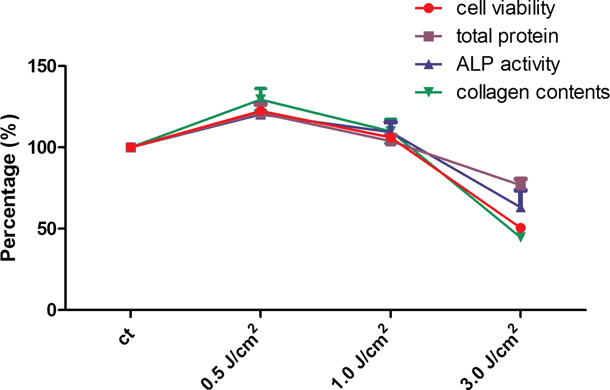
In vitro with AlClPc / NE in culture of osteoblasts in the presence of light. Laser dosage of 0 (control), 0.5, 1.0, and 3.0 J/cm2.
Bone-like nodule formation
The experiments concerning bone-like nodule formation evidenced formation of calcified nodules, quantified by means of three parameters, namely, control (ct), treatment with AlClPc/NE at a light dose of 0.5 J/cm2, and treatment with AlClPc/NE at a light dose of 3.0 J/cm2. As observed in Fig. 2B, irradiation at 0.5 J/cm2, elicited increased mineralization of bone-like nodules, compared with ct (Fig. 2A). However, Fig. 2C reveals that radiation at 3.0 J/cm2 led to a large reduction in the number of nodules, thus reinforcing the cytotoxic condition.

In vitro study of osteoblasts culture stained with alizarin red in the presence of light and AlClPc / NE.
Discussion
As far as laser therapy is concerned, it is known that this type of treatment has been successfully employed over the past years and its applications are countless. Its effect has already been proved in the case of orthodontic treatment, 38 bone defects, 39,40 and bone fractures, 41 and after implant placement. 42
The biostimulation assays conducted here demonstrated increase in the number of viable cells with high levels of total protein and collagen content as well as ALP activity at a laser dose equal to 0.5 J/cm2. This suggests that cellular biostimulation occurred because of the combination of a photosensitive drug (AlClPc/NE) with a laser energy dose. Because AlClPc is hydrophobic, only the formulation incorporated into the NE was studied here. 39
Indeed, experimental studies have shown that lower doses have no effect and that very high doses have an inhibitory toxic effect on cell cultures. An example of such studies is the work of Van Breughel et al., 43 who tested the effect of laser light output (HeNe laser; wavelength 633 nm) on human fibroblast cultures.
Using laser stimulation, researchers have observed the differentiation of mesenchymal stem cells into osteoblasts in a three-dimensional biomatrix. 44 Another study has shown that low-intensity laser therapy has a positive effect on the bone healing of diabetic rats. 45
Finally, a recent work has described that the use of low-intensity laser (830 nm) at 1.91 J/cm2, without association with a photosensitizer, stimulates in vitro mineralization through increased insulin-like growth factor (IGF-I) and bone morphogenetic protein production, which occurs as a result of Runx2 expression and ERK phosphorylation in mouse osteoblastic MC3T3-E1 cells, associated with an increase in the calcium content of the cell culture. 46
Currently, there are not many studies using laser therapy associated with a photosensitizer for the stimulation of bone cell growth.
As described by other authors, 14 a possible mechanism for the biostimulation of healing by low-level laser light is the absorption of light energy by mitochondria, which increases the energy of the osteoblast, thereby stimulating release of chemical mediators. In addition, biostimulation (in the visible region) could be associated with the irradiated light, leading to the generation of minimum levels of ROS. The latter species, in turn, play an important role in the triggering of many cellular processes. On the other hand, high concentrations of ROS cause cell death, probably by ATP depletion and lipid peroxidation.
Conclusions
Unlike biostimulation, this technique seeks to increase cell growth and this is obtained only by using a low light dose (<1.0 J/cm2). Therefore, whether biostimulation of osteoblastic cell cultures by PDT or the cytotoxic effect of this therapy occurs only depends upon the light dose, and the results can be completely reversed.
Footnotes
Acknowledgments
This work was partly supported by Conselho Nacional de Desenvolvimento Cientifico e Tecnológico (CNPq), Fundação de Amparo à Pesquisa do Estado de São Paulo (FAPESP), and Coordenação de Aperfeiçoamento de Pessoal de Nivel Superior (CAPES). F.L.P. and D.C.Z. received Ph.D. and M.S. scholarships from FAPESP and CNPq, respectively. We thank Cynthia Maria de Campos Prado Manso and Priscila Cerviglieri for linguistic advice.
Author Disclosure Statement
No conflicting financial interests exist.