
Abstract
Introduction
However, a dramatic emergence of antibiotic resistance has been observed in almost all pathogenic bacteria, and nowadays there is a consensus that antibiotics should be judiciously prescribed. 7 Because of the inevitable growth of multi-drug-resistant bacteria, it is important to continually seek alternative antibacterial therapeutic agents, especially ones to which bacteria will not develop resistance
Photodynamic therapy (PDT) consists of the use of photosensitizing drug (PS), along with visible light in order to excite the PS. 8 The excited PS changes the energy status inducing the formation of reactive oxygen species (ROS), which are highly toxic to the cell. 9 This technique could enable the targeting of microorganisms directly at the site of infection, thus overcoming the problems associated with antimicrobials.
The clinical application of PDT in treatment of periodontitis has been tested for nonsurgical management of aggressive forms of the disease. In previous work, PDT and nonsurgical periodontal treatment showed similar clinical outcomes after 3 months, and both led to statistically significant reductions in tumor necrosis factor-α (TNF-α) and receptor activation of nuclear factor κB ligand (RANKL) levels 30 days following treatment. 10 In another study, only bleeding on probing was significantly decreased when compared to other parameters, after 6 months. 11 However, it has also been reported that PDT used as an adjunctive in nonsurgical periodontal treatment improves clinical outcomes. 12
The key issues to be addressed are the effectiveness of the treatment in destroying sufficient numbers of disease-causing pathogens, and reduction of the inflammatory response in order to avoid bone loss. Since studies concerning the effectiveness of PDT as an adjunctive in the treatment of periodontal disease are few and inconclusive, 10 –12 here we focus attention on inflammatory conditions following the use of PDT in the gingival tissues of animals with ligature-induced periodontal disease.
Methods
Animals
Thirty-six male Wistar rats (250–350 g) were used in the study. The animals were kept in plastic cages, with free access to food and water. Prior to the surgical procedures, all animals were allowed to acclimatize to the laboratory environment for a period of 5 days. All experiments were conducted in accordance with national health guidelines for the welfare of experimental animals, and were approved by the Ethical Committee of the University of Uberaba (protocol # 002/2008).
Animal experiment design
Experimental periodontitis was induced by a ligature placement. More specifically, using general anesthesia by intramuscular administration of ketamine (1.0 ml/kg) a ligature was placed and immobilized around both mandible first molars of each animal. 13 The ligatures were removed from all animals after 7 days of experimental periodontal disease induction. The molars of all animals were subjected to SRP with manual #13–14 mine five curettes (Hu-Friedy Co. Inc., Chicago, IL), using 10 distal–mesial traction movements in the buccal and lingual aspects. The furcation and interproximal areas were scaled with the same curettes using cervico-occlusal traction movements. All SRP operations were performed by the same experienced operator. Next, the animals were randomly assigned to one of the following groups (Table 1): (a) animals without ligature receiving administration of eosin (0.8×10⁻⁷ mol/L [n=6]); (b) animals without ligature receiving administration of eosin (0.8×10⁻⁷ mol/L) followed by light irradiation (PDT) (n=6); (c) animals with ligature (n=6); (d) animals with ligature receiving administration of eosin (0.8×10⁻⁷ mol/L [n=6]); (e) animals with ligature receiving administration of eosin (0.8×10⁻⁷ mol/L) followed by light irradiation (PDT) (n=6); and (f) animals with ligature receiving light alone (n=6). The animals were killed by anesthetic overdose 14 days after the induction of periodontal disease.
(+) indicates presence and (−) absence.
Drugs and light source
Eosin (Merck KGaA, Darmstadt, Germany) was used at a final concentration of 0.8×10⁻⁷ mol/L, dissolved in 2% (wt/vol) Carbopol 940 (Pharma Special, Santana de Parnaíba, SP, Brazil). The light source used was a light emitting diode (Radii Cal, SDI, Bayswater, Victoria, Australia), with a wavelength range of 440–480 nm and a power density of 0.69 W/cm2 during 60 sec, totaling 41.4 J applied in a spot area of 0.8 cm2.
PDT treatment
After the SRP procedures, 100μl of the eosin (0.8×10⁻⁷ mol/L, dissolved in 2% [wt/vol] Carbopol 940) was administered for 1 min around the region where the ligature was placed, using a swab, followed by light irradiation for 1 min (where appropriate). The light source was positioned ∼0.5 cm from the affected tooth.
Histological analyses
The right and left jaws were dissected, fixed in 10% buffered neutral formalin for 48 h and decalcified in a solution of 10% ethylenediamine tetraacetic acid (EDTA) for 3 months. The jaws were then briefly washed in running tap water, dehydrated, and embedded in paraffin wax. Each sample was sliced into 6-μm sections in sagittal directions. The sections were mounted on glass slides, and stained with hematoxylin and eosin (HE) for the evaluation of bone resorption. The area of bone loss in the furcation region was determined histometrically, using an image analysis system (ImageJ) as previously described. 14
Myeloperoxidase (MPO) activity measurement
Neutrophil infiltration to the gingival tissues of the rats was evaluated using the MPO kinetic-colorimetric assay, as previously described. 13 Samples of gingival tissue were collected in 50 mM K2HPO4 buffer (pH 6.0) containing 0.5% hexadecyl trimethyl ammonium bromide (HTAB), and stored at −80 °C until used. The samples were homogenized (Kinematica Polytron PT3100, Littau-Luzern, Switzerland), and then centrifuged at 13,000 g for 4 min. The resulting supernatant, devoid of debris, was subjected to MPO activity assay, employing an ELISA reader (RT-2100C Microplate Reader, Zhejiang, China) operated at 450 nm, with three readings acquired within 1 min. Briefly, 10 μL of sample was mixed with 200 μL of 50 mM phosphate buffer at pH 6.0, containing 0.167 mg/ml o-dianisidine dihydrochloride (Sigma-Aldrich, St. Louis, MO) and 0.0005% hydrogen peroxide. The MPO activity of the samples was compared with a neutrophil standard curve. The results were presented as MPO activity (number of neutrophils per mg gingival tissue).
Cytokine measurement assay
Gingival tissue samples were weighed, and then homogenized in 150 μl of protease inhibitor cocktail (Sigma-Aldrich, St. Louis, MO). Levels of TNF-α were determined by ELISA using protocols supplied by the manufacturer (R&D Systems, Minneapolis, MN). The results are expressed as pg/mg of tissue.
Statistical analysis
Data were expressed as mean and SD (standard deviation). Statistical comparisons among groups were made using analysis of variance (ANOVA) followed by the Bonferroni test. Significance was accepted when the p value was ≤0.05.
Results
Effect of PDT on inflammatory bone loss induced at the furcation of the first molar
The association of eosin with LED irradiation on ligature-induced periodontal bone resorption was assessed by histomorphological evaluation at the first molar furcation region, and involved the staining of decalcified sections with HE. Animals without ligatures, and treated with either eosin or PDT, showed no bone resorption during the experimental period (Fig. 1A and B). On the other hand, animals with ligatures that received no treatment, or only treatment with eosin, showed a substantial level of bone resorption that was statistically significant compared with the animals without ligatures (Fig. 1C and D). In addition, the animals with ligatures that had been treated with PDT showed significantly reduced bone resorption (Fig. 1E; p<0.05). Ligatured animals treated with light alone showed decreased bone resorption, although in this case it was not statistical significant (Fig. 1F). The values of the resorption area for all groups are shown in Fig. 1G.
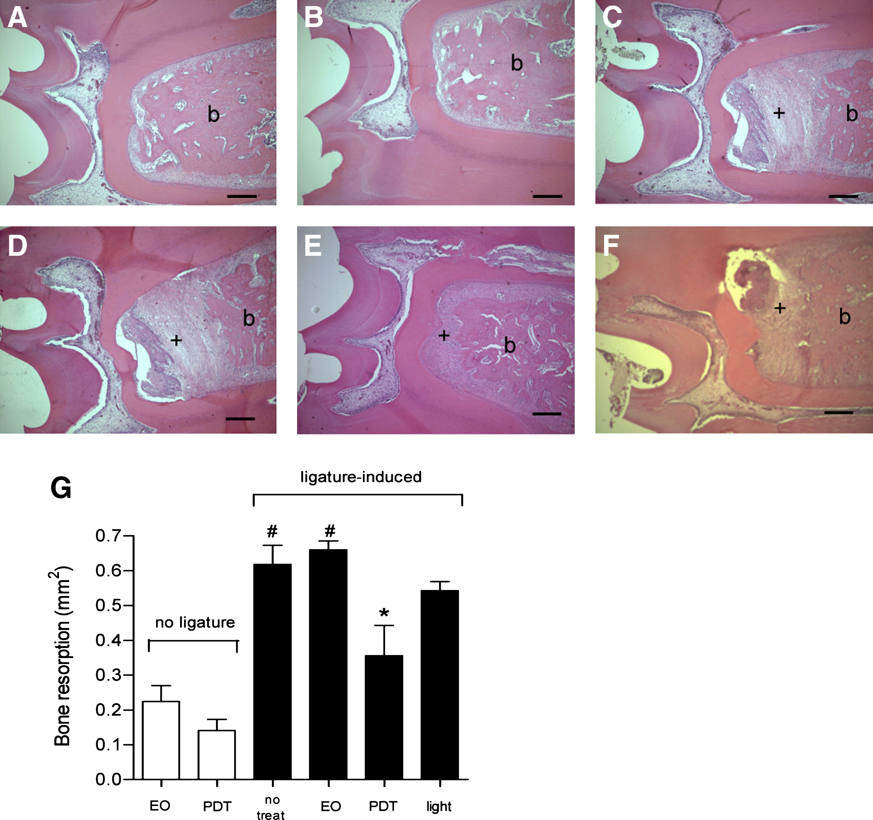
PDT decreases alveolar bone resorption. Histology at the furcation of first molars sampled from rats killed 7 days after ligature removal (staining with HE):
PDT treatment reduces neutrophil migration
The infiltration of neutrophils into the gingival tissues of the rats was investigated using the MPO activity assay. The groups without ligatures, and treated either with eosin alone or with PDT, showed little neutrophil migration into the gingival tissue. On the other hand, the groups consisting of ligature-induced animals that had not received any treatment, or that had been treated with eosin alone, showed strong neutrophil migration (p<0.05). Furthermore, the animals treated with PDT showed a statistically significant reduction in the numbers of infiltrating neutrophils (p<0.05), compared with the ligatured animals that had not received any treatment, or that had been treated with eosin alone (Fig. 2). However, neutrophil infiltration was higher for the animals treated with PDT, compared with either non-ligatured animals that received no treatment or non-ligatured animals that were treated with eosin alone (p<0.05). There was a (non-significant) decrease in neutrophil migration for the ligatured animals that had received light only, compared to ligatured animals that had not received any treatment, as well as those that had been treated with either eosin alone or with PDT (p>0.05).
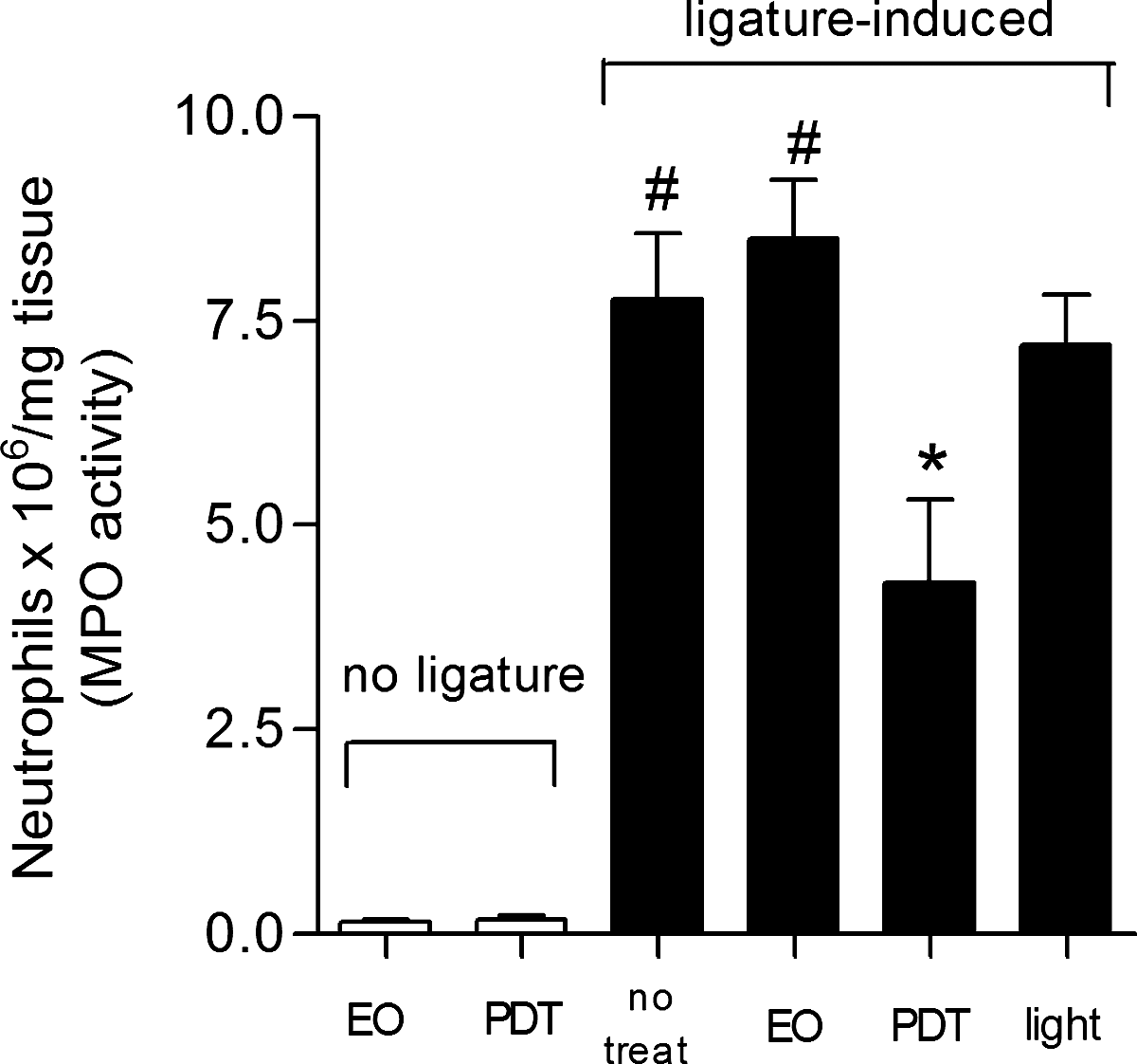
PDT decreases neutrophil migration to the gingival tissue. Myeloperoxidase (MPO) activity present in the gingival tissue homogenates was measured in order to estimate the relative numbers of infiltrating neutrophils in the gingival tissue. Results are shown as mean MPO activity and SD. # p<0.05 compared to non-ligatured animals; *p<0.05 compared to animals with ligature-induced periodontitis without treatment, and compared to animals with ligature-induced periodontitis treated with eosin (ANOVA followed by Bonferroni's test).
Effect of PDT on neutrophil chemotactic cytokine
The possible interference of PDT on the release of TNF-α was investigated in an attempt to clarify whether the downregulation of neutrophil migration, promoted by the treatment, was related to a decrease in the release of the neutrophil chemotactic mediator. The groups without ligatures, treated with eosin alone or with PDT, showed only small amounts of TNF-α in the gingival tissues. However, the groups of ligature-induced animals that had either not been treated, or that had been treated with eosin alone, showed increased TNF-α release (p<0.05). As is shown in Fig. 3, rats treated with PDT presented lower levels of TNF-α in the gingival tissue compared with ligature-induced rats treated with eosin alone (p<0.05). Interestingly, the ligatured animals that had received light treatment alone presented statistically significant decreased levels of TNF-α, compared to ligature-induced rats that were either untreated or treated with eosin alone (p<0.05).
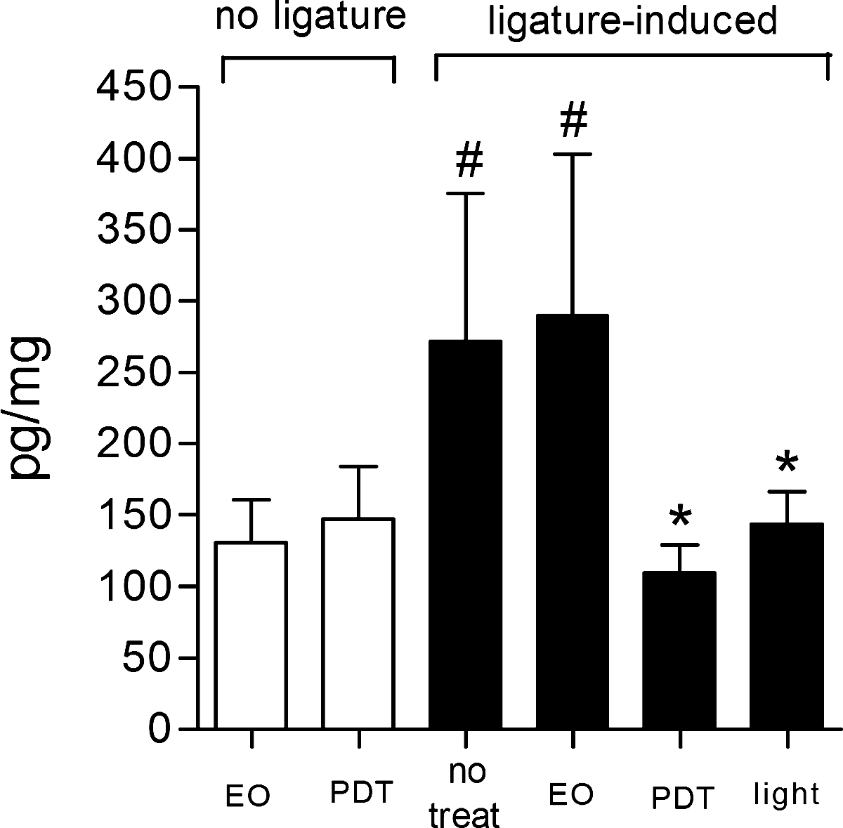
PDT decreases TNF-α release in gingival tissue. ELISA assays were used to measure the amounts of cytokine in gingival tissue homogenates. Results are shown as mean and SD. # p<0.05 compared to non-ligatured animals; *p<0.05 compared to animals with ligature-induced periodontitis without treatment, and compared to animals with ligature-induced periodontitis treated with eosin (ANOVA followed by Bonferroni's test).
Discussion
There have been an increasing number of studies concerning the antimicrobial effects of PDT as a periodontal adjunctive. The association of a photosensitizer with a specific light source induces the production of highly reactive oxygen species such as superoxide ions and hydroxyl radicals. 9 Studies have shown encouraging results using the principles of PDT against microorganisms involved in periodontitis. 15,16 Although an earlier study indicated that adjunctive PDT appeared to provide additional benefit in the treatment of chronic periodontitis, in terms of improved PD and clinical attachment level (CAL), 17 the small number of reported studies precludes any firm recommendations concerning its routine use.
To address these uncertainties, we conducted an animal study investigating the use of PDT therapy and one of its possible mechanisms of action in the periodontal tissue by measuring the inflammatory response after PDT. In the present study, it was demonstrated that the use of PDT inhibited bone loss, induced by ligature placement, in a rat model of experimental periodontitis. The results indicated that the effect of PDT in suppressing bone loss might, at least in part, be because of reduced expression of inflammatory cytokines, as well as lower neutrophil migration.
Histometric analysis indicated that there was less bone resorption, in agreement with a study conducted by de Almeida et al. 18 In this previous study, the authors demonstrated by radiographic examination that there was significantly less bone loss in a PDT group, compared to ligature-induced animals receiving no treatment, at 5 and 15 days postoperatively, but that there was no significant difference in bone loss at 30 days. Different from the present work, the technique used involved low-level laser therapy, with methylene blue. 18 Also, PDT was effective as an SRP adjunctive treatment for bone loss reduction in induced experimental periodontitis when compared to conventional nonsurgical treatment, both in normal rats and in systemic dexamethasone-inhibited animals using toluidine blue O as photosensitizer. 19
There have so far been few studies demonstrating the clinical efficacy of the use of PDT in the treatment of periodontal disease. In recent work, the authors compared the short-term performance of a single session of PDT and of a conventional ultrasonic debridement (UST) in persistent pockets of the disease in maintenance patients. For UST, the mean PD was reduced from 5.3 to 4.5 mm, whereas for PDT it was reduced from 5.3 to 4.7 mm, so there was no difference between the two treatment procedures. Microbial counts were significantly reduced, by ∼30–40%, immediately after debridement, but returned to baseline values after 3 months, irrespective of treatment. 20 In this case, PDT was not superior to conventional mechanical treatment of persistent pockets of disease. In other work, SRP and PDT had similar effects on crevicular TNF-α and RANKL levels in patients with aggressive periodontitis, 21 whereas PDT did not aid conventional non-surgical periodontal therapy in patients with diabetes. 22 It is important to point out that there has been considerable variability in the light sources as well as the photosensitizer dyes used in the studies published to date, which could explain the divergent results.
The role of virulence factors of bacteria in pathogenesis of periodontal disease is well known. Thus, the accumulation of bacterial products stimulate the gingival epithelium to induce the expression of adhesion molecules and pro-inflammatory cytokines and chemokines. In addition, the blood vessels that surround the affected tissue become highly permeable and express adhesion molecules, and a gradient of chemoattractant signals act in the selective recruitment of leukocytes to periodontal tissues. 23 Neutrophils migrate through the junctional epithelium and into the gingival sulcus, 24 and are the predominant infiltrating cells under these conditions, producing high levels of cytokines. Importantly, the animals with ligature-induced disease, that received PDT treatment, showed decreased numbers of infiltrated neutrophils, as well as lower levels of TNF-α. A previous study demonstrated that following exposure of P. gingivalis to a low-energy He-Ne laser (632 nm) and toluidine blue O (TBO), both lipopolysaccharide activity and IL-1 secretion from human peripheral mononuclear cells were significantly reduced. 25 This is an important result, as the presence of the ROS generated by PDT did not induce any inflammatory condition, but instead induced a reduction in the inflammatory mediators associated with lower neutrophil migration. It has also been shown that the use of PDT with methylene blue produced modest effects on osteoblast cells after 24 h, without any signs of apoptosis, suggesting that PDT can inactivate endodontic pathogens without affecting host cell viability. 26
The present work has highlighted the differences between the results obtained using either PDT or light alone. Interestingly, when compared with both ligatured control groups (without treatment or treated with eosin alone), the treatment with light alone significantly decreased the release of TNF-α, although there was no statistically significant reduction of bone resorption or neutrophil migration (in contrast to PDT). Some studies have reported the effect of light on cells and tissues, an effect known as “photobiomodulation”. Torres et al. 27 demonstrated that laser photobiomodulation had a positive influence on the healing of bone defects, and that it could be successfully used to improve bone quality around dental implants, enabling the early wearing of prostheses. 28 However, different from the present work, these earlier studies evaluated bone healing, whereas here the inhibition of bone destruction was investigated. It has also been reported that photobiomodulation significantly suppressed immune cell activation and cytokine/chemokine expression in a rat spinal cord injury model, 29 which is in agreement with the results presented here.
Because tissue destruction is the result of a complex interaction between the periodontal bacteria and the host's immune and inflammatory responses, avoidance of exacerbation of the immunoinflammatory response is an important strategy. Because we have demonstrated that PDT using eosin and LED light decreases the inflammatory markers avoiding the bone resorption, additional studies should be performed to demonstrate the efficacy of PDT as an adjunctive in the treatment of periodontal disease.
Footnotes
Author Disclosure Statement
No conflicting financial interests exist.