
Abstract
Introduction
Mast cells can be found all over the body, mainly on the dermis and superficial tissues, and are distributed along the microvascular bed, close to the basal membrane of endothelial cells and nerves. 3 Mast cell degranulation may start following physical stimulation, or by anaphylotoxins, immunoreactions modulated by IgE, bacterial cytokines and toxins, and others. 2
Mast cells, as secretory cells, will react to the increase of ATP levels caused by laser light. Increased mast cell activity may occur as a result of the imbalance of the level of Ca+, activation of serine esterase, the need of extra energy, and intracellular changes of cyclic adenosine monophosphate, leading to cellular degranulation. 4
Extrusion of mast cells granules and the release of their contents cause the appearance of some bioactive substances on the extracellular matrix, which may trigger the inflammatory reaction. These substances are known as primary mediators and include histamine, serotonin, chemotactic proteins and peptides, such as the eosinophil chemotactic factor-α, neutrophil chemotactic factor, and enzymes. Other factors such as leukotriene B4 may be released by mast cells following their stimulation. 5,6 The most common inductor of mast cell degranulation is the C5a released by the activation of the complement cascade. 7
Previous reports evidenced that photo-irradiation with both red and near infrared (λ630-1000nm) lasers or LEDs influences biological phenomena in both in vivo and in vitro models. 8 –11 Early reports have shown that phototherapy benefits microcirculation. 12,13 A previous study 13 mentioned that both laser and LED light causes vasodilatation in vivo. It was suggested that the effect of the laser light on microcirculation is caused by the stimuli of the degranulation of mast cells and consequent increase of cellular permeability. 14
The lack of reports on the effects of LED light on mast cells prompted us to assess the effect of LED phototherapy (λ630nm or λ850nm) on mast cell count in a rodent model.
Methods
Following approval by the Animal Experimentation Ethics Committee of the School of Dentistry of the Federal University of Bahia, 60 young adult male Wistar rats weighing an average of 250 g were obtained from the animal house of the School of Veterinary Medicine of the Federal University of Bahia, and were kept at the Animal Experimentation Laboratory of the School of Dentistry of the Federal University of Bahia. The animals were kept in individual plastic cages lined with wood chips and maintained at 22°C in a day/night light cycle. The animals were fed a standard laboratory diet Nuvilab (Nuvital, Paraná, Brazil) and had water available ad libidum.
After a regular quarantine period, the animals were randomly distributed into three groups as follows: I, Control; II, IR-LED (Fisioled, MMOptics, São Carlos, SP, Brazil), λ850 nm, 150 mW (0.15W),=1 cm2, 21.9 J/cm2, 73 sec; and III, Red- LED, λ630 nm, 150 mW (0.15W),=1 cm2, 21.9 J/cm2, 73 sec (Table 1). When each animal was under intramuscular general anesthesia (ketamin, 0.12 mL/100 g and xylazine, 0.06 mL/100 g), the tongue was pulled out with a tweezer and LED light delivered by a probe in a contact manner to the dorsum of the tongue. In the control group the procedure was simulated.
Animal death and removal of the specimens occurred after (T0), 20 (T1), 45 (T2), and 60 min (T3) following irradiation. The specimens were routinely processed to wax, cut, stained with toluidine blue and then underwent mast cell counting with a light microscope (Axiolab®, Zeiss, Thornwood, NY). Total, degranulated, and non-degranulated cells were counted on two consecutive fields (x 200) adjacent to the basal layer of epidermis. The results were statistically analyzed using Minitab15 software (Minitab, Belo Horizonte, MG, Brazil). Statistical analysis was performed by paired t-test and ANOVA with significance level set at 5%.
Results
A summary of the results may be seen in Table 2 and Figs. 1 –3.

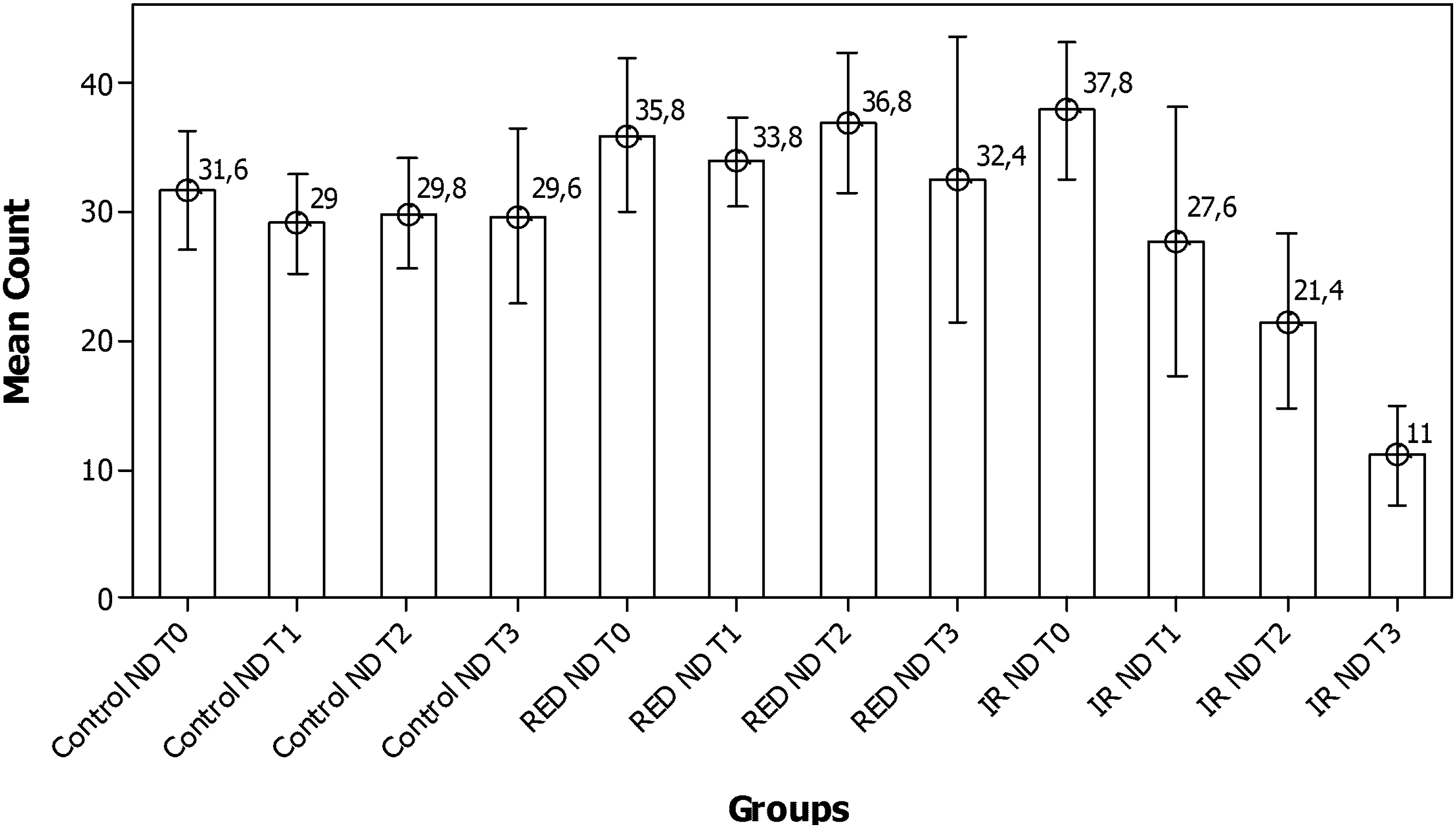
Mean total number of mast cells observed during the experimental time.
Mean number of degranulated mast cells observed during the experimental time.

Mean number of non-degranulated mast cells observed during the experimental time.
T, total number; D, degranulated; ND, non-degranulated; T0, immediate; T1, 20 min; T2, 45 min; T3, 60 min.
The results of the present study showed significant difference between control (p=0.021, ANOVA) and red-LED (p=0.030, ANOVA) groups regarding the number of degranulated mast cells, where a significantly greater number of these cells were noted at T2 and T3 respectively. IR-LED irradiated subjects showed significant differences in the total number (p<0.001, ANOVA) and the number of non-degranulated mast cells (p<0.001, ANOVA). Significant difference was also noticed regarding the number of degranulated mast cells (p<0.001, ANOVA). A significantly greater number of degranulated cells was noted at T3. Time also influenced our results. Both LED irradiated subjects showed significant difference when compared with control subjects in the total number (p<0.001, ANOVA), as well as the number of degranulated mast cells (p<0.001, ANOVA), and the number of non-degranulated ones (p<0.001, ANOVA). When comparing the two groups of LED irradiated subjects, significantly difference was noticed regarding the total number of cells (p<0.001, paired t-test) and the number of degranulated mast cells (p<0.001, paired t-test) with a greater number of these cells noted on IR-LED. On the other hand, red-LED irradiated subjects showed a significantly greater number of non-degranulated mast cells (p=0.001, paired t-test).
Discussion
Photobiological responses are the result of photochemical and/or photophysical changes produced by the absorption of non-ionizing electromagnetic radiation. In a visible region, when a photon is absorbed by a molecule the electrons are raised to a higher energy state and this excited molecule must then lose its extra energy. This may occur by re-emitting a photon of longer wavelength (e.g, less energy) as in fluorescence or phosphorescence, or the molecule can lose energy by giving off heat, or it can lose energy by undergoing photochemistry on mitochondrial cytochromes. The absorption of radiation in an infrared region results in molecular rotations (rotation of the whole molecule about some axis) and molecular vibrations (the stretching or bending of bonds resulting in the displacement of atomic nuclei relative to each other, but not affecting the equilibrium positions of nuclei). 15
The biological effect of low-level visible laser is through photochemistry (probably the photo-activation of enzymes), and the biological effect of infrared radiation is through molecular rotations and vibrations. Visible light initiates, probably by photo-activating enzymes in the mitochondria, a cascade of molecular events leading to a photo-response, and infrared radiation produces the same final response, but initiates the response at the membrane level (probably through photophysical effects on CA++ channels) at about halfway through the total cascade of molecular events that leads to biostimulation. 15
Mitochondrial cytochromes have been postulated as photo-acceptors for red to near-IR light energy and reactive oxygen species have been advanced as potential mediators of the biological effects of this light. The mechanism of photobiomodulation by red to near-IR light at the cellular level has been ascribed to the activation of mitochondrial respiratory chain components, resulting in initiation of a signaling cascade that promotes cellular proliferation and cytoprotection. 16 A comparison of the action spectrum for cellular proliferation after photo-irradiation with the absorption spectrum of potential photoacceptors led Karu 16 to suggest that cytochrome oxidase is a primary photoreceptor of light in the red to near-IR region of the spectrum.
Mast cells, such as other secretory cells, seem to react to an increased ATP level resulting from laser irradiation. The increased activity of the mast cells may be caused by a series of reactions that include disturbance of the balance of calcium ions, activation of serine esterase, and changes in the intracellular adenosine 3’:5’-cyclic monophosphate triggering degranulation. 4
There are previous reports showing that laser light increases mast cell degranulation on both rodent 17,18 and human oral mucosa. 19,20 However we were not able to find reports of the effect of LED light on these cells.
Despite studies focusing on mast cell and laser, a previous report using the human oral mucosa model irradiated with visible laser light (670 nm, 8 J/cm2, 5 mW, 4 min) 19 did not find a difference between irradiated and non-irradiated subjects regarding the total number of mast cells. The authors suggested that this finding might be related to the immediate removal of the tissue. Another study, using an animal model, found a significant increase of the total number of these cells 2 h after irradiation (820 nm, 21.6 J/cm2, 800 mW/cm2, 27 sec). 18 As these cells do not proliferate locally, this increase might be attributed to the direct migration of neighbouring cells to the irradiated site being this stimulated by the laser light. In the present study, we found significant differences in both irradiated groups, signaling a different behavior of the mechanism of induction of coherent and non-coherent light sources of these cells.
Numerous stimuli trigger mast cell degranulation, such as bacterial toxins, physical agents, poisons, biological peptides, polymers, and acetylsalicylic acid. 1 In the present study, the degranulation of mast cells occurred in the non-irradiated tongues of the control animals. Probably, this was caused by simulation of the procedure, a physical stimulus. No significant difference was seen in control group regarding different stages, and the different values found may be the result of the variability of the animals.
It is known that both absorption and penetration of the light depends upon the wavelength. The light affects surface and deep tissues and presents high absorption and low penetration. The effect of visible and invisible laser light (680 nm and 780 nm, 50 mW, 8 J/cm2, 36 Hz) on both the total number and on degranulated mast cells in human oral mucosa has been previously assessed. 14 The results of the study showed an increase of 27% (red laser) or 26% (IR laser) on the degranulation of mast cells when compared to controls, but not between the two wavelengths. Using red and IR LED light we also found significant influence of the LED light on the degranulation of mast cells. However, we found significant difference between the two wavelengths on the degranulation of mast cells (p<0.001). Subjects irradiated with IR-LED showed a significantly higher level of degranulation than the one seen on red-LED irradiated ones.
In this study, it is possible that time influenced the degranulation of these cells as the number of degranulated mast cells on irradiated animals increased significantly when compared to controls. Increased levels of degranulation may have an impact on the inflammatory response, as mediators of the inflammation are released following the degranulation of the mast cells. A previous study suggested that increased mast cell degranulation might be caused by increased ATP production caused by the light. 21
LED light also played a role in the density of mast cells, and time seems to have contributed for this event.
Low-energy laser irradiation has documented benefits in promoting the healing of hypoxic, ischemic, and infected wounds. In comparison to lasers, LED technology generates negligible amounts of heat. It is clinically proven to be safe, and has achieved non-significant risk status for human trials by the Food and Drug Administration. 22
The mast cell has a pathogenetic role in continuous allergic airway disease, and histamine has a role in the induction of the symptoms of allergic rhinitis. 23 Hay fever is induced especially by histamine, a mediator released in association with the degranulation of mast cells. Then, the decrease in mast cell numbers implies a similar tendency in the release of histamine. 24 In a study using metachromatic techniques to verify the action of HeNe laser on the nasal mucosa of rabbits, a progressive degranulation and/or decrease of mast cells with frequently repeated irradiation, was demonstrated, likewise on the tongue of the mouse. 25,26 In this study, both red and IR-LED light caused increased mast cell degranulation, and, comparing the two groups of LED irradiated subjects, a greater number of these cells was noted on IR-LED (p<0.001, paired t-test). However it should be further investigated.
Conclusion
In conclusion, both red and IR-LED light caused increased mast cell degranulation, and IR LED light resulted in a greater number of mast cells.
Footnotes
Author Disclosure Statement
No conflicting financial interests exist.