
Abstract
Introduction
A large percentage of cases do not require any medical intervention, as many of them disappear spontaneously during the child's preschool years. Therefore, they are not a further handicap, either aesthetic or psychological, to the child's future life. 2 According to a study by Garzon et al., patients with vascular anomalies are twice more likely to be women as men. 3 In 65.3% of cases, hemangioma affects children. A positive family history increases the risk of hemangioma formation approximately twofold. Among the significant risk factors belongs is a low birth weight (for every 500 g decrease in birth weight, the risk of developing anomalies increases by 40%). Premature birth, older maternal age, pre-eclampsia and placenta previa are also associated with a higher incidence of tumor. 4 However, it is generally assumed that occurrence of anomalies is mainly sporadic. They are most common in the white race, where the prevalence is ∼10–12%. The prevalence in the African-American population is 1.4%, and in Japanese and Taiwanese infants it is up to 0.8–1.7%. 5,6
In 1982, Mulliken and Glowacki proposed their new classification of vascular anomalies based on biological behavior and typical histology of endothelial cells. 7 The general category of vascular anomalies is divided into biologically active tumors, which are represented by hemangiomas, and biologically inactive vascular malformations, which are further subdivided into fast flow (arteriovenous malformations) and slow flow (capillary, venous, and lymphatic malformations). In 1996, the classification was subsequently accepted by the International Society for the Study of Vascular Anomalies (ISSVA).
Therapy is not simple. In many patients no treatment is ever necessary because hemangiomas are well known for their tendency toward spontaneous involution. Therefore, prolonged clinical observation is the most indicated procedure. Complete involution does not necessarily mean complete disappearance of a vascular lesion; 40–50% of patients may have such residual lesions as scar formation, fibrous-fatty tissue, telangiectasias, or atrophic skin. 8 However, a significant minority of hemangiomas do require treatment. Large hemangiomas causing a cosmetic handicap or those threatening the patient's life with complications such as ulceration, airway obstruction, eye damage, or inability to ingest require medical intervention. There are two main possibilities, as well as others, some of which are actively used and others that are under research. Among the main therapeutic processes belong surgical therapy and conservative therapy. The latter one is based on the corticosteroids. Cryotherapy was used in the past. Tumors were frozen by monoxide nitrogen. Gradual separation of tumor was assumed. The results were satisfactory but, unfortunately, only temporary. Cryotherapy has frequently been criticized because relapses have often occurred. With the modern modality of therapy, laser therapy, based on the selective destruction of larger vessels, the curative effect can be achieved with little or no damage to overlying skin. Because of the limited depth of penetration, the laser system is allowed only for the treatment of superficial anomalies or residual telangiectasias. The other option, sclerotherapy, is predominantly used for lymphatic malformation. Unfortunately it has been shown to cause destruction and scarring of normal adjacent tissue. 8 Therapy with unselective β blocker, propranolol, which is based on apoptosis of endothelial cells, is still under the research. Use of interferon α brings the risk of neurological side effects. 8 –10 However, each treatment option has limited therapeutic benefit with its own side-effect profile and risks.
The argon laser was the first to be used for dermatological patients namely with port wine stains (PWS) in the late 1960s and early 1970s. 11,12 This laser emits continuous-wave (CW) light in multiple wavelengths with two major bands at 488 nm and 514 nm. The light is preferentially absorbed by hemoglobin but is also absorbed by the other dermal components, including melanin and collagen. Because CW energy delivery outcomes depended on dwell time of the laser, uncontrolled heating of tissue often resulted in significant dermal scarring and permanent hypopigmentation. The high incidence of hypertrophic scarring in children under age 12 has made this laser nearly obsolete in the treatment of vascular malformations. A variety of different lasers and light sources other than the pulsed dye laser (PDL) are useful in the treatment of vascular lesions: continuous and millisecond pulsed lasers with wavelengths between green and yellow (potassium titanyl phosphate [KTP-generating the second harmonic from Nd:YAG laser] 532 nm and PDL 585–600 nm), 13,14 millisecond-pulsed high energy near infrared lasers (755-nm alexandrite, 800–810-nm diode, 1064-nm Nd:YAG). 15 Broadband light sources (intense pulse light [IPL]) have increasingly been used for the treatment of acquired vascular lesions. 16,17 The carbon dioxide laser plays a marginal role in the laser treatment of vascular lesions, its application being limited to the treatment of superficial lymphatic malformation. 18 Also Er:YAG laser can be used for superficial hematological malformations. 19
Despite limitations, the laser treatment remains the treatment of choice today. Pulses of light are used to selectively heat and destroy the abnormal blood vessels. The wavelength is chosen to be selectively absorbed by hemoglobin, and pulse duration (exposure time) is determined in order to confine heat to the blood vessels during the pulse, leading to selective destruction. The ultimate goal of laser treatment is the heating of the vessel wall through absorption by hemoglobin. A laser pulse with an exposure time that is shorter or equal to the thermal relaxation time of the target, and sufficient fluence, will cause photocoagulative damage in the target. 20 With pulse durations exceeding the thermal relaxation time of the target structure, more heat diffuses outside the vessels during the exposure, leading to unwanted thermal damage to the surrounding tissue structures. In contrast, very short pulses with high peak power can result in intravascular cavitations, potentially leading to vessel rupture and hemorrhage-purpura. 21
The aim of our study is to evaluate hemangioma treatment using four different types of lasers: alexandrite, Er:YAG, CO2, and PDL.
Methods
Laser irradiation sources
Four laser systems were used (Table 1). Solid state laser systems, alexandrite and Er:YAG, are pumped by the xenon flash-lamp. The active media alexandrite (chromium doped chrysoberyl [Cr3+:BeAl2O4]) or Er:YAG (Erbium doped yttrium aluminum garnet [Er3+:Y3Al5O12]) are located together with the flash-lamp in the ceramic cavity, which transfer an optical energy from the flash-lamp to the crystal. The generated radiation wavelengths are 755 and 2940 nm for alexandrite and Er:YAG laser, respectively.
DCD, dynamic cooling device.
Rhodamin G dye laser active medium is the solution of organic dye Rhodamin G in ethylenglykol. The tube with the dye solution is located in the reflecting ceramic cavity together with the flash-lamp, which is used for pumping. The generating radiation wavelength of the Rhodamin G is 595 nm.
CO2 gas laser uses transversal excitation for the pumping of the carbon dioxide molecules. The radiofrequency (RF) power supply (pumping source) provides voltage to the electrodes, which produce an electrical discharge perpendicular to the laser resonator. Resulting laser emission has a wavelength in the far infrared range: 10.600 nm.
Patients and methods
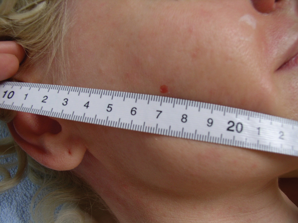
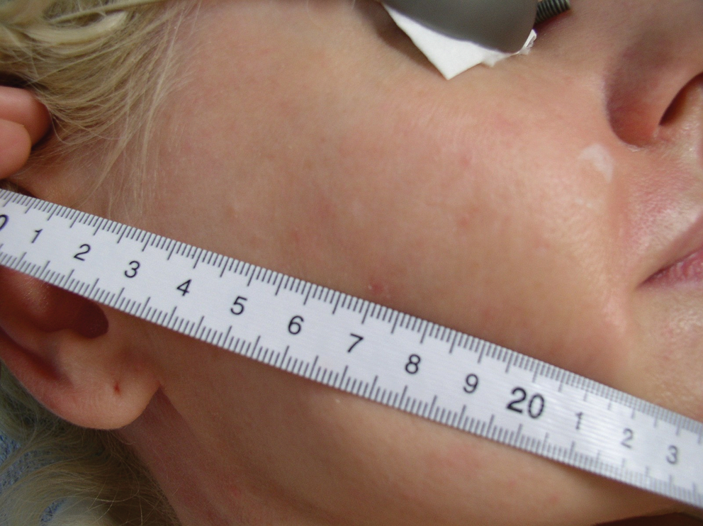
We retrospectively reviewed 869 consecutive patients with hemangiomas (3 mm in diameter, Fig. 1) located in cosmetically sensitive areas treated in the Ave Laser Centre between January 1, 2000 and December 31, 2010. The patients including in our study were divided into four groups according to the type of laser used: alexandrite laser (n=85, 65 women and 20 men), CO2 laser (n=78, 58 women and 20 men), Er:YAG laser (n=105, 87 women and 18 men), and PDL laser (n=601, 453 women and 148 men). All patients were treated in one session without anesthesia application. The ablative systems vaporized the tissues until the hemangioma was removed (Figs. 1 and 2). The non-ablative systems used one shot, which destroyed the hemangioma blood vessels (Figs. 3 and 4).

Boy with hemangioma 3 mm in diameter before laser treatment.

Patient from Fig. 1 after non-ablative laser therapy (PDL, 595 nm, pulse duration 1.5 ms, spot size 5 mm, fluence 11 J/cm2; dynamic cooling device system with cryogen spray [DCD]– 30/20).
Girl with hemangioma 3 mm in diameter before laser treatment.
Patient from Fig. 3 with small scar after ablative laser therapy (Er:YAG, 2940 nm, pulse duration 300 μs, time 1 sec, spot size 2 mm, power 200 W).
Patients in all groups were divided into age categories that were defined as follows: <30: >20 years, ≤30 years; from 31 to 40: >30 years, ≤40 years; from 41 to 50: >40 years, ≤50 years; from 51 to 60: >50 years, ≤60 years; and from 61 to 70: >60 years, ≤70 years. The exact distribution of patients according to their age is summarized in Tables 2 –5.
The retrospective study was conducted according to American Dental Association (ADA) recommendations. Patients were requested to provide informed consent to the clinical examination and regular follow-ups, including photographic records by means of the informed consent form in accordance with the Declaration of Helsinki.
Statistical evaluation
For the treatment efficacy analysis, the following factors were evaluated: therapeutic effect (yes vs. no), loss of pigment (yes vs. no), and appearance of scar (yes vs. no). To determine whether there is a statistically significant difference in treatment efficacy between four observed types of lasers: alexandrite, CO2, Er:YAG, and PDL, Kruskal-Wallis ANOVA was used. To determine whether these three factors (therapeutic effect, loss of pigment, and appearance of scar) are dependent upon patients' age and gender, Kruskal-Wallis ANOVA and Mann-Whitney U-test were performed, respectively. All the tests were considered to be statistically significant at the significance level of p<0.05. For the statistical analysis STATISTICA version 9 (Statsoft Inc.) was used.
Results
The frequency and percentage of observed curative effect, loss of pigment, and appearance of the scar in patients treated with alexandrite, CO2, Er:YAG, and PDL lasers divided into groups according to age are summarized in Tables 2, 3, 4, and 5, respectively.
Frequency and percentage of observed curative effect, loss of pigment, and appearance of scar in patients treated with alexandrite laser are evaluated in Table 2. Treatment effect occurred in only 9.41% (8 patients out of 85). Loss of pigment was observed in 32.9%; appearance of scar was detected in 8.24%.
Treatment was successful using CO2 laser system (97.44%) (Table 3); but loss of pigment was seen in 52.56% and the presence of scar was also frequent (76.92%). These results were statistically significant.
Table 4 evaluated the therapy effect of Er:YAG laser. This system is also very effective: 99.05%. Loss of pigment occurred in 18.10%; appearance of scar occurred in 70.48%. These results were also statistically significant.
PDL laser is the most effective method: 100% (Table 5). The loss of pigment was not observed, and 0.83% (5 patients out of 596) had scars after treatment. This result was statistically significant.
Results (p values) of Mann-Whitney U-test, used to determine whether there exists a statistically significant difference in observed variables (curative effect, loss of pigment, and appearance of scar) between men and women, are given in Table 6 and confirmed the results shown in Tables 2 –5. U-test was performed first for all patients regardless of the laser used, and then for each type of laser separately. Statistically significant results are highlighted in bold. From Table 6 it is seen that statistically significant difference exists in loss of pigment between men and women treated with Alexandrite or Er:YAG laser, and in loss of pigment in all patients regardless of the type of laser used.
Statistically significant results are highlighted in bold.
In Table 7 p values of Kruskal-Wallis ANOVA used to compare the observed curative effect, loss of pigment, and appearance of scar among age categories of patients were shown. Statistically significant results were highlighted in bold. This statistical method was used to detect differences among age categories in observed variables, again both for all types of the lasers together and then separately for each laser. Further examination of the data using multiple comparison tests found statistically significant difference only in appearance of scar in the Er:YAG group of patients between age categories: from 31 to 40 vs. from 51 to 60 (p=0.009) and from 31 to 40 vs. from 61 to 70 (p=0.0200).
Statistically significant results are highlighted in bold.
Kruskal-Wallis ANOVA was also used to determine whether there was a statistically significant difference in the response to laser treatment (therapeutic effect, loss of pigment, and appearance of scars) among the lasers, regardless of age and sex of patients. The results (Kruskal-Wallis test statistic H and p value) are summarized in Table 8. Statistically significant results were again highlighted in bold. With the multiple comparisons, we have demonstrated statistically significant differences for the next observed variables.
Statistically significant results are highlighted in bold.
Curative effect: alexandrite vs. CO2 (p=0.000), alexandrite vs. Er:YAG (p=0.000), and alexandrite vs. PDL (p=0.000);
Loss of pigment: alexandrite vs. PDL (p=0.000), CO2 vs. Er:YAG (p=0.000), CO2 vs. PDL (p=0.000), and Er:YAG vs. PDL (p=0.018);
Appearance of scar: alexandrite vs. CO2 (p=0.000), alexandrite vs. Er:YAG (p=0.000), CO2 vs. PDL (p=0.000), and Er:YAG vs. PDL (p=0.000).
From Table 2 to Table 8 it is evident that the therapeutic effect of all the lasers except alexandrite was very high: almost 100%. In the CO2 and the Er:YAG laser groups, a high percentage of side effects was observed as well. Exposure to these lasers caused loss of pigment and scar formation in many cases. The best therapeutic effect, with only minor side effects has been achieved with the PDL laser.
Discussion and Conclusion
Although hemangiomas are well known for their natural history of spontaneous involution, we can never be sure that close follow-up and anticipatory guidance (so-called “active non-intervention”) is the best approach. This uncertain prognosis may arouse anxiety in parents. This is the reason why early intervention may be indicated. Laser radiation seems to be very an effective tool for hemangioma therapy. Batta et al. 22 conducted the first prospective and randomized controlled study in uncomplicated haemangiomas in 121 children. Treatments were performed with an FPDL (585 nm, 0.45 ms, 3–5 mm, 6–7.5 J/cm2). Sixty children were treated and 61 children served as control. After 1 year, 30% of hemangiomas in the treatment group had resolved completely in contrast to 5% in the control group. We also confirmed that PDL (595 nm; 1.5 ms; 7 mm; 9–11 J/cm2) had the optimal effect without scars.
Er:YAG or CO2 laser radiation is useful for small superficial lesions. 15 The stronger statistically significant difference in loss of pigments and the presence of scars was observed. It is known that Er:YAG laser radiation's high water absorption allows very precise ablation of tissue to depth <30 μm per single shot. 23,24 This also allows a single pass light to peel through the multi-pass deeper peels. This is clearly evident in results for darker skin type patients. The adjacent thermal damage caused by scattering is limited to <5 μm per shot. Common procedure requires only local anesthesia during the treatment, or there is even no need for anesthesia at all. In comparison with pulsed CO2 laser treatment, the lack of residual thermal damage in cases of Er:YAG treatment brings significantly faster healing with less patient discomfort. An average recovery time in this case takes ∼7–10 days (in comparison with 10–14 days in the case of CO2 treatment).
Alexandrite laser is not effective for these purposes because of maximal absorption in melanin and not in hemoglobin. 25
Footnotes
Acknowledgment
This research has been supported by a Grant of the Czech Ministry of Education, IGA MZCR 9991-4 Grant.
Author Disclosure Statement
No conflicting financial interests exist.