
Abstract
Introduction
The argon fluoride excimer laser (hereafter referred to as the ArF excimer laser) operates via a photochemical process that generates less heat. The short wavelength (193 nm) of the ArF excimer laser provides sufficient energy to break covalent bonds in nanoseconds, which makes non-thermal material processing possible. 14,15 This heatless characteristic enables the ArF excimer laser to be used for microscopic processing of polymers in industry 16,17 and for corneal surgery for vision correction in medicine. 18,19 Improved properties of irradiated surfaces have also been reported in the field of polymer chemistry. 20 –22 In dentistry, application of the ArF excimer laser to enamel and dentin preparation has been reported. 14 In contrast to the heat-generating Er:YAG laser, ArF excimer laser irradiation of dentin should not decrease TBS between dentin and composite resin. However, the effects of the ArF excimer laser on dental hard tissue have been examined, mainly with respect to morphology, 15,23 –25 with only one report of the effects of the laser in pretreatment for bonding restoration based on investigation of the contact angle of the irradiated dentin surface. 26
The aim of the present study was to evaluate the TBS between composite resin and human dentin submitted to ArF excimer laser irradiation in combination with conventional surface pretreatment. The treated dentin surface was also observed using transmission electron microscopy (TEM) to investigate bonding interface. We hypothesized that the TBS between dentin and composite resin would not decrease after ArF excimer laser irradiation of dentin.
Materials and Methods
An EX5 Excimer Laser (GAM Laser, Orlando, FL) was used in the study. The beam size was 6×3 mm, the wavelength was 193 nm, and the pulse length was 10 ns. A self-etching primer system (Clearfil Mega Bond Primer and Bond, Kuraray Medical, Tokyo, Japan) was used for pretreatment and Clearfil AP-X (A3; Kuraray Medical) was used as the composite resin. This study was approved by the Ethics Committee of the Faculty of Dentistry, Tokyo Medical and Dental University.
Condition of ArF laser irradiation
In the present study, the parameter of ArF laser irradiation were fixed as irradiation time 10 sec, pulse repetition rate 20 pps and emission voltage 15 kV. A schema of the laser irradiation is presented in Fig. 1. The laser beam was focused on the dentin surface of specimens using a condenser lens (focal distance 30 cm, PCX-25-U-152.6, Lambda Research, Littleton, MA). The distance between lens and target was fixed at 30 cm. In the pilot study, energy per pulse was measured at the target using a laser power meter (PE25-SH-V2, Ophir, UT) and the area of the abraded spot of dentin was measured using a scanning electron microscope (S-4500, Hitachi, Japan); energy per pulse was divided by the spot area and the energy density per pulse 217 mJ/cm2 was obtained. The dentin abrasion depth 50.2 μm was also measured using a scanning electron microscope.
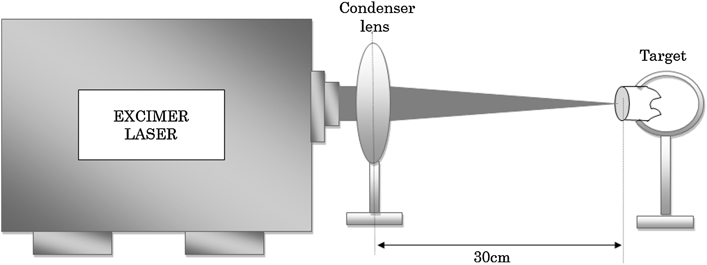
A schema of the laser irradiation.
TBS testing
All steps of the experiments we will describe were performed by one operator. Twenty molars without caries were used in TBS tests. The teeth were stored in distilled water at 4°C immediately after extraction until use. Each tooth was sectioned perpendicular to its long axis using a diamond saw (Isomet 1000, Buehler, IL) to expose a flat dentin surface. The surface was polished with #1000 wet abrasive papers (Noritake, Aichi, Japan) and cleaned using an ultrasonic washing machine (AU-16C, Hirosei, Hiroshima, Japan) for 30 sec. The teeth were randomly assigned to five groups and the dentin surfaces were treated as follows: Group 1: ArF excimer laser irradiation, priming treatment, and bonding treatment; Group 2: priming and bonding; Group 3: ArF excimer laser irradiation and bonding; Group 4: ArF excimer laser irradiation; and Group 5: no treatment (control).
For ArF laser irradiation, irradiation with the condition described previously was repeated as moving an irradiated spot until the entire dentin surface was uniformly covered. For primer treatment, the primer agent was applied on the dentin surface for 20 sec. For bonding treatment, the bonding agent was applied on the dentin surface and cured for 10 sec using a halogen lamp (Tokuso Power Lite, Tokuyama, Tokyo, Japan). Composite resin was built up on the treated surface to a height of 4 mm and was cured using a halogen lamp for 40 sec. After storing in distilled water for 24 h, rod-shaped specimens with a 1×1 mm adhesive interface were prepared for TBS testing using a diamond saw. The specimen was attached to a universal testing machine (AGS-500B, Shimadzu, Kyoto, Japan) using a special jig and was subjected to TBS measurements at a crosshead speed of 1 mm/min in air at 23°C. Ten specimens were used per condition. Differences in TBS were analyzed by one-way ANOVA and a Games-Howell multiple comparison test (p<0.05) using SPSS software v. 17.0 (SPSS, Chicago, IL).
TEM observation of cross sections of treated dentin surfaces
For each experimental group, one additional dentin sample was treated as described previously. After the surface treatment, specimens were prefixed in 2.5% glutaraldehyde, washed with 0.1 M phosphate buffer, postfixed in 1% osmium tetroxide, dehydrated with 50-100% alcohol, and EPON™ embedded. Next, the treated dentin surface was sectioned with a thickness of 80–90 nm and the cross section was observed by TEM (H-7100; Hitachi, Tokyo, Japan) with an acceleration voltage of 75 kV.
Results
TBS tests
Table 1 shows the TBS for each group. The TBS of all treated samples was greater than that for control (untreated) samples (p<0.05). TBS for samples with bonding treatment was greater than that for samples without bonding (p<0.05). Regardless of ArF irradiation, there were no differences in TBS when bonding treatment was included in the procedure.
SD, standard deviation; D, difference according to the result of the one-way ANOVA and a Games–Howell multiple comparison test. The different letters indicate significant difference between tensile bonding strengths (p<0.05).
TEM observations
A hybrid layer was observed at dentin surfaces treated under conditions including priming. The collagen matrix was sectioned and dispersed in the hybrid layer in Group 1 (Fig. 2), whereas collagen in the hybrid layer was dense and connected with unaffected dentin in Group 2 (Fig. 3). In contrast, the hybrid layer could not be seen at the treated surface in Group 3 (Fig. 4). Instead, a multilocular phase was observed at the dentin surface (Fig. 4). In Group 4, an altered-texture layer of 500-nm thickness was present at the irradiated surface and a horizontal crack was observed inside this layer (Fig. 5). In Group 5, an altered-texture layer and a horizontal crack could not be observed (Fig. 6).
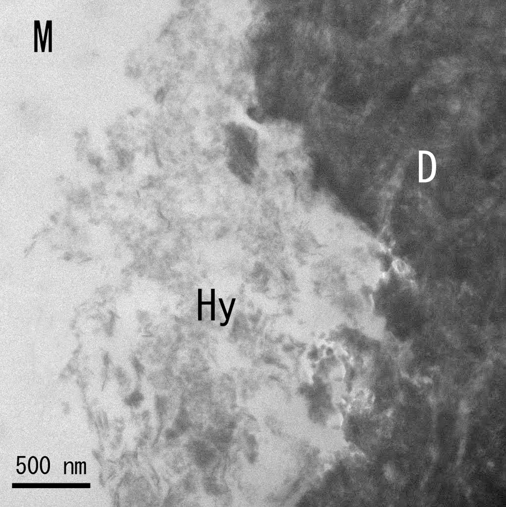
Representative TEM image of a pretreated dentin cross section of Group 1 (original magnification×30,000). Hy, hybrid layer; M, resin; D, unaffected dentin.
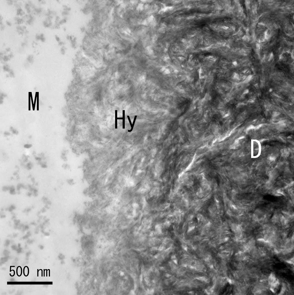
Representative TEM image of a pretreated dentin cross section of Group 2 (original magnification×30,000). Hy, hybrid layer; M, resin; D, unaffected dentin.
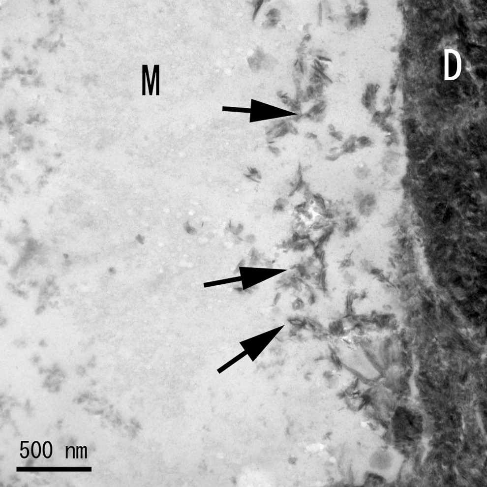
Representative TEM image of a pretreated dentin cross section of Group 3 (original magnification×30,000). M, resin; D, unaffected dentin; arrows, multilocular phase.
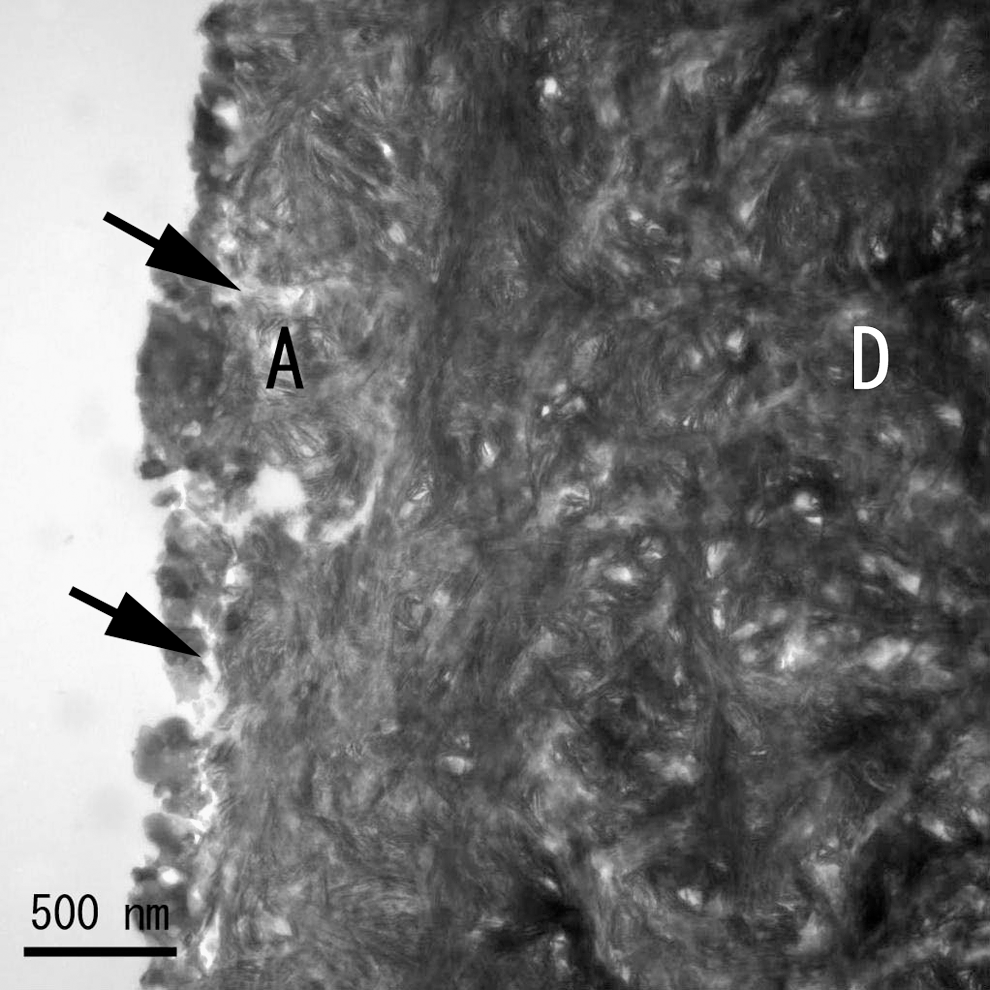
Representative TEM image of a pretreated dentin cross section of Group 4 (original magnification×30,000). A, altered-texture layer; D, unaffected dentin; arrows, horizontal crack.
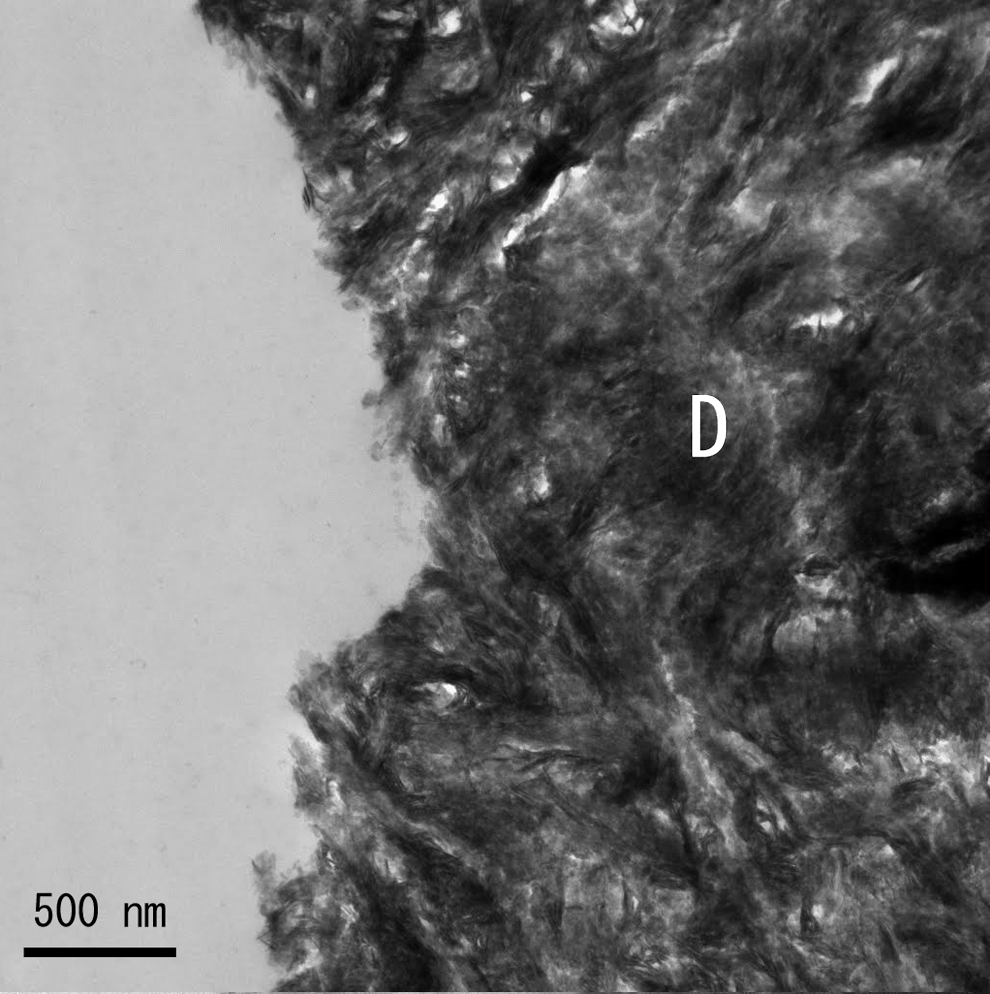
Representative TEM image of a pretreated dentin cross section of Group 5 (original magnification×30,000). D, unaffected dentin.
Discussion
In our previous investigation of the contact angle of an ArF excimer laser-irradiated dentin surface, wettability increased with the energy density of the laser beam. 26 Therefore, the irradiated conditions under which the energy density was the greatest were used in this study. It was assumed that the TBS of samples with no surface treatment would be small enough to cause fracture of the specimens during preparation. Therefore, we adopted the protocol of Shono et al. 27 which minimizes loading on the specimen during preparation. Also, the TBS between resin and dentin has been found to be more sensitive to the technique of operators than to the material properties. 27 Therefore, in the present study, one operator performed all the steps of the experiment.
Our results indicated that the TBS of samples with ArF excimer laser irradiation followed by conventional priming and bonding treatment did not differ from that for samples with only conventional treatment. Therefore, we accept our hypothesis that the TBS between dentin and composite resin would not decrease after ArF excimer laser irradiation of dentin. For Er: YAG laser-irradiated dentin, specific structural features at the bonding interface caused by heat have been reported, including reduced thickness of the hybrid layer caused by higher acid resistance of the irradiated dentin surface and horizontal cracks formed by thermal shock. 28,29 Using TEM, we found that the ArF excimer laser produced an altered-texture layer of 500-nm thickness on the irradiated dentin surface of samples in Group 4, despite the heatless property of the laser. In processing polymethylmethacrylate with a KrF excimer laser, chemical damage caused by a laser of smaller intensity than the ablation threshold was found at a subsurface of ablated area. 30 Such laser-affected but non-ablated dentin was observed as the altered-texture layer in the present study. Thus, dentin components in this layer are partially decomposed by the photochemical process of the ArF excimer laser. The altered-texture layer was not observed at the dentin surface with laser irradiation followed by conventional treatment because the layer was decalcified in the priming process. Instead, a split and dispersed collagen matrix in the hybrid layer formed under this condition, indicating chemical decomposition of collagen matrix in the altered-texture layer caused by ArF eximer irradiation. Despite such split and dispersed collagen matrix in the hybrid layer, TBS of the specimen with laser irradiation followed by conventional treatment was similar to that only with conventionally treatment. Moreover, TBS following ArF excimer laser irradiation only was greater than that for a non-treated dentin surface, while a combination of ArF excimer laser irradiation and bonding treatment resulted in an identical TBS to that for conventionally treated dentin, even though a hybrid layer was not observed. Therefore, the bonding process using ArF excimer laser irradiation is substantively different from that of conventional bonding using micro-mechanical interlocking promoted by dentin hybridization. In polymer chemistry, photo-chemical process caused by ArF eximer laser irradiation has been known to improve quality of material surface for bonding to other material increasing surface free energy. For example, a hydrophobic material such as polytetrafluoroethylene has increased hydrophilicity after ArF excimer laser irradiation, which can be used to process polytetrafluoroethylene with other material. 21,22 In dentistry, Ishida et al. 26 demonstrated an increase in the surface free energy of dentin after ArF excimer irradiation by measuring the contact angle. Such an increase of surface free energy produced close contact between the dentin and resin monomer, resulting in a TBS with ArF excimer laser irradiation and bonding that is identical to that of conventionally treated dentin. The effect of the photochemical process reached a deeper layer than that of primer decalcification, and monomer in primer reacted with the photochemically processed dentin under the hybrid layer. As a result, despite the insufficient hybrid layer with split and dispersed collagen, the TBS of conditioning with ArF excimer laser irradiation followed by conventional treatment is identical to that with conventional treatment. Therefore, the photochemical process caused by ArF excimer laser and the resulting altered-texture layer of irradiated dentin surface would have a positive effect on dentin bonding to composite resin.
In the present study, the TBS between composite resin and human dentin did not decrease after ArF excimer laser irradiation to dentin. From the result, one may say that use of ArF excimer laser for caries removal and cavity preparation would be applicable from the standpoint of following adhesive restoration. Moreover, the possibility of laser etching using ArF excimer laser was also suggested, because the TBS of samples with ArF excimer laser irradiation and bonding was similar to that of priming and bonding. On the other hand, although the ArF excimer laser is thought to generate less heat, structures similar to horizontal cracks were observed in the altered-texture layer of the irradiated dentin surface treated with laser irradiation only. It is possible that a transitory thermal shock produced the crack, or that dentin components separated by irradiation (debris) reattached on the irradiated surface and formed a structure similar to a horizontal crack. The multilocular phase observed in resin with laser irradiation and bonding supported the presence of such debris. The debris on the bonding interface is disadvantageous for reliable bonding. Therefore, further investigation of the altered-texture layer is necessary to determine optimal parameters for irradiation to improve the bond strength between resin and dentin after ArF excimer laser irradiation.
Conclusions
The result of this in vitro study showed that TBS between ArF excimer laser-irradiated dentin and composite resin was identical to that after conventional treatment when the bonding procedure followed laser treatment. TEM showed a sectioned and dispersed collagen matrix in a hybrid layer after laser irradiation, priming, and bonding, but no hybrid layer after only laser irradiation and bonding at the dentin surface. This suggests that the bonding mechanism of dentin after ArF excimer laser irradiation differs from that of conventional bonding in which dentin hybridization is important.
Footnotes
Acknowledgments
This research was partially funded by the Japan Society for the Promotion of Science (JSPS) and Grants-in-Aid for Scientific Research (KAKENHI, B; 2124901). We are grateful to Masayuki Kanai (Aishi College of Dental Technology) for providing instrumentation.
Author Disclosure Statement
No competing financial interests exist.