
Abstract
Introduction
It is known that distention of the periodontal ligaments will result in the differentiation of mesenchymal cells either into osteoblasts or fibroblasts. Within 2 days of the application of the force, both osteoclasts and osteoblasts will start the remodeling of the bone. Disposition of the matrix will occur at the traction side and reabsorption occurs at the compression side. 12,13 These phenomena characterize a selective removal of the bone in some areas, and new bone formation in others, causing the movement of both the tooth and support tissues. 14
At the pressure side, the fibrous supporting tissue is gradually replaced by a newly formed tissue. 15,16 In addition, the extracellular matrix and collagen present at the periodontal ligament degenerate, degrade, and are restructured. 17
There is evidence that the type of injury at the pressure side depends upon the type of the applied force. This change is known as “hyalinization”, because of the glassy aspect histologically observed. Hyalinization of the periodontal ligament interrupts tooth movement. Hyalinization may be considered a sterile necrosis at the pressure side of the periodontal ligament that occurs at early stages of the orthodontic movement. 13,18 The formation of extensive areas of hyalinization may delay significantly the orthodontic treatment, chiefly if the applied force is repeatedly used before a complete repair of the area. 19 The cells found on hyaline areas are unable to differentiate into osteoclasts. Tooth movement will halt until the reabsorption of the alveolar bone occurs, the hyaline tissue is eliminated, and a new periodontal ligament is formed. 13
The appearance of necrotic tissues is an important component of the orthodontic tooth movement, and it is not only observed at initial stages of the treatment but also later on when small hyaline areas are present. 20 The presence of hyalinization in later periods may account for the clinically observed differences in the rate of tooth movement among patients. 21
Previous reports have suggested that the use of laser light during orthodontic treatment causes an increase in the rate of tooth movement. This may be attributed to an increased proliferation of the cells of the periodontal ligament, improved blood supply, and increased activity of both osteoblasts and osteoclasts. 3,7,22 –28
The effect of laser phototherapy (LPT) is not only present on soft tissues but also on bone. It is known that laser light increases osteoblastic activity and reduces the number of osteoclasts. 29 Other studies have suggested that the use of laser light presents a positive effect on bone healing, reduces pain, and quickens bone repair. 30,31 LPT seems to accelerate bone repair, and the newly formed bone seems to be of good quality. 25,32
We have recently reported the use of LPT on orthodontic movement. We found that LPT-irradiated specimens showed a significantly higher number of osteoclasts cells when compared to their controls at 7 and 19 days, as well as a significant increase in the number of osteoblasts between days 7 and 13. The amount of collagen matrix was significantly reduced between days 7 and 13 at both the pressure and tension sides on controls, but not on LPT-treated animals. LPT-treated subjects showed significantly higher deposition of collagen matrix at the pressure side at both the 13th and the 19th days. On the tension side, a significant increase in the amount of collagen matrix was observed on nonirradiated specimens between days 7 and 19. We concluded that LPT caused significant histologic changes on the alveolar bone during induced tooth movement, including on the number of both osteoclasts and osteoblasts, and on collagen deposition on both pressure and tension areas. 3
This study aimed to assess the effects of LPT on the hyalinization observed during orthodontic movement in rodents.
Materials and Methods
Following approval by the Animal Experimentation Ethics Committee of the School of Dentistry of the Federal University of Bahia, 30* healthy Rattus norvegicus young adults male Wistar rats, averaging 3 months of age and weighing between 250 and 300 g, were obtained from the Animal House of the Faculty of Veterinary Medicine of Federal University of Bahia, and kept at the Animal Experimentation Laboratory of the School of Dentistry of the Federal University of Bahia in individual plastic cages lined with wood chips and maintained at 22°C in a day/night light cycle. The animals were fed a standard laboratory diet (Labina®, Purina, São Paulo, Brazil) and had water available ad libidum. After a regular quarantine period, the animals were randomly distributed into two major groups with 15 animals in each. Group I acted as a control and was not irradiated. Animals in group II were treated with laser light as described subsequently. The animals were subdivided into three subgroups (n=5) according to the timing of the animal death (7,13, and 19 days).
In both groups, orthodontic movement was effected using the apparatus devised by King et al. 33 The apparatus was installed on the upper arch of each animal. The force was applied using a 0.010 inch wire (Morelli®, Sorocaba, SP, Brazil) fixed to both extremities of one NiTi coil (Reflex® Closed Coil Springs, light, 150 g, 3 mm, TP Orthodontics, Inc., La Porte, IN, Fig. 1). The apparatus was installed with the animal under intramuscular general anesthesia (12 mL/100 g of Quetamine, Vetaset®, São Paulo, SP, Brazil plus 6 mL/100 g Xylasine, Coopazine®, Coopers, São Paulo, SP, Brazil). 34 With the aim of fixing the wire anteriorly, a hole was drilled with a round bur (KG Sorensen, São Paulo, SP, Brazil) between the two central incisors. This was to provide anchoring of the device. The teeth were then conditioned with 37% phosphoric acid (Alpha Acid® – DFL, Rio de Janeiro, RJ, Brazil). A force of 40 g/F was adjusted to the system with a dynamometer (25–250 dial type – HALDA® Halmstad, Sweden). The wire was marked on the lingual face and fixed with composite resin (Fill Magic Ortodôntico com Flúor, Vigodent®, Rio de Janeiro, RJ, Brazil). Lower incisors were reduced in size to avoid damage to the apparatus during feeding. The first upper molar tooth was removed to eliminate mechanical interference (chewing) that could interfere with the desired mesial movement of the left upper first molar tooth.
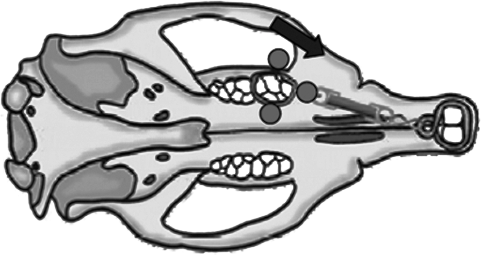
Diagram of the irradiation protocol [Reproduced from Habib, F.A.L., Gama, S.K., Ramalho, L.M.P., et al. (2010) Laser-induced alveolar bone changes during orthodontic movement: a histological study on rodents. Photomed. Laser Surg. 28,823–830.]
LPT was performed using a diode laser [Laser Unit®, Kondortech, São Carlos, SP, Brazil, λ790 nm, round shaped beam, 40 mW, continuous wave (CW), diameter=2 mm (0.0314 cm2), 1.273 W/cm2, time=2×112 sec+1×275 sec (total time 499 sec), 2×142.6/4.48 J+1×350/11 J, 635.2 J/cm2/ 20 J/session, Table 1]. The treatment was performed every other day during the experimental time. The number of sessions varied according to the timing of the animal's death, and the session dose (20 J) was divided into three points: 4.5 J applied at both mesial and distal, and 11 J on the buccal side, the last being applied extra-orally because of the anatomical impossibility of precisely delivering the dose intra-orally. 3 On this application point, the dose of 9 J was increased 20% to compensate losses caused by transcutaneous application of the light. This percent was calculated by using an 8–10% energy loss caused by reflection, and 10% to compensate the loss of energy during the penetration of the light. 35
Animal death occurred by overdose of general anesthetics (36 mg/100 g of Quetamine, Vetaset®, São Paulo, SP, Brazil plus 18 mL/100 g Xylasine, Coopazine®, Coopers, São Paulo, SP, Brazil) at days 7, 13, and 19 after installation of the apparatus. The choice of this timing aimed to get closest to the timing used in human studies, observing the modeling difference between humans and rodents. The apparatus was then removed, and the maxilla taken, cleaned and cut for histologic examination. 3 The specimen was kept in 10% formalin solution during 24 h, decalcified by 5% formic acid, and routinely processed to wax. The slides were stained with hematoxylin and eosin (HE), and underwent histologic analysis performed by an experienced and calibrated pathologist in a double-blind manner (Axiolab®, Zeiss,Thornwood, NY). Descriptive and semi-quantitative analyses were performed according to criteria used previously. 3 The criteria used for semi-quantitative analysis were as follows: Absent, Discrete (presence of one area of hyalinization at the pressure zone), Moderate (presence of two areas of hyalinization associated with the pressure zone), and Intense (presence of more than two areas of hyalinization associated to the pressure zone).
Statistical analysis
Statistical analysis was performed using Kruskal–Wallis or Fisher's test, because of the small sample size, and significance level was set at 5%.
Results
At day 7, on control specimens, hyalinization was discrete in 60% of the cases, moderate in 20%, and absent in 20%, and was mainly located at both mesial and distal apical regions. Laser-treated subjects showed moderate hyalinization in 80% of the cases, and it was absent in the remaining 20%. Typical aspect of the hyalinization in both control and experimental groups may be seen in Fig. 2A and B.
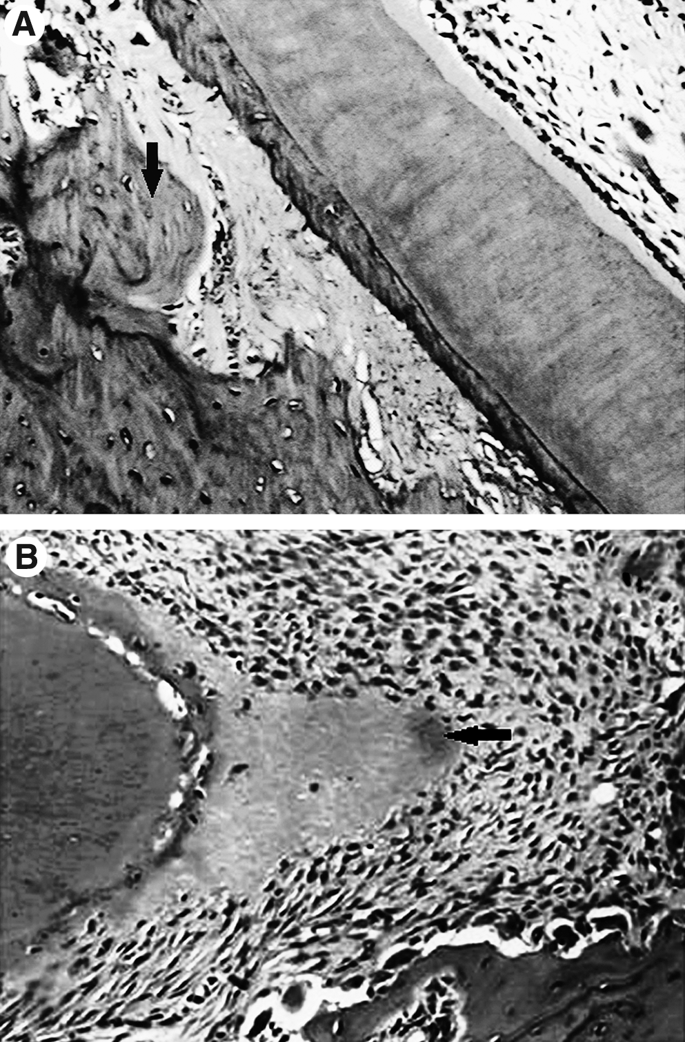
Photomicrographs showing hyalinization (arrows) at the 7th day on (
At the day 13, hyalinization was absent in 60% of the specimens and discrete in 40%, and was mainly seen at the mesial side of the distal root and at the distal side of the mesial root. In laser-irradiated specimens hyalinization was discrete in 40%, intense in 20%, and absent in 40% (Fig. 3A and B). At the end of the experimental period, control specimens, hyalinization was absent in 80% of the cases and discrete in the remaining 20%, and was mostly located ay the distal surface of the palatine root. On irradiated subjects, hyalinization was moderate or absent in 40% and discrete in 20% (Fig. 4A and B). Figure 5 shows a percent distribution of the results.
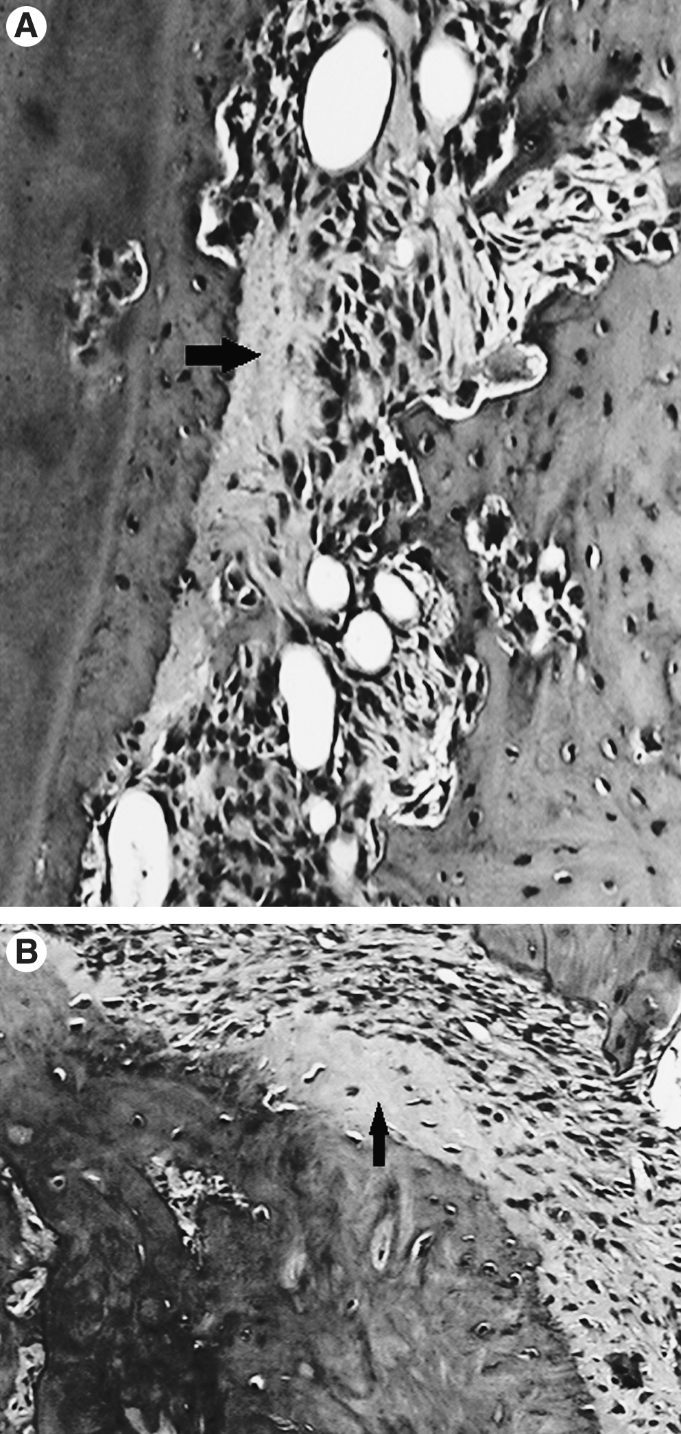
Photomicrographs showing hyalinization (arrows) at the 13th day on (
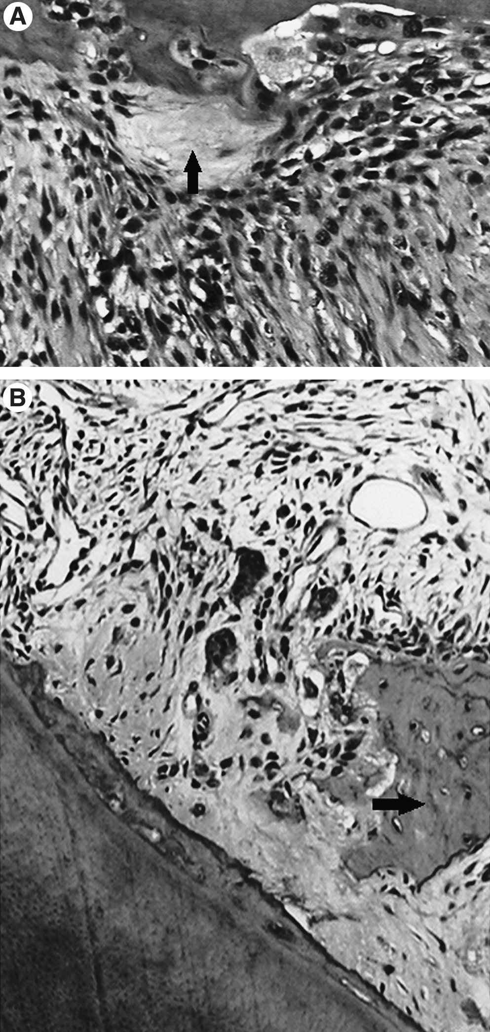
Photomicrographs showing hyalinization (arrows) at the 19th day on (
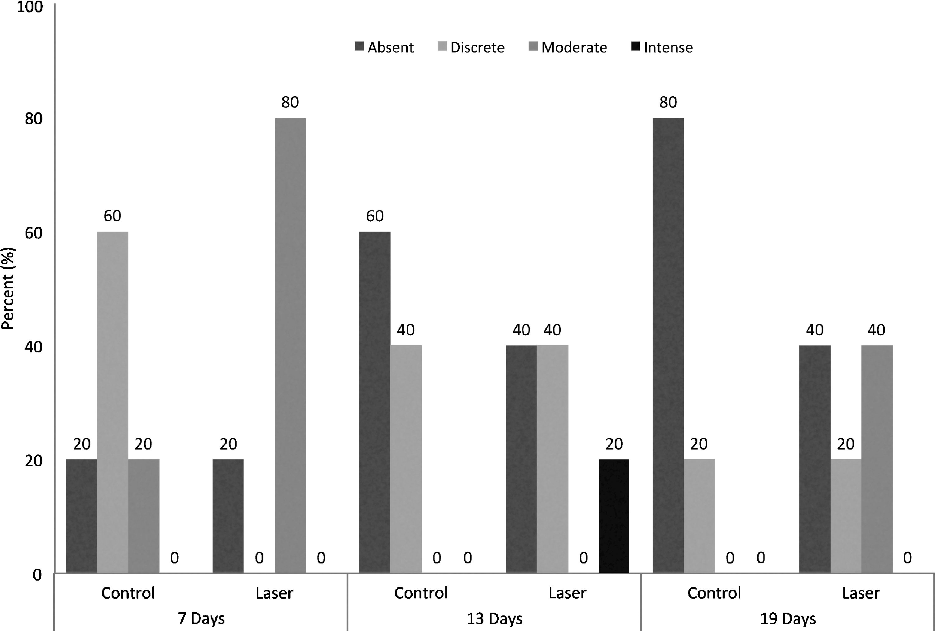
Summary of the histomorphometric study during the experimental time.
Statistical analysis showed that hyalinization was significantly reduced from day 7t to day 19t (p≤0.001) on controls (Fig. 2). The same was observed on irradiated subjects between days 7 and 13 (p=0.015). Comparing controls and irradiated animals evidenced that laser-treated animals showed significantly more hyalinization than did the controls at both the 7th (p=0.007) and 19th days (p=0.048).
Discussion
Many previous studies aiming to reduce the timing of the orthodontic treatment using several approaches, such as the use of chemical mediators, hormones, or drugs. 3,25,36 –40 The animal model used in previous studies has been, mainly, the rat, because of its quicker metabolism when compared to humans. 41,42 Previous reports in the literature have shown that LPT is capable of inducing histologic modifications during orthodontic movement. 3,22,43 –45
As a therapeutic agent, LPT has both local and systemic effects. 46 In the present study, we used both irradiated and nonirradiated subjects, not disregarding the systemic effect of the light as in some studies. 25,47 However, many studies have used contralaterals as controls. 23,43,45,48 The protocol of irradiation used in the present investigation had been used previously. 3,23,25,43,48
In the present study, we used a initial force of 40 g/F applied in a constant manner as suggested by a previous report. 49 We used upper incisors as anchors because of the size of their roots and their characteristic uninterrupted eruption.
Laser application was performed intra-orally at the palatine and extra-orally because of anatomical constraints. A previous study has used a fiber measuring 0.6 mm to deliver the light to teeth. 25
Areas of hyalinization impair the activity of the osteoclast, preventing them from performing reabsorption of the bone. It is well accepted that the shorter the duration of the presence of hyalinization during remodeling of the bone, the more quickly will tooth movement occur. 12 –14,34,50
As hyalinization may be considered a sterile necrosis at the pressure zone of the periodontal ligament observed during the initial stages of the orthodontic movement, 13,18 the formation of extensive hyaline areas might cause an important delay for the tooth movement. In the present study, we found a significantly reduced expression of hyalinization after 19 days. On irradiated subjects, hyalinization was increased at day 7 with significant reduction at day 13. It was evident that the significant increase in the level of hyalinization observed in irradiated subjects at early stages of the movement would favor an increase in the rate of the movement later on. It is well known that cells present at hyalinized areas are not able to differentiate into osteoclasts, vital cells for the reabsorption of the alveolar bone and for the development of a new periodontal ligament. 13
Most reports on tooth movement in orthodontics evidence that hyalinization is progressive, and that it is reduced over the course of the treatment time. It is considered a complication, as it delays the onset of bone reabsorption that starts after reorganization of the cellularity of the area of necrosis. Therefore, the larger the hyalinization area, the longer the time for tooth movement will be. 21 Our findings are indicative that, on clinical grounds, the use of LPT during orthodontic treatment would accelerate the process by stimulating cellular events that occur on the bone during the treatment.
Conclusions
It is possible to conclude that the use of laser light caused histologic alterations during the orthodontic movement, characterized by increased formation of areas of hyalinization at early stages, and late reduction when compared to nonirradiated animals.
Footnotes
Acknowledgment
The authors gratefully acknowledge the Conselho Nacional de Desenvolvimento Científico e Tecnológico (CNPq) for the financial support.
Author Disclosure Statement
No competing financial interests exist.
*
Sample size was calculated using G Power Software.