
Abstract
Introduction
The conventional axiom for surface preparation of the resected root end has been to produce a smooth root surface by different types of burs. 3 However, little information exists regarding whether smooth root ends heal more quickly than do rough root ends. Weston et al. 4 investigated the effect of the surface topography of resected root ends on periodontal ligament (PDL) fibroblast attachment and found no significant difference in cell attachment to the root ends prepared with various instruments.
Lasers have been used for root end resection. 5,6 This technique presents many advantages over burs, including hemostasis 7 and the absence of discomfort and vibration. 8 The erbium (Er):YAG laser stands out for offering good cutting capacity for the root end without signs of thermal damage. 9 Komori et al. 10 showed that Er:YAG laser irradiation promoted clean resected surfaces with evidence of dentinal tubules without a smear layer and no signs of thermal damage.
Oliveira et al. 11 suggested the neodymium (Nd):YAG laser as a promising tool to optimize apicoectomy surgery by sealing the dentinal tubules and decreasing permeability. The Nd:YAG laser could reduce apical microleakage associated with failure of periapical surgery. Laser irradiation has an antimicrobial effect, which is vitally important in apicoectomy surgery. 12 In addition, laser irradiation has been used in apicoectomy surgery for root end cavity preparation 13 because of effective removal of the smear layer 14,15 and exposure of collagen fiber on the root surface, 16,17 which could influence clinical healing by enhancing PDL cell attachment.
Cellular responses are greatly influenced by surface properties, including roughness and morphology. Different qualities of resected root ends may affect the orientation of surrounding PDL cells and tissue. 18 After root-end resection, the surrounding cells proliferate, migrate to the wound site, and synthesize new matrix components. 19,20 During laser irradiation, many changes occur in the dental hard tissue because of the mechanisms of hard tissue interaction, and produce surfaces with different textures and morphologies. 11,16 Regarding some studies, 21,22 laser irradiation produces biocompatible surfaces for periodontal cell viability. On the other hand, the effect of laser irradiation can be favorable or unfavorable depending on laser wavelengths and parameters. Resected root surfaces after treatment with burs, Er:YAG and/or Nd:YAG laser irradiation may have different effects for initial cell attachment. Therefore, the aim of this in vitro study was to evaluate fibroblast adhesion to dentin surfaces treated with Er:YAG or Nd:YAG laser irradiation by analyzing the morphology and roughness of dentin surfaces.
Materials and Methods
Specimen preparation
Thirty-five freshly extracted single-rooted human teeth with fully formed apices were collected after approval from the Ethical Committee of Tokyo Medical and Dental University (#535). For preparation of dentin disks, the root apex of each tooth was removed and dentin disks were prepared using a low-speed saw (IsoMet; Buehler Ltd., Lake Bluff, IL). A total of 90 dentin disks were prepared and washed three times with phosphate-buffered saline (PBS).
Laser devices
Both Er:YAG laser (Erwin AdvErl; Morita, Kyoto, Japan) and Nd:YAG laser (Manipulser; MANI, Tokyo, Japan) systems were used in this study. The parameters of the Er:YAG laser were as follows: wavelength of 2.94 μm, range of output energy settings from 30 to 350 mJ/pulse, maximum pulse repetition rate of 25 pps (Hz), and pulse duration of 200 μs. In this study, a straight tip with a diameter of 800 μm (P800FL; Erwin AdvErl) was used. Nd:YAG lasers produce a near infrared wavelength of 1.064 μm, with a pulse length of 150 μm delivered via a quartz optical fiber of 320 μm in diameter.
Treatment groups
A total of 90 samples were randomly divided into three groups of 30 each: • Group A (Control) samples were treated with a carbide bur under water irrigation for 60 sec by one operator. No laser irradiation was performed. • Group B (Er:YAG laser) samples were treated with Er:YAG laser irradiation with an energy output of 60 mJ/pulse (energy density: 5.97 J/cm2/pulse) and a pulse frequency of 10 pps. The contact tip was moved in a sweeping manner to irradiate the entire dentin surface. The working time required for this procedure in each sample was 60 sec and was performed by only one operator. • Group C (Nd:YAG laser) was irradiated by an Nd:YAG laser with an energy output of 60 mJ/pulse (energy density: 4.58 J/cm2/pulse), a pulse frequency of 10 pps, and a working time of 60 sec. Before laser irradiation, methylene blue dye was applied to the dentin surfaces to absorb the Nd:YAG laser beam efficiently. Then excess dye was removed by washing thoroughly after laser treatment, and the treatment was performed by one operator.
Following each treatment, the samples were thoroughly rinsed with saline solution. After rinsing, a sterilized cotton pellet was applied to dry each sample.
After laser treatment, 20 samples from each group were randomly chosen for further cell culture treatment, and the remaining 10 from each group were morphologically evaluated with a scanning electron microscope (SEM) and a confocal laser scanning microscope (CLSM).
SEM observation of dentin surface morphology
Five out of 10 samples from each group were randomly chosen for SEM observation to investigate the surface morphology. The dentin disks were fixed for 2 h with 2.5% glutaraldehyde solution and rinsed with 0.1 M phosphate buffer solution. The disks were then dehydrated in a series of graded ethanol solutions. After washing with 3-methylbutyl acetate, the disks were dried in a critical-point drying apparatus (HCP-2; Hitachi, Tokyo, Japan) with liquidized CO2. The samples were sputter coated with osmium using an NL-OPC80N plasma coater (Filgen, Nagoya, Japan) and observed under an SEM (S-4500; Hitachi) at an accelerated voltage of 15 kV and a magnification of ×1500.
CLSM observation
Five samples from each group (n=15) were analyzed for surface topography using a scanning laser microscope (1LM15W; Lasertec, Yokohama, Japan). To evaluate surface roughness, the arithmetic mean deviation of the roughness profile (Ra) was calculated by measuring the two-dimensional surface profile using the image analysis and measurement software LM Eye. Measurements were performed at three points for each specimen. Five specimens were prepared for each group, and the results were analyzed by one-way ANOVA and Fisher's protected least significant difference test at the 5% level.
Cell culture
Mouse NIH/3T3 fibroblast cells were seeded onto 24-well plates and cultured in Dulbecco's modified Eagle's medium (DMEM) (Wako, Osaka, Japan) containing 5% fetal bovine serum (Gibco, Invitrogen, Grand Island, NY) at 37°C in a humidified incubator under 95% air and 5% CO2. After the surface treatments, 20 disks from each group were placed into 24-well plates containing 1×10⁴ NIH/3T3 cells/well in 500 μL of DMEM and further incubated for 12 or 24 h.
SEM observation of cell morphology
The dentin disks were cultured with NIH/3T3 cells for 12 or 24 h. Ten samples from each group (n=30) were randomly chosen to evaluate the influence of the surface treatments in the Er:YAG laser, Nd:YAG laser, and control groups according to NIH/3T3 cell morphology by SEM. At the end of the two incubation periods, unattached cells on the dentin surfaces were removed by three rinses with sterile PBS. The samples with attached cells were then fixed, rinsed, postfixed, dehydrated, dried, and coated with gold in a manner similar to that used for SEM sample preparation. The cells were examined by SEM at an accelerating voltage of 15 kV and a magnification of ×1000.
Numbers of attached cells
Overall, 10 samples from each group were used for proliferation assays. The cell numbers were analyzed using the WST-8 assay (Cell Counting Kit-8; Dojindo, Kumamoto, Japan). After 12 or 24 h of culture, the dentin samples were washed with sterile PBS to eliminate unattached cells, and the adhered cells were removed from the dentin disks by incubation with 0.25% trypsin in EDTA. The resulting cell suspensions were placed in 96-well plates in 100 μL of DMEM (Wako, Osaka, Japan) together with 10 μL of a working solution containing WST-8 and incubated for 2 h at 37°C. The absorbances of the colored product were measured using a microplate reader (Well Reader SK601; Seikagaku, Tokyo, Japan). Four measurements were performed for each sample.
Statistical analysis
The number of attached cells in each group was calculated as the mean±standard deviation (SD). Differences between the groups were analyzed by one-way analysis of variance (ANOVA) together with a post-hoc Fisher's protected least significant difference test. Values of p<0.05 were considered to indicate statistical significance.
Results
SEM observation of dentin surface morphology
Group A (Control) basically displayed a smooth appearance, and the surfaces were covered with debris (Fig. 1A). In Group B (Er:YAG laser), photomicrographs revealed irregular and scaly morphologies with intense ablation of intertubular dentin, removal of the smear layer, and open dentinal tubules (Fig. 1B). Group C (Nd:YAG laser) exhibited partially irradiated craters produced by Nd:YAG laser irradiation (Fig. 1C). Inside the crater, globules probably composed of melted hydroxyapatite, microcracks, and minute fissures were found.
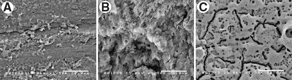
SEM images of the dentin surfaces after the treatments.
CLSM observation
Three-dimensional CLSM images of all surface morphologies are shown in Figs. 2, 3, and 4. In Group A, the dentin surface appeared relatively smooth with the smear layer as shown in Fig. 2. Er:YAG laser irradiation significantly changed the surface morphology. Figure 3 shows an optical image of Group B. The image revealed a homogeneous deep profile of the surface topography, and the smear layer was not visible (Fig. 3). In Group C, the dentin surface treated with Nd:YAG laser irradiation appeared as a flake-like structure, and the superficial part of the dentin surface was affected by laser irradiation (Fig. 4).
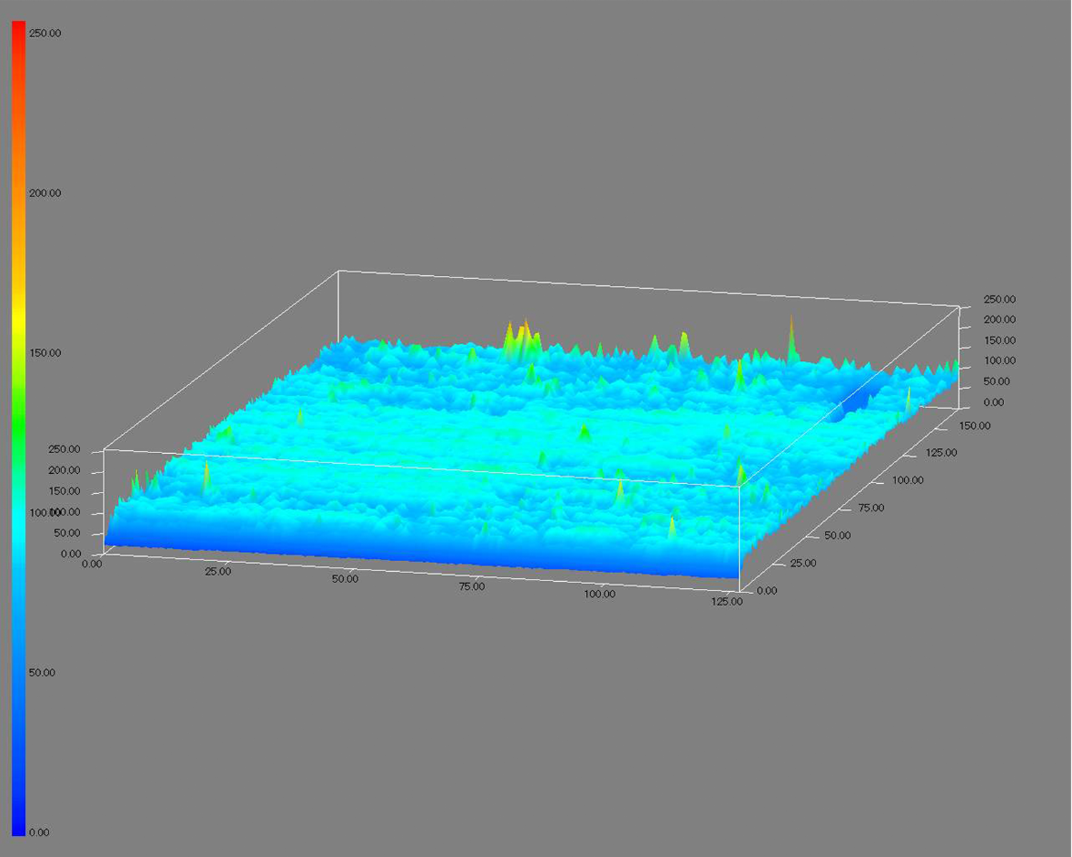
Three-dimensional confocal laser scanning microscope (CLSM) image of Group A. The dentin surface appears relatively smooth with the smear layer. Original magnification: ×50.
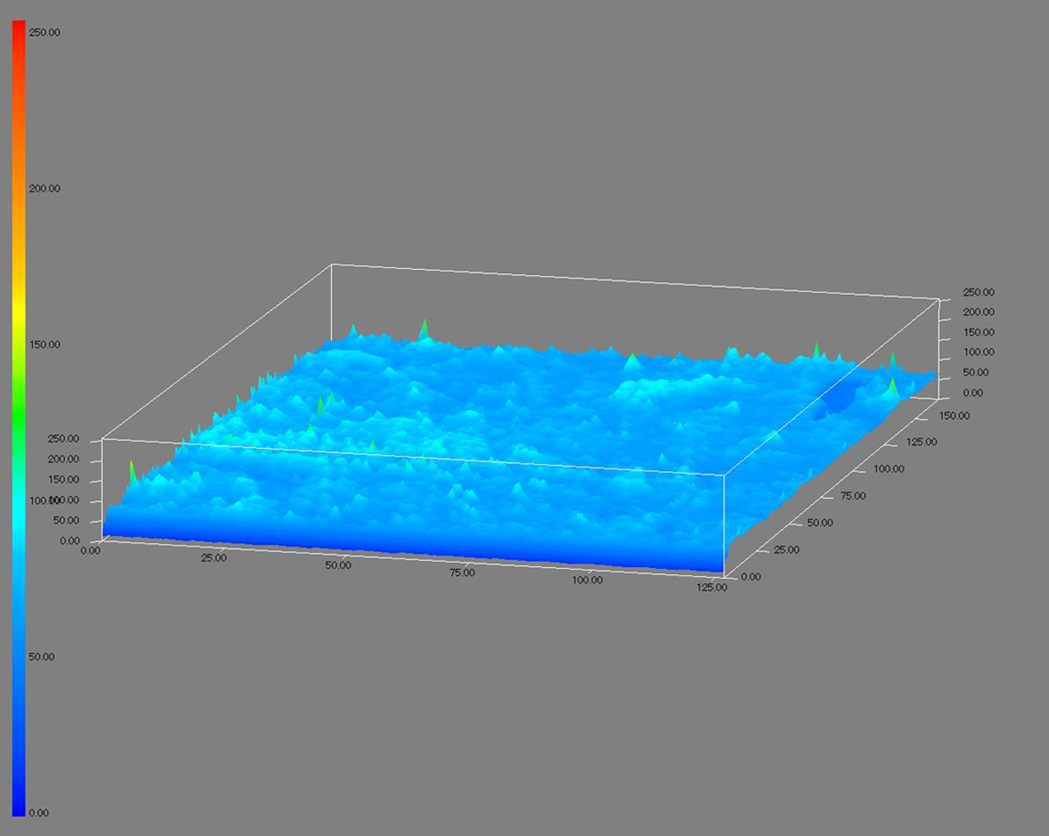
Three-dimensional confocal laser scanning microscope (CLSM) image of Group B. The image reveals a homogeneous deep profile of the surface topography, and the smear layer was not visible. Original magnification: ×50.
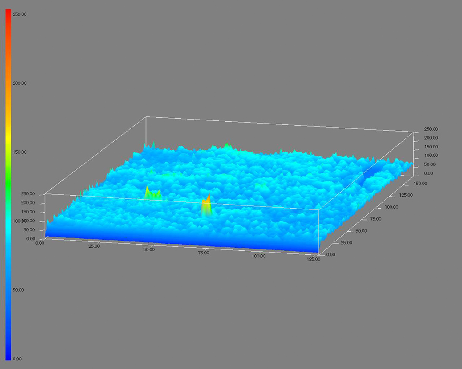
Three-dimensional CLSM image of Group C. The dentin surface appears as a flake-like structure, and the superficial part of the dentin surface was affected by laser irradiation. Original magnification: ×50.
To quantify the observed change in topography of all dentin samples, surface roughness analyses were performed using CLSM and statistical analysis (Fig. 5). Group A displayed smaller Ra values (5.75±1.62 μm) compared with those of laser treatment groups. Group B exhibited significantly higher Ra values (9.86±4.3 μm) than those of Groups A and C (7.41±0.84 μm).
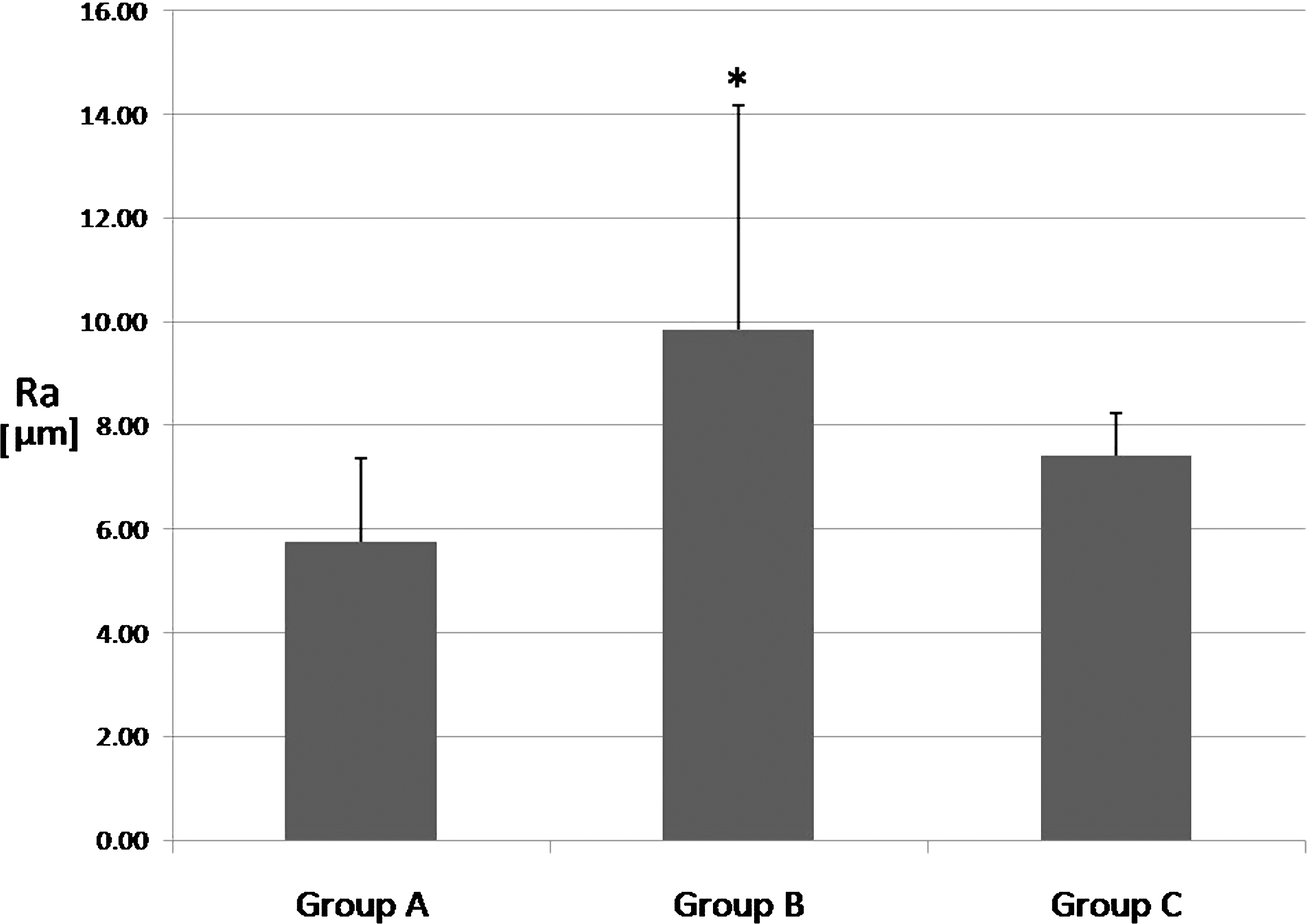
Measured roughness average, Ra, of the dentin surface. Group B exhibited significantly higher Ra values than those of Groups A and C. Statistically significant difference between the groups is indicated by an asterisk (p<0.05).
SEM observation of cell morphology
Representative SEM images of attached cells on the three different dentin surfaces after 12 and 24 h of culture are shown in Figs. 6 and 7, respectively. After 12 h of culture (Fig. 6), the attached cells started to adhere via thin filopodia to the surfaces of Groups A and C (Fig. 6A and C). In Group B, the cells attached to the dentin by expanded filopodia (Fig. 6B). After 24 h of culture (Fig. 7), the cells continued to proliferate and adhered to all dentin surfaces (Fig. 7A–C). No obvious differences in the cell morphologies were observed between the control group and the laser treatment groups after 24 h of culture. Group B presented many cells attached to the dentin surface (Fig. 7B).
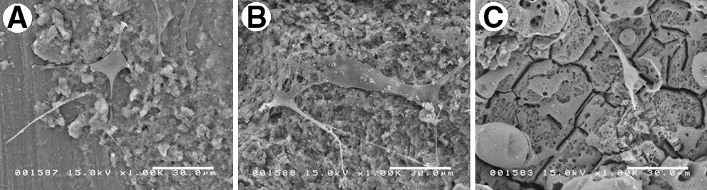
SEM images of attached fibroblasts after 12 h of culture.
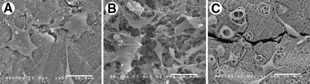
SEM images of attached fibroblasts after 24 h of culture. No obvious differences in the cell morphologies were observed between the control group
Attached cell numbers
The numbers of cells attached to the three different surfaces after 12 and 24 h of culture are shown in Fig. 8. After 12 h of culture, Group B showed a significantly high number of attached cells compared with Groups A and C. After 24 h of culture, the adhesion rate was highest on the surfaces in Group B, followed by Group A (Control). These groups differed significantly (p<0.05) from Group C, which exhibited the lowest number of attached cells.
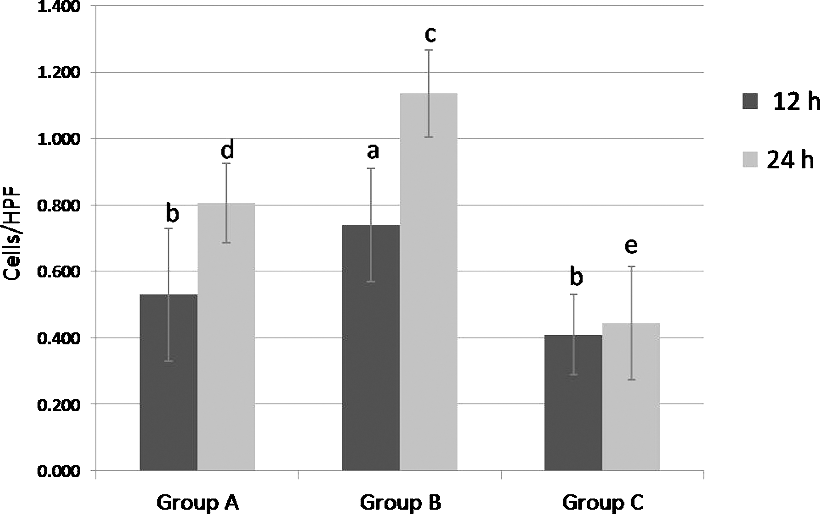
Numbers of fibroblasts attached to the dentin surfaces in the control and experimental groups. The ordinate represents the number of attached cells/high-power field (HPF) after 12 and 24 h of culture. All data are means±SD (n=4). Different letters indicate statistical significance (p<0.05).
Discussion
The critical point after apicoectomy surgery is the attachment of PDL fibroblasts to the resected root surfaces, because PDL fibroblasts may differentiate to cementoblasts or osteoblasts, and functionally oriented PDL is required for periodontal regeneration. Most studies using laser techniques for root-end resection mainly concentrated on root surface decontamination and sealing ability. 23 –25 In this study, we focused on the modifications induced by Er:YAG laser or Nd:YAG laser and the effects of the altered surfaces on cell responses.
In the conventional method, surface preparation of the resected root end is performed by various types of burs that produce surfaces covered with a smear layer, which represents the presence of microorganisms in the apical area. 5 However, little information exists about the effects of surface topography after using Er:YAG or Nd:YAG laser irradiation for surface preparation. In the present study, an energy output of 60 mJ/pulse and a pulse repetition rate of 10 pps were selected because too-low energy output does not significantly change the dentin surface. Feist et al. 21 reported that the surfaces treated with 60 mJ/pulse Er:YAG laser irradiation underwent faster adhesion and growth of cells than did surfaces treated with 100 mJ/pulse Er:YAG laser irradiation. It was necessary to determine the optimal irradiation condition of the Er:YAG and/or Nd:YAG laser for faster cell attachment to dentin surfaces.
The amount of energy absorbed depends upon the absorption coefficient of each molecule in the tissue. The Nd:YAG laser at a wavelength of 1.06 μm has much lower absorption in water and hydroxyapatite than does the Er:YAG laser. Because of this low absorption, the use of methylene blue is necessary to increase the absorption efficiency of the laser beam when the Nd:YAG laser is applied to the dentin surface. The better absorption allows for a more efficient photothermal effect on hard tissue. 26 This mechanism produces different dentin surfaces after different wavelength treatments.
The laser technique might have an advantage, because it has an etching effect and does not cause demineralization of peritubular dentin, and the dentinal tubules remain open with no widening. 27 In the SEM observation of this study, Er:YAG laser irradiation induced typical micro-irregularities with complete removal of the smear layer. This morphological pattern is consistent with previous studies using Er:YAG lasers 28,29 and can be attributed to the laser mechanism, which can cause dissolution of mineral components and fusion of amorphous particles, resulting in microscopic irregularities. Some studies indicated that the Nd:YAG laser can cause dentin surface melting followed by recrystallization and a rough surface. 11,30 In this study, SEM results revealed that irregular craters were observed in the Nd:YAG laser-irradiated surfaces, including melted areas and microcracks with partial removal of the smear layer. The melting and recrystallizing effects of Nd:YAG laser irradiation cause rough dentin surfaces and produce many cracks and fissures. 31
The laser-irradiated surfaces had generally rough surfaces compared with the nonirradiated surfaces (Figs. 2 –4). Moreover, Er:YAG laser-irradiated dentin surfaces gave the largest Ra value because of deep, rough surface formation (Fig. 5). Yamada et al. 26,32 reported that the lowest Ra value was obtained after Nd:YAG and CO2 laser irradiation, and the deepest cavities were formed after Er:YAG laser irradiation. Previous studies showed that the surface roughness of root surfaces following irradiation with an Er:YAG laser does not depend upon the angulation of the working tip, 33 and that the surface irregularities increased with increased power settings. 34 Increased power may also lead to production of a char layer on the root surfaces, melted minerals, and cavitation defects, 35 which hinder the cell attachment of PDL cells. 36
In the present study, the morphological roughness of the Er:YAG lased-surfaces enhanced the adhesion of fibroblasts, and a significantly higher number of attached fibroblasts were observed at 12 and 24 h. Moreover, cell morphology can be regarded as an indicator for the affinity of the cells; flat cells are firmly attached to the surface by means of numerous attachment extensions and lamellipodia, and round cells can be considered to be poorly attached. 37 In our study, spindle-shaped and well-attached fibroblasts were found in the Er:YAG laser group at 12 h based on SEM observation (Fig. 6B). Similar results were obtained by Feist et al., who studied the adhesion of fibroblasts on root surfaces treated by Er:YAG laser irradiation; the lased surface exhibited a significantly higher number of attached cells. 21 In addition, regarding the effect of laser-induced morphology, there has been the speculation that the surface alteration caused by Er:YAG laser irradiation may expose chemical substances in the dentin that are highly selective for chemotaxis of fibroblasts. 38 Some chemical agents reportedly caused alterations in the chemical structure of human dentin after laser irradiation. 34 These modifications induced by Er:YAG laser irradiation could be a direct consequence of root conditioning by exposure of certain extracellular constituents acting on the attachment mechanism of fibroblasts. 35
However, morphological alteration by Nd:YAG laser irradiation inhibited cell attachment because of its destructive effects. Nd:YAG laser irradiation altered the biocompatibility of the cementum surface, making it unfavorable for fibroblast attachment, 39 whereas Fayad et al. found morphological changes on the dentin surface after CO2 laser irradiation, including charring and melting of the dentin, and concluded that these changes may have played a role in the absence of PDL cell attachment. 36 Cyanamide and cyanate, which are cytotoxic chemicals, were detected on charred surfaces. These cytotoxic residues prevent cell attachment, migration, and proliferation. 40
Although numerous articles 21,23,29,37,41 have been published regarding biocompatibility of erbium family lasers, there is no comparison study on the effects of Er:YAG and Nd:YAG lasers on the attachment of PDL fibroblasts. It is necessary to establish an evidence-based approach to the use of lasers for apicoectomy surgery. In vitro experiments should be performed to clarify the cell-resected root-end surface interaction.
Conclusions
The results of the present study indicate that: (1) Er:YAG laser setup provided a biocompatible surface, inducing micro-irregularities; (2) Nd:YAG laser irradiation inhibited the proliferation of fibroblasts and reduced the number of attached cells; and (3) the surface structure treated by the Er:YAG laser with low power provided better conditions for the attachment of fibroblasts than did that treated by the Nd:YAG laser or carbide bur. This in vitro study suggests that the Er:YAG laser might be an alternative to surface preparation of the resected root end during apicoectomy surgery. Further laboratory and in vivo studies are required to confirm our results and better understand the effects of laser treatment on PDL cell mechanisms.
Footnotes
Author Disclosure Statement
No conflicting financial interests exist.