
Abstract
Introduction
This information indicates that the inner surface of the ceramic restoration must be roughened to optimize micromechanical retention of cement. Treatment of porcelain surfaces enhances the surface area and creates microporosities on the porcelain surface, increasing the potential for mechanical retention of cement. 1,5 Various techniques have been reported to mechanically facilitate resin–ceramic bonding in order to increase the bonding strength of the resin cement to the inner surface of the ceramic. Abrading the inner surface of a restoration with air abrasion or etching with aluminum oxide (Al2O3) particles or hydrofluoric acid (HF) and application of a silane coupling agent is a well-known and recommended procedure for enhancing bond strength. 6
Ceramics are characterized by an amorphous glassy matrix that consists of a haphazard network of cross-linked silica in a tetrahedral arrangement, embedded in varying amounts of undissolved feldspar and leucite crystals. HF reacts with the glassy matrix and forms hexafluorosilicates; once glassy matrix has been partially removed, the crystalline structure is exposed, creating a surface relief with tunnel-like undercuts. Consequently, the surface of the ceramic is decontaminated and roughened, aiding micromechanical retention on its surface. 7,8 Sandblasting produces a rough irregular surface and facilitates micromechanical retention by increasing the surface area and energy available for the adhesion of resin cements. Fine alumina oxides under pressure (which are used in this method) decrease surface tension, enabling optimal wetting of silane-coupling agents. 3,9
Laser treatments for dental materials have recently been investigated, especially for fusing ceramics to tooth surfaces, and laser treatment of dental ceramics has been performed in a few studies. 1,3,9 –16 However, little information is available regarding the effects of laser irradiation on the surface roughness and shear bond strength of ceramics. Er:YAG, Nd:YAG, and CO2 lasers have been used for this purpose. 10 The Er:YAG laser can be used on the inner surface of ceramics because of its good interaction with dental structures, and is therefore a potential alternative method for repairing ceramics. 12,15
For evaluation of dental microstructures by scanning electron microscopy (SEM), samples should be dehydrated, covered with gold sputtering, and placed in a vacuum environment. On an SEM image, information on topographic characteristics is restricted because interpretations can only be made by comparison. 17 In addition, SEM imaging does not provide three-dimensional quantitative measurements of surface morphology. 18 In contrast, atomic force microscopy (AFM) characterizes the surface morphology of many different materials with near-atomic resolution, requires minimal preparation of specimens, and is capable of evaluating the three-dimensional surface using a very small probe that follows the profile of the surface. 18 AFM provides not only images but also quantitative information (dimensions, profile, roughness, periodicity) related to the surface. Because of its mechanism of image formation, there is no need for staining, dehydration, thin film covering, or a vacuum environment. 17
The purpose of this study was to compare the surface roughness (Ra in μm) of two different ceramics treated with: (1) sandblasting (Group SB); (2) sandblasting + Er:YAG laser (Group SB-L); (3) Er:YAG laser sandblasting (Group L); (4) 5% hydrofluoric acid (Group HF); and (5) 5% hydrofluoric acid + Er:YAG laser (Group HF-L). The null hypothesis was that sandblasting, acid etching, and Er:YAG laser irradiation would increase surface roughness compared with untreated surfaces.
Methods
Specimen preparation
To obtain 50 lithium disilicate-based core ceramic discs (diameter, 10 mm; thickness, 1 mm), IPS Empress 2 wax patterns were prepared and invested in IPS Empress 2 Speed investment. The wax was eliminated in a burnout furnace pre-heated to 850°C with an alumina plunger for 90 min. The IPS Empress 2 ingots were softened at 920°C and were automatically pressed into the mold in a furnace (EP 600; Ivoclar-Vivadent).
After pressing and cooling to room temperature, the specimens were divested with 125-μm glass beads at 4-bar pressure, ultrasonically cleaned in a special liquid (Invex liquid; Ivoclar-Vivadent) for 10 min, washed in running water, and dried. They were then treated with airborne particle abrasion with 50-μm Al2O3 at 2-bar pressure.
To obtain 50 feldspathic ceramic discs (diameter, 10 mm; thickness, 1 mm) metal molds with disc-shaped holes were used, and an impression of the metal mold was made with Silicone Putty (Virtual vinylpolysiloxane impression material, Ivoclar, Schaan, Liechtenstein). The refractory die material (Vitadur Vest Rövetman; Vita Zahnfabrik H Rauter GmbH & Co. KG, Bad Säckingen Germany) was then poured into the Silicone Putty. Veneering porcelain powder (Vita VM9 Powder; VITA Zahnfabrik H. Rauter GmbH & Co.) was mixed with the manufacturer-supplied condensing liquid and condensed using the vibration blotting technique. The slurry obtained was blotted with tissue to eliminate excess water and then condensed into the mold. The prepared disks were fired in a programmable vacuum porcelain furnace (Vita Vacumat 4000 Premium T; Vita Zahnfabrik) in accordance with the firing programs provided by the manufacturer. No glaze was applied to the ceramic surface of the discs.
The bonding surfaces of all 100 porcelain discs were polished using silicon carbide paper (800 grit) under water cooling, and then polished with OptraFine Assortment (Ivoclar, Schaan, Liechtenstein) to standardize them. The surfaces were cleaned with ethanol and dried carefully in air before surface treatment. After the finishing procedures, the discs were subjected to ultrasonic treatment (Biosonic JR; Coltene Whaledent) in 99.5% acetone to remove any surface residues and dried. After these procedures, the ceramic discs were randomly divided into five groups (n=10), based on the surface treatments to be applied.
Group C (untreated control)
All untreated polished discs served as controls and the surface roughness of the ceramic discs was evaluated before surface treatments were applied.
Group SB (sandblasting)
Ceramic surfaces were abraded with 50-μm Al2O3 particles (Korox; Bego, Bremen, Germany) at a pressure of 2.8 bar, from a distance of 10 mm perpendicular to the treated surface for 20 sec.
Group SB-L (sandblasting + Er:YAG laser)
Ceramic surfaces were abraded using the same parameters as for group SB. After sandblasting, an Er:YAG laser (Fotona; At Fidelis, Ljubljana, Slovenia) was used to irradiate the ceramics. A contact hand piece (R14) (1.3 mm in diameter) with an integrated spray nozzle was placed perpendicular to the ceramic surface at a 1-mm distance, and the entire ceramic area was manually scanned with water cooling. The laser parameters were as follows: 500 mJ (pulse energy); 10 W (power); MSP mode (100 μs pulse length); 20 Hz (pulses per second), 37,68 J/cm2 (energy density).
Group HF-L (acid etching + Er:YAG laser)
Ceramic discs were etched with 5% HF acid (IPS Ceramic Etching Gel; Ivoclar Vivadent, Schaan, Liechtenstein) for 20 sec; the gel was rinsed off with water for 20 sec and then dried with oil-free compressed air for 20 sec. Similar procedures were performed for feldspathic ceramic discs at 60 sec for each procedure. Er:YAG laser irradiation was performed using the same parameters as for Group SB-L.
Group L (Er:YAG laser)
Ceramic surfaces were irradiated with an Er:YAG laser using the same parameters as for Group SB-L.
Group HF (Acid etching)
Ceramic discs were etched with 5% HF acid using the same procedure as for Group HF-L with two different ceramics.
Evaluation of surface roughness
Surface roughness of the porcelain discs was evaluated using a profilometer (Mitotoyo Surf Test SJ 201 P/M; Mitutoyo Corp, Takatsu-ku, Japan) before and after surface treatment. To measure the roughness profile value in micrometers, a diamond stylus (tip radius, 5 μm) was moved across the surface under a constant load of 0.75 mN with a speed of 0.5 mm/sec and a range of 350 μm. The instrument was calibrated using a standard precision reference specimen. Three traces were recorded for each specimen at three different locations in different positions (parallel, perpendicular, and oblique) giving nine tracings per sample. The average of these nine mean surface roughness measurements was used as the score for each sample. The scores were entered into a spreadsheet (Excel; Microsoft, Seattle, WA) for calculating descriptive statistics.
AFM evaluation
One additional specimen from each group was evaluated under AFM (NTEGRA Solaris, NTMDT, Russia). Digital images were obtained in air. A 0.01- to 0.025-Ω cm gold-doped (Au-doped) silicon tip (40 μm) was used in non-contact mode. Changes in vertical position provided the height of the images and were registered as bright and dark regions. A constant tip-sample “tap” was maintained through a constant oscillation amplitude (set-point amplitude). Five 25×25 μm digital images were acquired for each surface and recorded with a slow scan rate (1 Hz).
Statistical analyses
Surface roughness data did not follow a normal distribution; therefore, a non-parametric statistical analysis was performed for data comparisons. Surface roughness (Ra) values were analyzed using a Wilcoxon signed rank test to compare the surface treatments applied on lithium disilicate-based and feldspathic ceramics. Mann–Whitney U and Kruskal–Wallis tests were used for pairwise comparisons among the ceramic groups (α=0.05).
Results
The surface roughness values and box-plot diagrams of the ceramic groups for each of the surface treatments are presented in Table 1 and Figs. 1 and 2. Wilcoxon signed rank test results showed that all groups except for Groups L and HF-L (Empress 2) had rougher surfaces than the untreated groups (p<.05) (Table 2). Among the Empress 2 ceramic groups, groups SB and SB-L had higher values and group HF-L had lower values. A Mann–Whitney U test showed that there was no significant difference among groups HF-L, L, and HF (p>0.05). Group SB had the roughest surface for both lithium disilicate-based core and feldspathic ceramic discs (p<0.05).
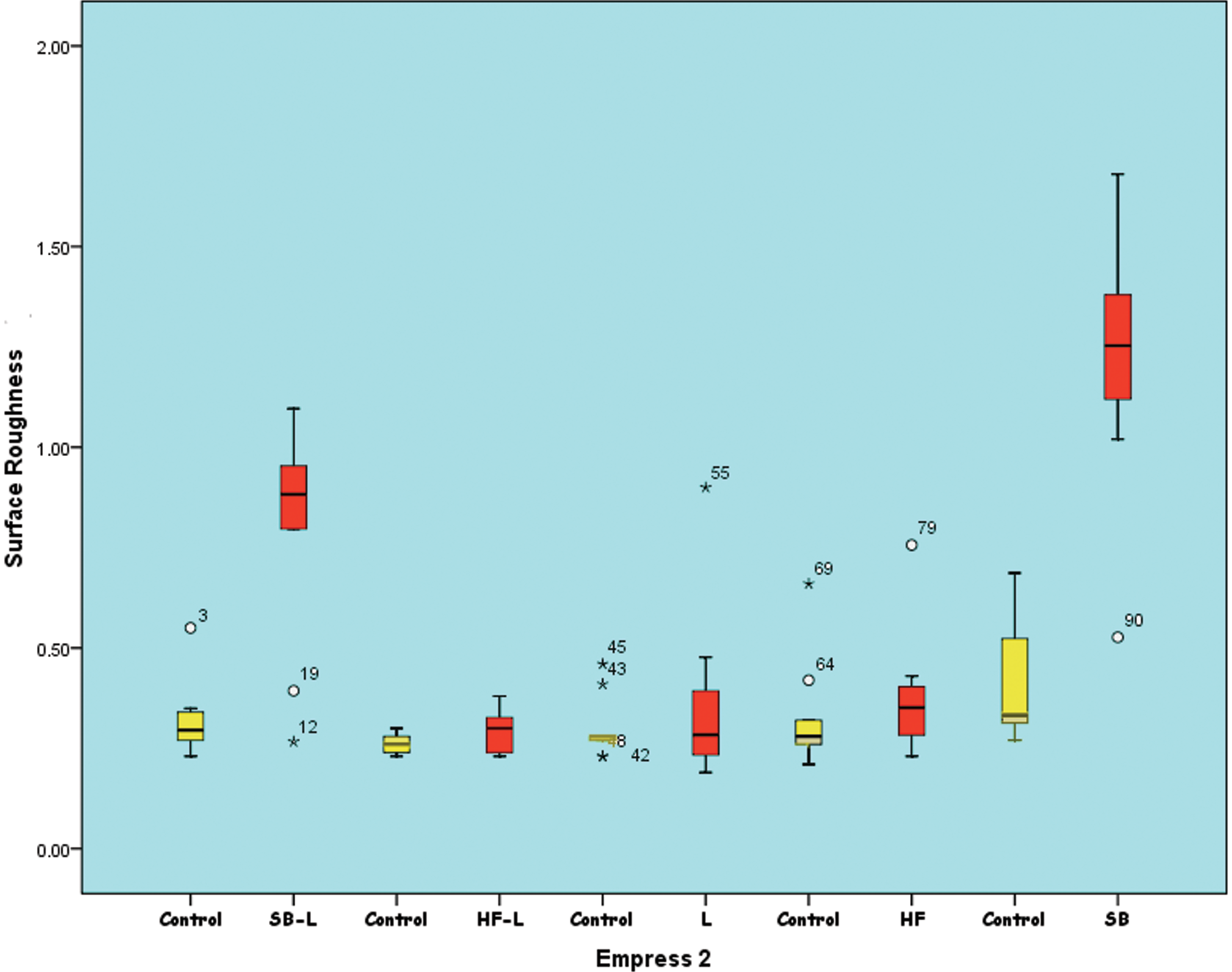
Box-plot diagrams of the surface roughness (Ra in μm) of Empress 2 ceramics according to the surface treatments applied. The median is shown by a horizontal line within the box. The minimum and maximum values are illustrated by the upper and lower strokes. (O) Marks outliers.
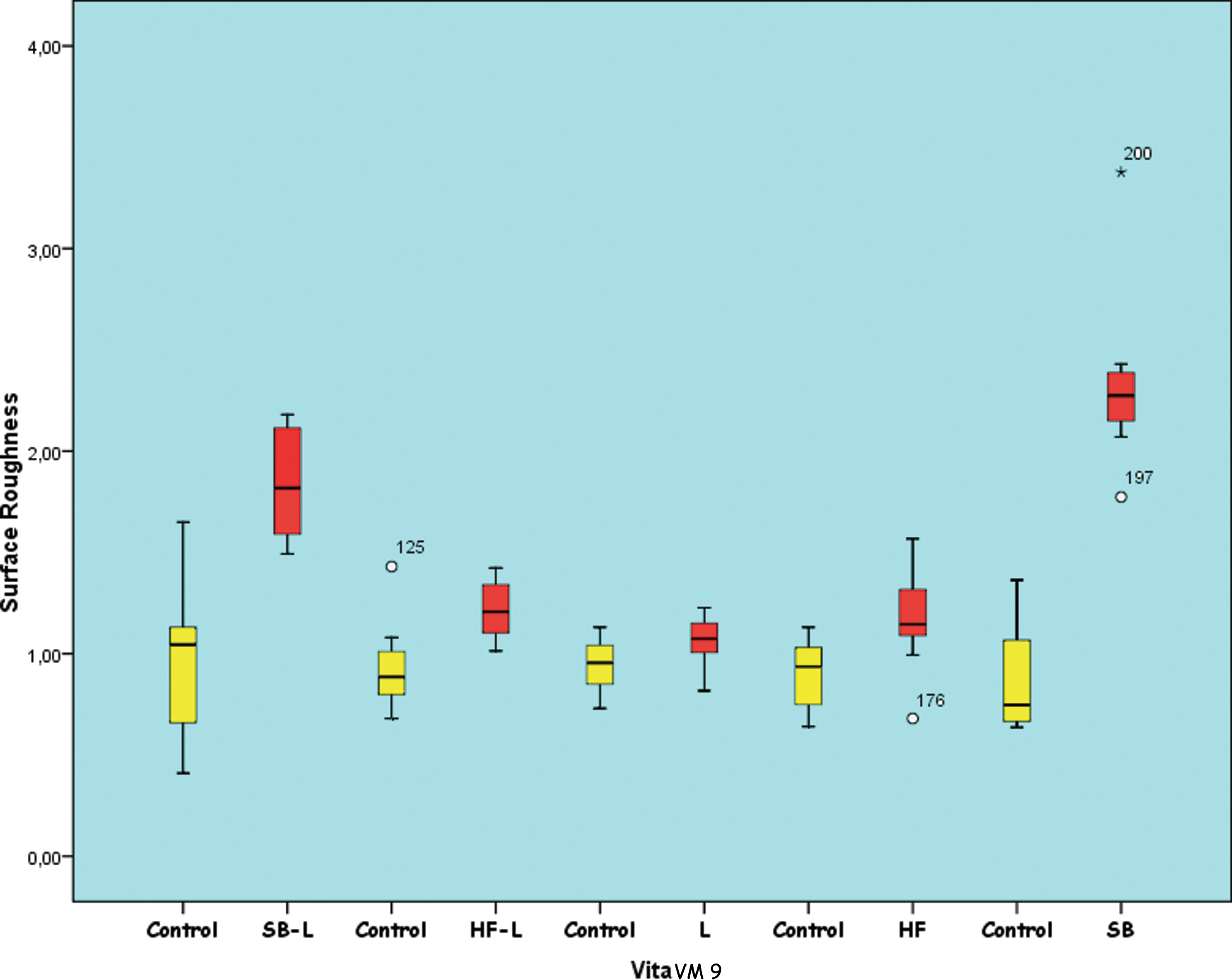
Box-plot diagrams of the surface roughness (Ra in μm) of feldspathic ceramics according to the surface treatments applied. The median is shown by a horizontal line within the box. The minimum and maximum values are illustrated by the upper and lower strokes. (O) Marks outliers.
p<0.05.
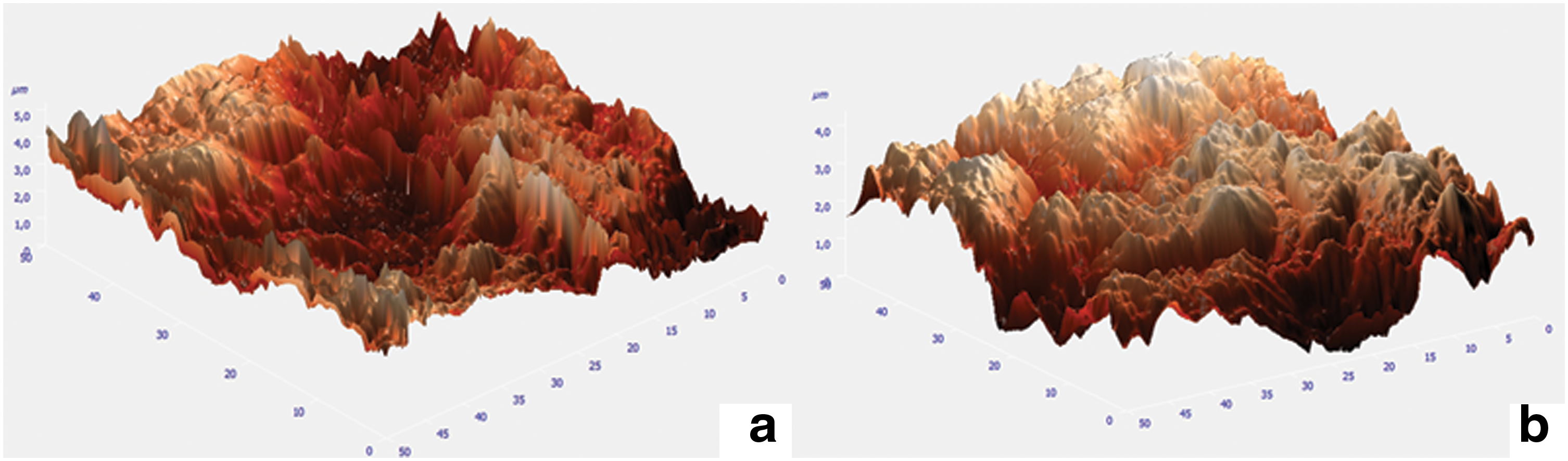
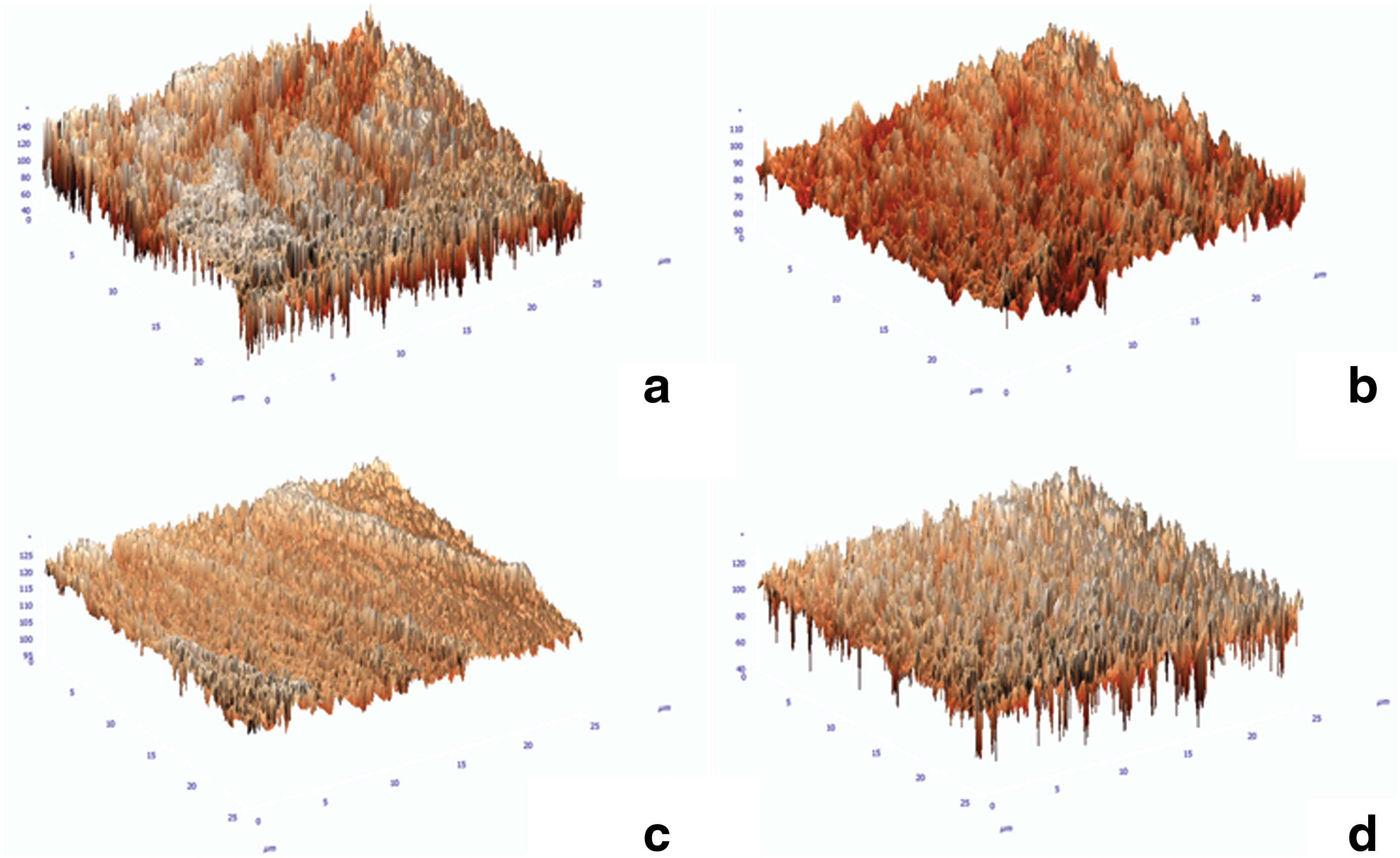
Representative AFM images of Empress 2 and Vita VM9 porcelain surfaces treated with the different techniques are presented in Figs. 3, 4, and 5. The surface treatment procedures generated similar topographies, except for surfaces treated with the Er:YAG laser (Group L). Group L surfaces showed moderate irregularity with peaks and valleys, and less roughness than was achieved with sandblasting and HF acid (Figs. 4c and 5c). Group SB had the most distinct sharp peaks compared with those in the other groups (Fig. 3).
Atomic force microscopy (AFM) images of sandblasted ceramics (SB).
Atomic force microscopy (AFM) images of Empress 2 ceramics.
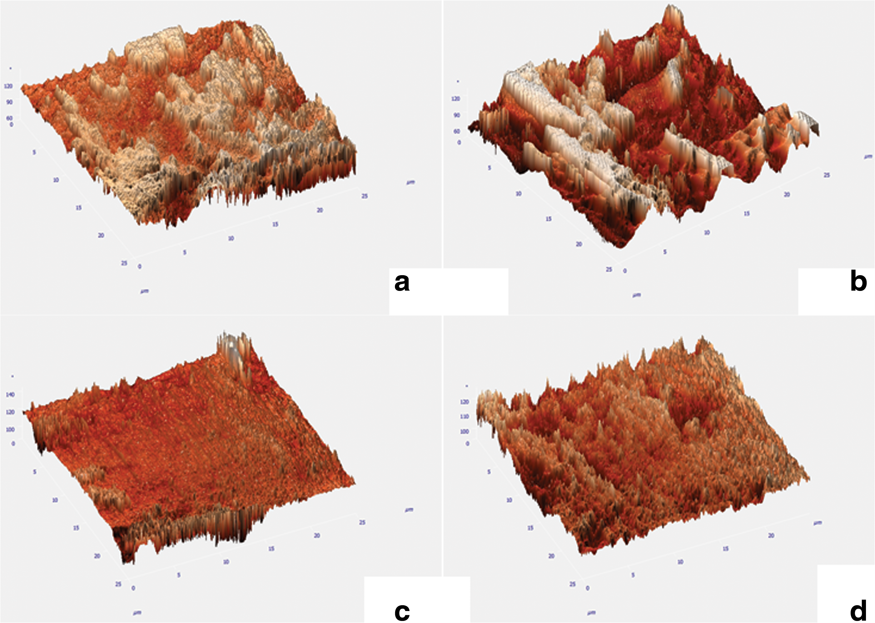
Atomic force microscopy (AFM) images of Vita ceramics.
Discussion
Micromechanical interlocking and chemical bonding roughen and clean the surface for adequate activation and hence play a major role in the bond strength between resin cement and the inner surface of a ceramic restoration. 1,3,10,13,15,19,20 Various surface treatment procedures for achieving a micromechanically retentive porcelain surface have been used clinically. 9 –11,14,15 This study presented an alternative combination of an Er:YAG laser with Al2O3 sandblasting and HF acid etching. Although many studies have investigated the effects of lasers on hard tissues, 21 –29 , their effect on porcelain surfaces has not been clear. In this study, AFM images of surfaces treated with laser irradiation revealed a shallow irregular surface. We hypothesized that these irregularities would enhance mechanical retention between the resin composite and porcelain surface. The null hypothesis was partially accepted: sandblasting and HF acid etching increased the surface roughness compared to untreated surfaces, but laser irradiation alone did not.
Sandblasting produces a rough irregular surface with an increased surface area, and enhances the wettability of the ceramic and the composite resin. However, excessive sandblasting induces chipping or a significant loss of porcelain material and is not recommended for cementation of silica-based and feldspathic ceramic restorations. 20,30,31 Among the surface treatments available for ceramic surfaces, sandblasting with Al2O3 has been widely used to provide micromechanical retention in several types of ceramics. 1,9,32 –34 Kara et al. 3,14 showed that air abrasion provides the highest surface roughness in low-fusing and lithium disilicate-based core all-ceramic materials. AFM images of air-abraded lithium disilicate-based core specimens showed a non-uniform surface with distinct sharp projections dotted with pores. The study by Kara et al. also indicated that on SEM images, laser irradiated surfaces appeared to be relatively smoother than sandblasted surfaces. 14 In the current study, sandblasting was applied before laser irradiation to simulate clinical circumstances, as it is a conventional procedure performed in commercial laboratories. In the present study, sandblasted porcelain specimens showed the highest surface roughness, and AFM images had more distinct sharp peaks than those of the other groups.
Er:YAG laser irradiation of a ceramic surface can remove the glass phase of the ceramic and create a rough surface. 35 Furthermore, Er:YAG laser irradiation increases the micromechanical retention of resin. 10,35 However, Subaşı and Inan 36 used 400 mJ pulse energy and found significantly lower surface roughness values than air abrasion. Similarly, in this study, Er:YAG laser-irradiated porcelain specimens showed lower surface roughness values even though higher laser energy parameters were used (500 mJ, 10 W). On the other hand, Ersu and colleagues 13 reported that surface roughness and shear bond strength values were higher in CO2 laser-irradiated In-Ceram® SPINELL, In-Ceram ALUMINA, and In-Ceram ZIRCONIA ceramics than in those treated with sandblasting and HF acid etching, but SEM analyses of the surfaces irradiated with the CO2 laser revealed microcracks. Gokce et al. 35 reported that the shear bond strength of Empress 2 specimens after Er:YAG laser irradiation at 300 mJ was higher than that of surfaces irradiated with 600 and 900 mJ. The differing compositions and reflectance of ceramic materials might have affected these results. A hydroxyapatite powder could be applied to stain the ceramic surface to enhance energy absorption and create a favorable surface. 15
HF acid selectively dissolves glassy or crystalline components of ceramics and creates a microporous irregular surface, thereby roughening the surface area and facilitating penetration of the resin into the etched ceramic surface. 6,37 Kara et al. 14 reported that treatment of low fusing ceramics with 5% HF acid etching produced same roughness values with Er:YAG laser. IPS Empress 2 specimens have a high crystalline content and exhibit significantly higher bond strengths than those of IPS Empress specimens, independent of surface conditioning. 3,20,38 Akyil et al. 10 determined that the bond strengths obtained from groups exposed to HF acid etching after each laser irradiation were lower than those in groups exposed to HF acid etching alone. This might be because HF acid etching was not effective on ceramic surfaces, as laser irradiation might have led to the development of a heat-damaged layer on the ceramic surface. 10,35 Given this risk, the reverse procedure (HF+laser) was used in this study. Despite this, the surface roughness values of Empress 2 ceramics exposed to Er:YAG laser irradiation after HF acid etching were lower than those of ceramics exposed to Er:YAG laser irradiation alone. A heat-damaged layer on the ceramic surface might also have been responsible for the reduction in the values of Group SB-L compared with Group SB.
Differences in the composition and microstructure of all ceramic restorations might affect the surface texture and bond strength between the ceramics and resin cement. Further studies are required to evaluate the effects of different power settings and different laser applications on ceramic surfaces to obtain optimum bond strength and roughness values.
Conclusions
Based on the results obtained and within the limitations of this in vitro study, the following conclusions can be drawn.
Sandblasting created a rougher surface than the other surface treatment methods (p<0.05). There were no significant differences in surface roughness after HF acid etching, Er:YAG laser irradiation, or the combination of HF and Er:YAG irradiation (p<0.05). AFM images of these laser-irradiated surfaces showed superficial irregularities with peaks and valleys, and less roughness than was visible after sandblasting and HF acid etching. Groups subjected to sandblasting had more distinct sharp peaks than the other groups.
Footnotes
Author Disclosure Statement
No competing financial interests exist.