
Abstract
Introduction
During recent decades, various photobiostimulatory effects of low-level laser therapy (LLLT) have been reported. The use of this therapy has been accepted and adopted in many medical and dental fields. 7 –9 In particular, the acceleration of bone regeneration by LLLT has been the focus of recent research 10 –12 because it may have potential benefits in clinical therapy in orthopedics and dentistry.
We previously reported LLLT stimulated bone or bone nodule formation in vivo 13,14 or in vitro 15 –18 via increased expression of insulin-like growth factor (IGF)-I 16 and bone morphogenetic proteins (BMPs). 18 Therefore, it was hypothesized that LLLT may enhance the stability of mini-implants via peri-implant bone formation, which is stimulated by IGF-I and/or BMPs.
Therefore, the aim of this study was to investigate the stimulatory effects of LLLT on bone healing and on the stability of mini-implants placed in bone, in an attempt to improve the success rate of mini-implants and to abbreviate the latent period in adolescents. To examine the effect of LLLT on the stability of titanium mini-implants placed in young rat tibiae, the stability of the mini-implants was measured using the Periotest (Siemens AG, Benssheim, Germany), and changes in peri-implant bone volume were examined using in vivo microfocus CT (R_mCT@; Rigaku, Tokyo, Japan). To determine the mechanism responsible for the effect of LLLT on the stability of mini-implants, the gene expression of BMP-2, the most potent growth factor in bone formation, 19 using peri-implant bone tissue from the rat tibiae, was investigated.
Materials and Methods
Animals
Thirty 6-week-old male Sprague–Dawley (SD) rats (body weight: 160±20 g) were used in this study. The stability of mini-implants (n=14) was measured, and total RNA was extracted from peri-implant bone samples (n=16) to evaluate BMP-2 gene expression. Peri-implant bone volume was measured in nine male Fischer rats (aged 6 weeks; body weight: 120±20 g) using R_mCT and scanning electron microscopy (SEM; TM-1000; Hitachi Science Systems, Ibaraki, Japan) (Table 1). Fischer rats were used for this measurement because they are smaller than SD rats and fit better in the small R_mCT chamber. The experiments were approved by the Animal Experimentation Committee of Nihon University School of Dentistry, Japan (No. P09D029).
Implantation procedure
Seventy-eight mini-implants were placed in the rat tibiae throughout the cortical bone to the trabecular bone level. Sixty mini-implants with 1.4 mm diameters, 7.3 mm total lengths, 3.3 mm head lengths, and 4.0 mm thread lengths (Mogi Shokai CO.,LTD., Tokyo, Japan) were chosen for the experiment in which stability was measured (n=28) and total RNA was extracted (n=32). Eighteen mini-implants with 1.5 mm diameters, 3.5 mm total lengths, 1.0 mm head lengths, and 2.5 mm thread lengths (Keisei Medical Industrial CO., LTD., Tokyo, Japan) were used for R_mCT and SEM observation (Table 1) because the shorter implants minimized the appearance of metal artifacts in R_mCT images. After each rat was anesthetized with an intraperitoneal injection of sodium pentobarbital (30 mg/kg body weight, Somnopentyl; Schering-Plough, Munich, Germany), an incision was made along the tibial crest, and the surface of the tibia was exposed. 3 A hole was then drilled with a bone drill (maximum applied torque 1.3 Ncm, 625 rpm) under physiological saline flow at a point 7.0 mm inferior to the knee joint, as defined by a line drawn perpendicular to the medial tibial surface. A pilot hole was made with a 1.0 mm diameter (for 1.4 mm diameter mini-implants) or 1.1 mm diameter (for 1.5 mm diameter mini-implants) drill, following previous studies that have used pilot holes with diameters of 71–73% of those of the mini-implants to enhance implant stability. 20 A mini-implant was inserted into each hole using a hand driver (Fig. 1). The fascial and superficial tissue layers were repositioned and sutured with 4-0 silk sutures (Ethicon; Johnson & Johnson, New Brunswick, NJ). To prevent postoperative infection, tetracycline hydrochloride paste (Showa Yakuhin Kako, Tokyo, Japan) was applied to the surgical site. The mini-implants in the right tibiae were then subjected to LLLT once daily for 7 days, and those in the left tibiae were used as nonirradiated controls (Fig. 2).
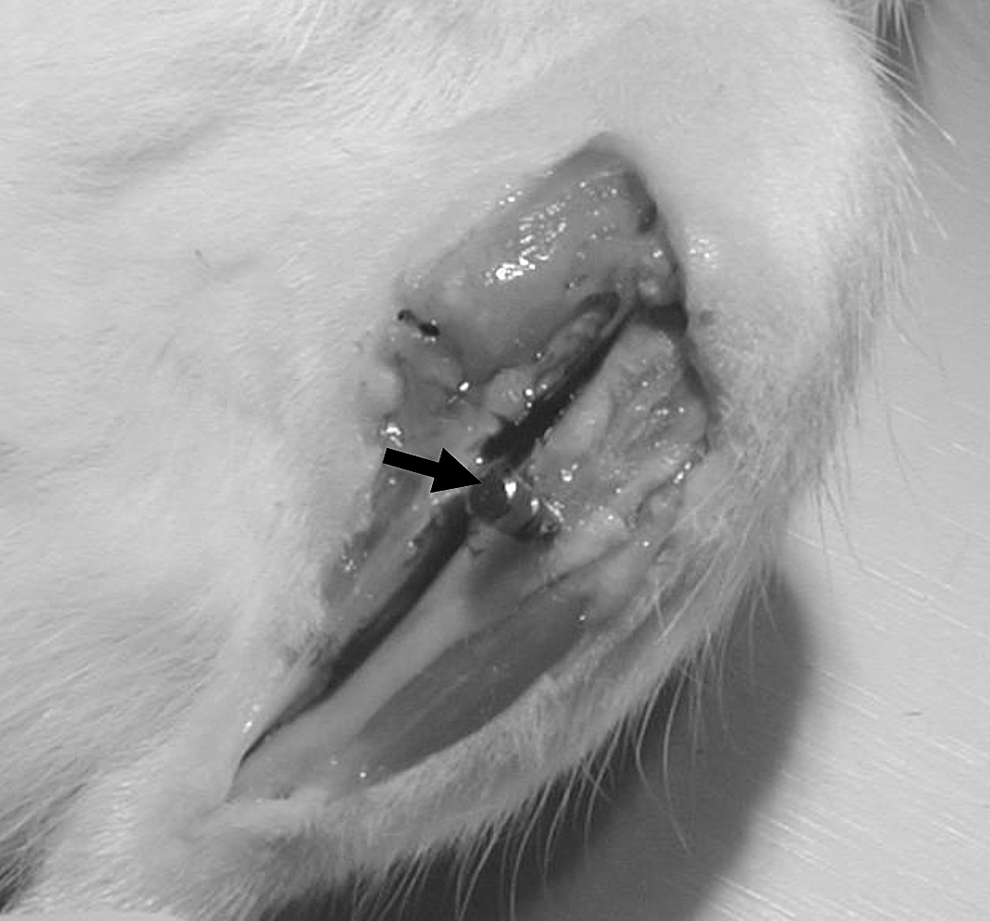
The mini-implants (1.4 mm diameter, 7.3 mm length, arrow) was placed in rat tibiae.
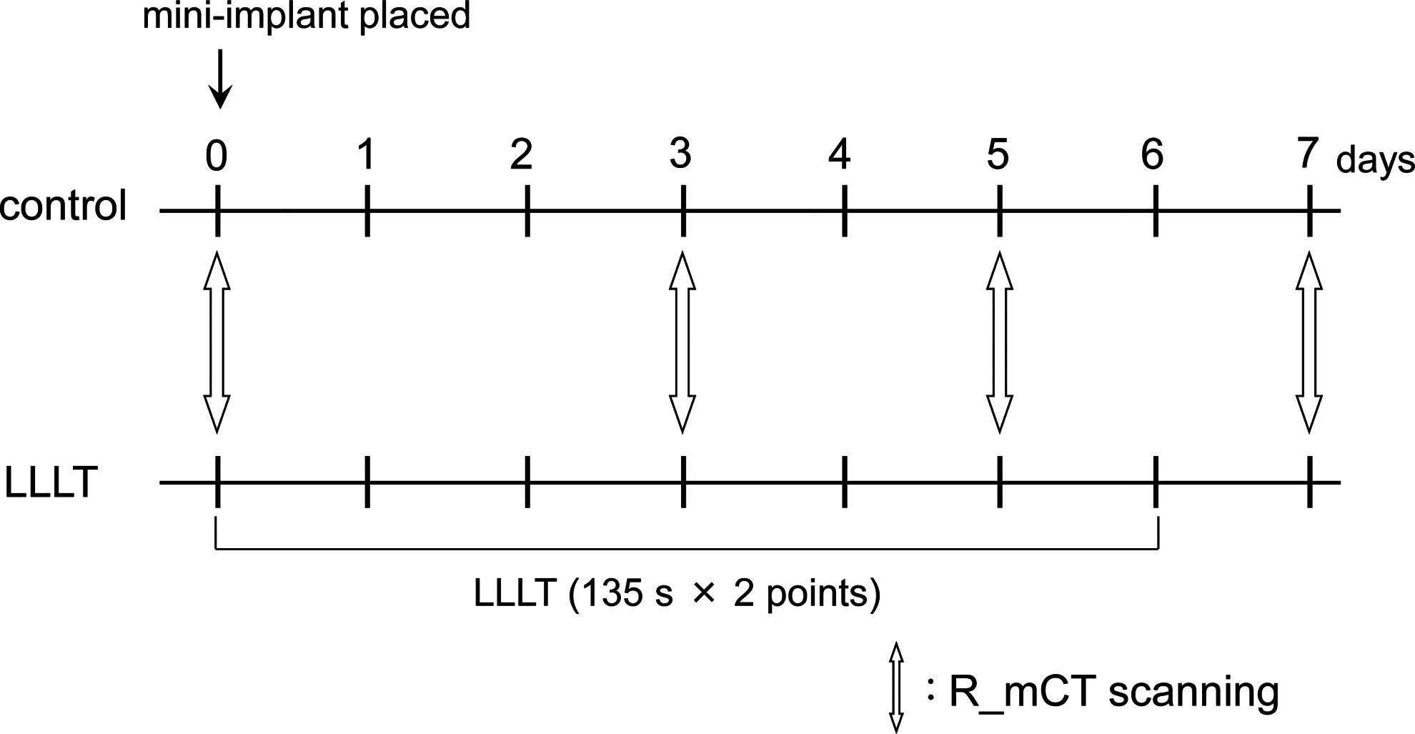
Schedule of low-level laser therapy (LLLT) treatment and R_mCT scanning.
Laser irradiation
A low level gallium-aluminium-arsenide (Ga-Al-As) diode laser (Panalas-1000; Matsushita, Inc., Osaka, Japan) was used in this study. LLLT (830 nm, continuous emission, 200 mW, 195 J/cm2, 135 sec ×2 points; the mesial and distal sides of the mini-implant, 54 J per session) was initiated immediately after surgery and performed once daily for 7 days (days 0–6, Fig. 2). An optical fiber with a tip defocused by a concave lens to ensure the uniformity of irradiation within circular areas (4.2 mm diameters) delivered the laser beam and the irradiation was administered percutaneously by placing the lens 1 mm from the tissue. The total energy corresponding to 270 sec of exposure was 54.0 J, which is similar to the dose used in the previous studies. 13,14
Stability measurement
After all procedures, the rats were killed using sodium pentobarbital on days 7 (n=7) and 35 (n=7), and the rat tibiae with mini-implants were dissected. All tibiae were buried in plaster to ensure rigid fixation and to facilitate the measurement of stability, and the stability of all mini-implants was measured immediately using the Periotest according to our previous study. 20 In accordance with the manufacturer's instructions, the measurement was performed by holding the Periotest handpiece parallel to the long axis of the tibia and striking the head of the mini-implant with the tip of the handpiece at a point 2.0–3.0 mm from the mini-implant head. The Periotest value (PTV) of each mini-implant was calculated by averaging five repeated measurements. All measurements were performed by the same examiner to eliminate intra-examiner error.
R_mCT analysis
R_mCT was performed using ×6.7 magnification, a voxel size of 30 μm3, a tube voltage of 90 kv, and a tube current of 100 μA. i-view-R image reconstruction software (J. Morita Mfg. Corp., Kyoto, Japan) was used. The field of view was 14.4 mm3, the number of pixels was 4803, and the scanning time was 2 min. The rats were anesthetized with sodium pentobarbital (30 mg/kg body weight) and placed on the stage, and R_mCT examination was performed once daily on days 0, 3, 5, and 7 (Fig. 2). Bone volume was measured on voxel images using software developed for that purpose (Kitasenjyu Radist Dental Clinic, i-View Image Center, Tokyo, Japan). The area in which bone volume was measured extended from the spearhead of the mini-implant in a rectangular parallelepiped (1.5×3.0×3.0 mm) because the lateral surface of the tibia was included when the measurement area was defined as a cube (Fig. 3). To assess the presence of newly formed bone within the measurement area, bone mineral density (BMD) phantoms of 300, 400, 500, 600, 700, and 800 mg/cm3 (Ratoc System Engineering Co., Ltd., Tokyo, Japan) were scanned by R_mCT under the same conditions used for peri-implant bone scanning, and BMD was calculated from the X-ray absorption value of R_mCT. The X-ray absorption threshold was determined to be 400 mg/cm3 because the BMD of all rat tibiae exceeded this value. The number of voxels that exceeded the threshold were counted by the measurement software. The voxel volume was measured at each examination under the same conditions. Then, changes in voxel volume were calculated by subtracting the volume measured on the operative day (day 0) from each subsequent value.
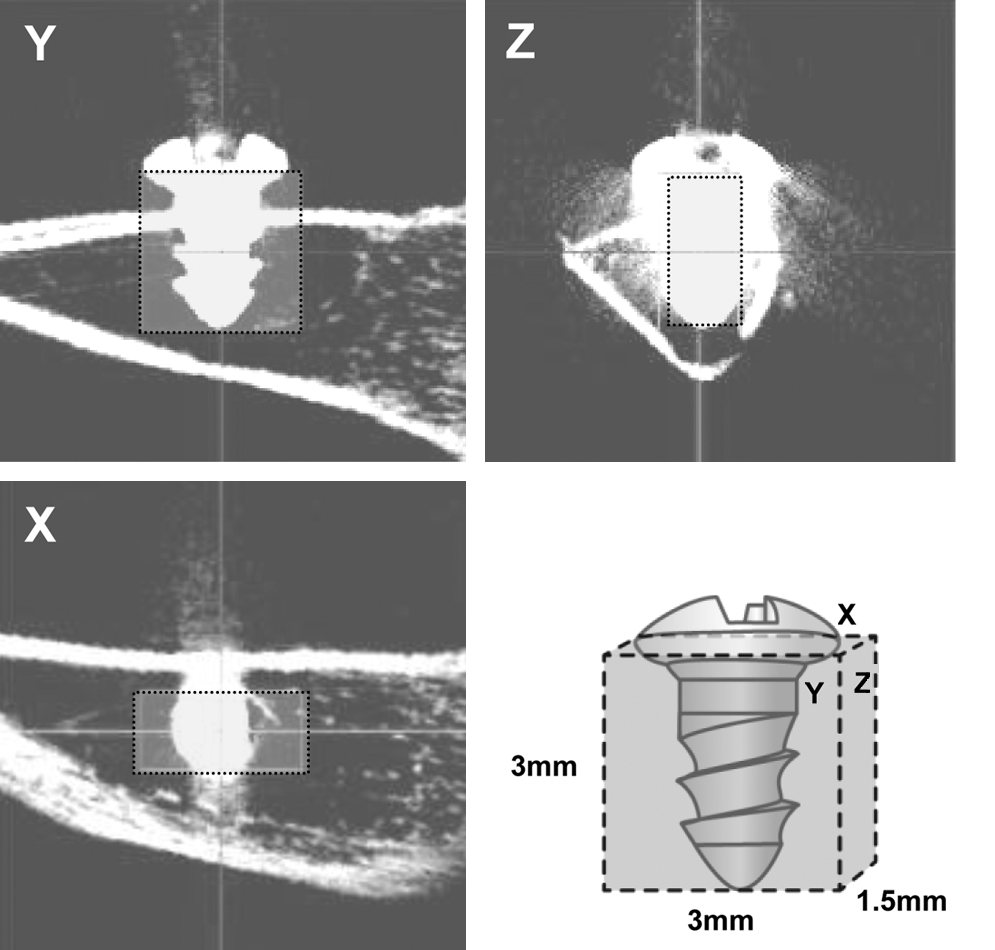
Designation of the bone volume measurement area.
RNA preparation and real-time reverse transcription polymerase chain reaction (RT-PCR) analysis
To elucidate the molecular mechanisms of the stimulation of bone formation by LLLT, BMP-2 expression in irradiated and nonirradiated peri-implant bone was analyzed by RT-PCR. At 1, 3, 5, and 7 days after implantation, rats were killed using sodium pentobarbital, and the mini-implants were extracted using a hand driver. Peri-implant bone in the drilled regions with 0.15-mm margins was then dissected freshly using a dental surgical drilling unit (1.7 mm diameter) with a trephine (Micro Tech. Corp., Tokyo, Japan) under constant cooling with sterile water. Total RNA was isolated from the peri-implant bone tissue using a commercially available kit (RNeasy Mini Kit; Qiagen, Valencia, CA). Aliquots containing equal amounts of total RNA were subjected to real-time RT-PCR. RT of target RNA and amplification of complementary DNA (cDNA) were achieved in a single-tube reaction following the amplification program described by the manufacturer (PrimeScript® RT Reagent Kit; Takara Bio, Otsu, Japan). The 2-μL cDNA mixtures were subjected to real-time RT-PCR using SYBR Green I dye. Real-time RT-PCR was performed in a 25 mL reaction mix containing 1×SYBR1 Premix Ex TaqTM (TaKaRa, Tokyo, Japan) and 0.2 mM of specific primers (sense and antisense), as shown in Table 2. The primers were designed based on sequence data obtained from the web site of the National Center for Biotechnology Information using the Primer3 software (Whitehead Institute for Biomedical Research, Cambridge, MA) and checked using the Basic Local Alignment Search Tool. RT-PCR was performed in a thermal cycler (Smart Cycler II System; Cepheid, Sunnyvale, CA), and the data were analyzed using Smart Cycler software (ver. 2.0). RT-PCR conditions were 40 cycles of 95°C for 5 sec and 60°C for 20 sec. The specificity of the RT-PCR products was verified by conducting a melting curve analysis between 60°C and 95°C. All RT-PCR assays were performed in triplicate, and the levels of messenger RNA (mRNA) expression were calculated and normalized to the level of GAPDH mRNA at each time point.
SEM observation
SEM observation was conducted at 0, 3, 5, and 7 days after mini-implant placement. After the rats were killed using sodium pentobarbital, the tibiae were resected at the knee joint, fixed for 48 h in 10% neutral buffered formalin (Wako Pure Chemical Industries, Osaka, Japan), and then washed in clear water with ethanol dehydration and acetone degreasing. The tibiae were embedded in polyester resin (Rigolac 2004; Showa Highpolymer, Tokyo, Japan) at a constant temperature of 60°C for 8 h, and 10.0×10.0×6.0 mm blocks were then cut mesiodistally using a crystal cutter (Maruto Instrument Co., Ltd., Tokyo, Japan). These blocks were centered on the bone and oriented according to the long axis of the mini-implant. The surface of each specimen was ground with waterproof grinding papers (800, 1200, and 2000) and a hard grinding cloth with a liquid containing 1 μm of diamond particles. The bone surrounding the mini-implant was observed using SEM, and photographs were taken at ×25 magnification. The part corresponding to the Y-plane image on R_mCT was chosen as the observation area (Fig. 3).
Statistical analysis
All measurements and collection of data were made blindly without it being known if specimens were irradiated or nonirradiated. Statistical analyses were performed using SPSS software (ver. 16 for Windows; SPSS Inc., Chicago, IL). Student's t test was used to analyze differences between the LLLT and control groups. p<0.05 was considered to indicate statistical significance.
Results
Stability analysis
PTVs were significantly lower in the LLLT group than in the control group on days 7 (0.79-fold) and 35 (0.67-fold; p<0.05). PTVs were significantly lower on day 35 than on day 7 in both the control (0.55-fold) and LLLT (0.47-fold) groups (p<0.05; Fig. 4).
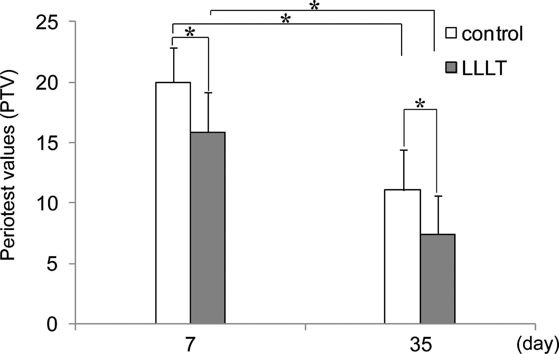
Effect of low-level laser therapy (LLLT) on the stability of mini-implants. The stability of mini-implants was measured using the Periotest. Periotest values (PTVs) on days 7 and 35 were reduced significantly by LLLT treatment. PTVs were significantly lower on day 35 than on day 7 in both the LLLT and control groups. *p<0.05. Values are expressed as mean±standard deviation for seven rats.
R_mCT and SEM findings
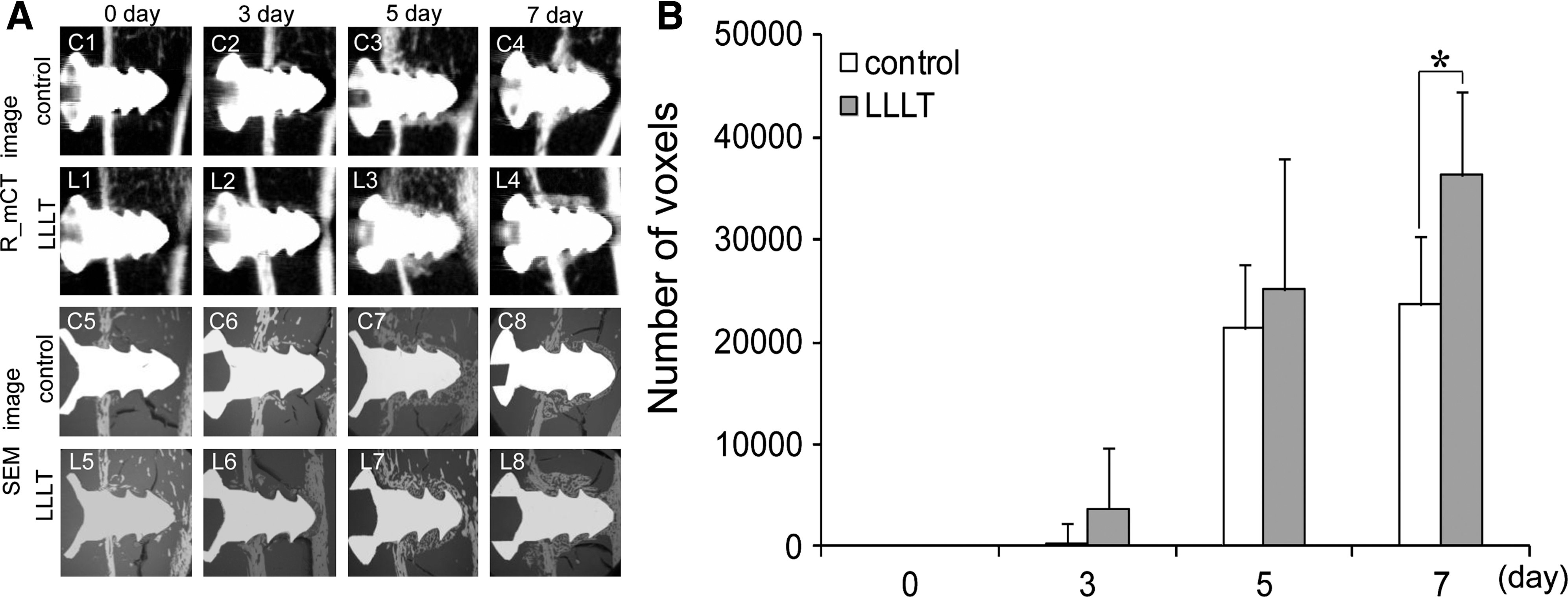
R_mCT images revealed peri-implant ossification over time. Newly formed bone tissue was observed around the mini-implants in both groups as early as 5 days after surgery, and bone formation appeared to be stronger in the LLLT group. SEM images showed findings similar to those of R_mCT images (Fig. 5A, C1–8 and L1–8).
(
Bone volume analysis
No significant difference in bone volume was observed between the control and LLLT groups on days 0, 3, or 5. However, bone volume was 1.53-fold greater in the LLLT group than in the control group on day 7 (p<0.05; Fig. 5B).
BMP-2 gene expression in peri-implant bone tissue
BMP-2 gene expression was 1.92-fold higher in the LLLT group than in the control group (p<0.05) as early as 1 day after LLLT. No significant difference in BMP-2 expression was observed between groups on days 3, 5, and 7 (Fig. 6).

Effects of low-level laser therapy (LLLT) on BMP-2 gene expression. Total RNA was isolated from peri-implant bone of rat tibiae and analyzed by real-time reverse transcription polymerase chain reaction (RT-PCR). Ratios of target genes relative to housekeeping genes (GAPDH) are reported. *p<0.05. Values are expressed as mean±standard deviation for four rats.
Discussion
Recent studies have reported various biostimulatory effects of LLLT, including wound healing, 7 fibroblast, 8 and nerve regeneration. 9 In particular, the acceleration of bone regeneration by LLLT has been a focus of recent research. 10 –12 We reported the stimulatory effects of LLLT on bone regeneration in the midpalatal suture during rapid maxillary expansion in rats 13 and used a quantitative in vitro bone-nodule formation assay to determine that these effects were caused by increased cellular proliferation, alkaline phosphatase (ALP) activity, osteocalcin expression, and calcium accumulation. 15
In an in vivo study of the effects of LLLT on dental implants, Khadra et al. 21 placed coin-shaped implants into rabbit tibiae and performed LLLT. They found that the tensile force of implants was significantly (1.4-fold) greater in the LLLT group at week 8.
Because the stability test is objective and easy to use for the evaluation of mini-implant prognoses 20 and because an inverse relationship (p<0.01) has been found between the bone–implant contact ratio and PTV, 20 we examined the effect of LLLT on the stability of mini-implants in the present study using the Periotest. At 7 and 35 days after the placement of mini-implants, PTV was significantly lower in the LLLT group than in the control group (Fig. 4). In agreement with the findings of Khadra et al., 21 this result suggests that LLLT contributed to the improvement of mini-implant stability.
Changes in bone volume using R_mCT to assess osseous support around the mini-implants were also examined. Osseous support at the implant–bone interface is commonly evaluated by histomorphometric analysis. However, bone histomorphometric analysis of the implant surface involves only the measurement of structural parameters in two-dimensional sections. 22,23 Recently, quantitative bone structure analysis using conventional microfocus CT (mCT) which is usually used for isolated samples but not for living animals, has been considered for the evaluation of implant stability. 24,25 The advantages of mCT analysis include its rapidity in comparison with histological tissue preparation, the ability to perform quantitative three-dimensional analysis, and the ability to reconstruct various views from the sections depending upon the purpose of analysis. Therefore, mCT provides a better understanding of healing processes in peri-implant bone. 24,25 R_mCT 26,27 used in the present study, consists of X–ray tube and sensor rotated around a secure object stage, which has more advantages and has enabled us to obtain bone images in living experimental animals without killing them, and to observe longitudinal changes in individuals in the same sample. 28 –31 However, few studies have quantitatively examined longitudinal changes in peri-implant bone. In the present study, R_mCT was used to measure changes in bone volume within the measurement area of peri-implant bone. R_mCT images revealed newly formed bone in the trabecular regions on days 5 and 7 in both groups, and bone formation seemed to be stronger in the LLLT group (Fig. 5A, C1–4 and L1–4). Furthermore, we compared CT and SEM images of the region of interest. The SEM images revealed findings similar to those observed in the R_mCT images (Fig. 5A, C5–8 and L5–8). Bone volume was significantly greater in the LLLT group than in the control group at 7 days after surgery (Fig. 5B).
These results suggest that LLLT enhanced new bone formation surrounding the implants. Stability test and R_mCT findings indicated that LLLT increased the stability of the mini-implants because of increased peri-implant bone formation.
Several studies have examined the effect of LLLT on bone healing around titanium dental implants. Dörtbudak et al. 32 revealed significantly higher osteocyte viability in peri-implant bone treated by LLLT, which positively affected the integration of dental implants. In an energy-dispersive X-ray microanalysis, Khadra et al. 21 observed a significant increase in calcium and phosphorus on LLLT-treated implant surfaces. Using Raman spectroscopy and SEM, Lopes et al. 33 observed that LLLT accelerated the maturation of bone surrounding dental implants in rabbit tibiae.
The present study used the same wavelength of the low-level laser apparatus as was used by Khadra et al. 21 and Lopes et al. 33 The energy density (195 J/cm2) used in the present study was higher than those used by Kahadra et al. 21 (23 J/cm2) and Lopes et al. 33 (21.5 J/cm2), because the irradiation points were only two in the present study. Because the Ga-Al-As diode laser is known to diffuse in the tissue, we emphasized the total energy more than the energy density and the total energy of 54 J per session was determined according to our previous studies that LLLT stimulated bone formation in mid-palatal suture during expansion 13 and tension side of tooth movement 14 in the rat. This dose was not very different from those used by Kahadra et al. 21 (27 J) and by Lopes et al. 33 (83 J). The effect of LLLT on peri-implant bone formation is thought to become apparent within a certain range in the total energy of LLLT.
Our results and those of other groups suggest that LLLT has a positive impact on the osseointegration of implants.
Recently, several in vitro studies have sought to identify the regulatory mechanisms involved in the effect of LLLT on bone. Kusakari et al. 34 reported that LLLT of osteoblast-like cells stimulated DNA and protein synthesis and elevated ALP activity. Hamajima et al. 35 reported that the increased expression of the osteoglycin gene by LLLT in the early proliferation stage of cultured osteoblastic cells might play an important role in the stimulation of bone formation in concert with matrix proteins and growth factors. Moreover, we reported that LLLT during the early formation stages of osteoblast-like cells isolated from fetal rat calvaria significantly stimulated cellular proliferation, ALP activity, and osteocalcin gene expression. 15 We further demonstrated that these phenomena were promoted by the increased production of IGF-I, BMPs, and transcription factors associated with osteoblast differentiation. 16 –18 However, it remains unclear whether in vivo experiments will yield similar findings.
In the present in vivo study, the effects of LLLT on BMP-2 mRNA expression were examined, because BMPs are considered to be the most important factors in bone formation. 19 We reported previously that LLLT stimulated BMP-2 gene expression more than it stimulated the expression of BMP-4, -6, or -7 in mouse osteoblast-like cells. 18 In the present study, BMP-2 expression of the cells around the mini-implant was 1.92-fold higher in the LLLT group than in the control group on day 1 after implantation. However, large standard errors prevented this difference between groups from being significant (p=0.07), even at the level of greatest difference (2.3-fold on day 3). Therefore, LLLT stimulated peri-implant bone formation via the increased gene expression of BMP-2 from surrounding cells in the early stages following LLLT.
Further studies are required to examine the expression of other growth and transcription factors related to bone formation and osteoblast differentiation, such as runt-related transcription factor 2 and Osterix, to elucidate the mechanism responsible for the stimulation of bone formation by LLLT.
Conclusions
LLLT enhanced the stability of mini-implants placed in rat tibiae and accelerated peri-implant bone formation via the increased gene expression of BMP-2 from surrounding cells. Therefore, it is most likely that when mini-implants are placed into the alveolar bone of adolescent patients, LLLT around the mini-implants enhances their stability.
Footnotes
Acknowledgments
This study was supported by a Grant-in-Aid for Scientific Research (No. 19592369, 21592613) and a grant of “Strategic Research Base Development” Program for Private Universities (No. S1001024) from the Ministry of Education, Culture, Sports, Science, and Technology of Japan (MEXT); Sato Fund and a grant from Dental Research Center, Nihon University School of Dentistry; and the Promotion and Mutual Aid Corporation for Private Schools of Japan.
Author Disclosure Statement
No conflicting financial interests exist.