
Abstract
Introduction
The current trend in minimally invasive dentistry involves alternative techniques for dental cavity preparation, such as laser irradiation 11 –17 and aluminum oxide air abrasive. 12,18 Such approaches replace the invasive technique of using high-speed dental burs. 12,17,19 , Cavity preparation using lasers takes more time than cavity preparation with rotary cutting instruments, but advantages include low noise and vibration and eliminating the need for local anesthesia in most cases. An additional advantage of laser cavity preparation is that a smear layer of debris is not produced on the surface of the prepared cavity. 15 Among the various laser types used in dentistry, the erbium: yttrium aluminum garnet (Er:YAG) laser is one of the most highly recommended. Its strong reputation results from the fact that its 2.94 μm wavelength emission coincides with the main absorption peaks of water (∼3.0 μm) and hydroxyapatite, thus resulting in good absorption in all biologic tissues, including enamel, dentin, and cementum. 12,13,15,17 Furthermore, the increase in temperature under Er:YAG laser irradiation to the pulp and tissues comes within acceptable limits (<3°C) with water spray for cooling. 16,20 Cooling also prevents cracking of enamel and dentin. 17
The evaluation of bonding durability is important, as the bond between the restorative material and the tooth substrate has a significant impact on the clinical success of restoration. 5,9 In an attempt to mimic the natural aging process of dental restoration, thermocycling protocols and the water storage (WS) of bonded specimens have been suggested as efficient methods to provide in vitro simulation of in vivo conditions. 11 Thermal cycling simulates the introduction of hot and cold extremes (between 5° and 55°C) in the oral cavity while showing the relationship of the linear coefficient of thermal expansion between tooth and restorative material. 21,22 On the other hand, in the WS aging procedure, the bonded specimens are stored in fluid at 37°C for a specific period, which may vary from a few months up to 4–5 years, or perhaps longer. Degradation of interface components by hydrolysis of resin or collagen may occur after WS. 22,23
The Er:YAG laser is a promising alternative method for cavity preparation; however, very little information has been published on the adhesion of one-step self-etch adhesives to the laser-irradiated dentin after aging procedures. Therefore, the objectives of this study were to compare the microtensile bond strengths (μTBS) of one-step self-etch adhesives to laser-irradiated and bur-cut dentin after WS and thermocycling. The null hypotheses were as follows: (1) there were no differences in μTBS between laser-irradiated and bur-cut dentin; and (2) artificial aging, including WS and thermocycling, decreases the bond strength of the one-step self-etch adhesive systems to dentin.
Methods
Seventy-two human maxillary third molar teeth that were free of caries, cracks, fractures, and restoration were selected. After extraction, the teeth were cleaned of surface debris and stored in 0.5% chloramine-T at 4°C for<1 month. Each tooth was mounted in cold-curing acrylic resin (Meliodent, Bayer Dental Ltd., Newbury, UK). They were then submerged in tap water to reduce any temperature increase caused by the exothermic polymerization reaction of the acrylic resin. While fully hydrated, each third molar was first cut just below the occlusal pit and fissure, perpendicular to the long axis of the tooth, by means of a slow-speed diamond saw (Isomet Low Speed Saw, Buehler Ltd., Lake Bluff, IL). The surface was ground with 600-grit silicon carbide paper (Carbimet Buehler, Buehler Ltd.) under running water for 30 sec to create a smear layer of clinically relevant thickness. The specimens were randomly assigned to two groups according to the cavity preparation methods.
Cavity preparation
Er:YAG laser irradiation (group I)
The laser system used in this study was the Er:YAG laser (Smart 2940D Plus, Deka Laser, Florence, Italy) with a 2.94 μm wavelength. Laser energy was delivered in pulse mode with a repetition rate of 10 Hz, energy at 200 mJ, output power of 2 W, and pulse duration of 700 μs. The time of irradiation was not standardized during cavity preparations. The distance of application was 10 mm, which was standardized by a custom-designed apparatus consisting of two parts: a holder to fix the laser handpiece in such a way that the laser beam was delivered perpendicular to the specimen surface at a constant working distance from the target site, and a semi-adjustable base, upon which an acrylic plate and fragment were attached. Furthermore, an occlusal cavity was prepared with a dimension of 4 mm×4 mm2 and 2 mm deep by Er:YAG laser on each tooth. The depth of the cavity was calibrated by measuring with a periodontal probe. Water irrigation at a rate of 5 mL/min also was used during lasing of the specimens.
Bur preparation (group II)
The cavities were prepared with a high-speed #1090 fissure diamond bur (Diatech Dental AG, Heerbrugg, Switzerland) under air-water spray coolant at a rate of 5 mL/min. The cavity was 4 mm×4 mm2 and 2 mm deep. New burs were used after every five preparations.
Specimen preparation
Three different one-step self-etch adhesives—Clearfil S3 (Kuraray Medical, Tokyo, Japan), AdheSE One (Ivoclar Vivadent, Schaan, Liechtenstein), and Adper Easy One (3M ESPE, St Paul, MN, USA)—were applied to the cavity. Details regarding the adhesive systems selected, such as manufacturers, composition, application technique, and batch number, are listed in Table 1. The selected manufacturers' recommended hybrid resin composites—Clearfil AP-X (Kuraray Medical, Tokyo, Japan), Tetric N-Ceram (Ivoclar Vivadent, Schaan, Liechtenstein), and Filtek Z250 (3M ESPE, St Paul, MN)—were applied following the incremental technique and light cured for 20 sec. All procedures and curing times were performed according to the manufacturers' instructions. Specimens were stored in distilled water at 37°C for 24 h and were divided into three subgroups: control (C), WS, and thermocycles (TC).
Bis-GMA, bisphenol A glycerolate dimethacrylate, MDP, 10-methacryloyloxydecyl dihydrogen phosphate; HEMA, 2-hydroxyethyl methacrylate.
Aging procedure
The specimens retrieved at 24 h of WS were used as controls. The specimens in the WS groups were aged in distilled water at 37°C in a water bath machine (BM 402; Nüve, Ankara, Turkey) for 6 months. The water was changed weekly in order to accelerate the degradation process. The specimens in the TC groups were subjected to thermocycling (10.000 cycles between 5° and 55°C) (Nova, Nova Ticaret, Konya, Turkey). The dwell time in the water bath was 30 sec, and the transfer time was 7 sec.
Microtensile testing
For μTBS testing, specimens were serially sectioned perpendicular to the bonding surface using the Isomet saw under running water in order to obtain rectangular sticks with a cross-sectional surface area of ∼1.0 mm2 (±0.2 mm). Each stick was attached to a custom jig of a universal testing machine (Lloyd LF Plus; Ametek Inc., Lloyd Instruments, Leicester, UK) using a cyanoacrylate adhesive (Model Repair II Pink, Dentsply-Sankin, Otawara, Japan) and subjected to a tensile force at a crosshead speed of 1 mm/min until failure occurred. The fractured sticks were removed from the testing apparatus, and the cross-sectional area at the site of failure was measured to the nearest 0.01 mm with a digital caliper (Altas 905; Gedore-Altas, Istanbul, Turkey). The μTBS was calculated from this measurement and expressed in MPa. Twenty bond strengths of each group were obtained, and the data were analyzed using a three-way ANOVA test. Independent samples t tests and post-hoc comparisons were performed at a 0.05 significance level.
Failure analysis
The fractured specimens were examined under a stereomicroscope (SMZ 800, Nikon, Tokyo, Japan) at 40× magnification, to evaluate the fracture pattern. Failure modes were classified into one of three categories: adhesive failure if debonding occurred between resin and dentin; mixed failure if the failure exhibited was partially adhesive and partially cohesive in bonding resin or in hybrid layer; or cohesive failure occurred in resin or in dentin. All observations were conducted by one person. In addition, the fractured surfaces for each group were examined using a scanning electron microscope (SEM, LEO 440, Zeiss, Oberkochen, Germany). Representative specimens were photographed at 5000× magnification after the μTBS test. SEM micrographs of bur-cut specimens and the Er:YAG laser-irradiated specimens are presented in Figs. 1, 2, and 3.
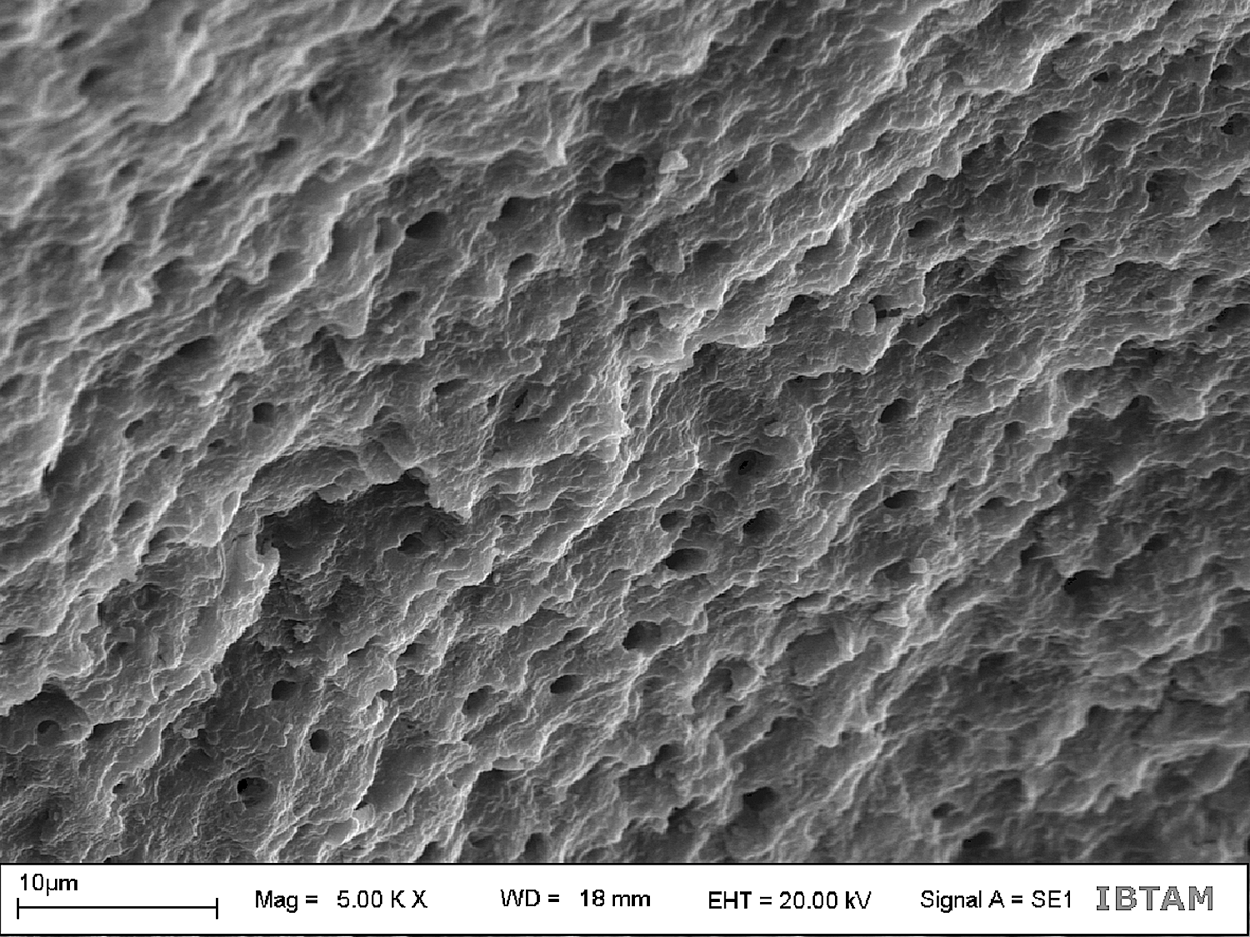
Scanning electron microscope (SEM) photograph of bur-cut dentin surface after microtensile bond strength (μTBS) test (original magnification: ×5000).
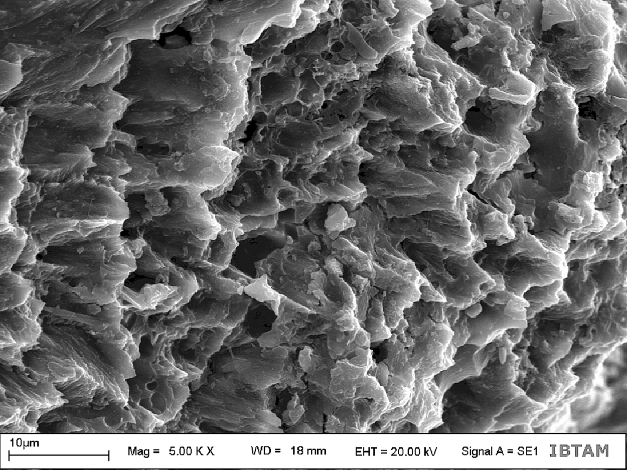
Scanning electron microscope (SEM) photograph of the Er:YAG laser treated dentin surface after microtensile bond strength (μTBS) test (original magnification: ×5000).
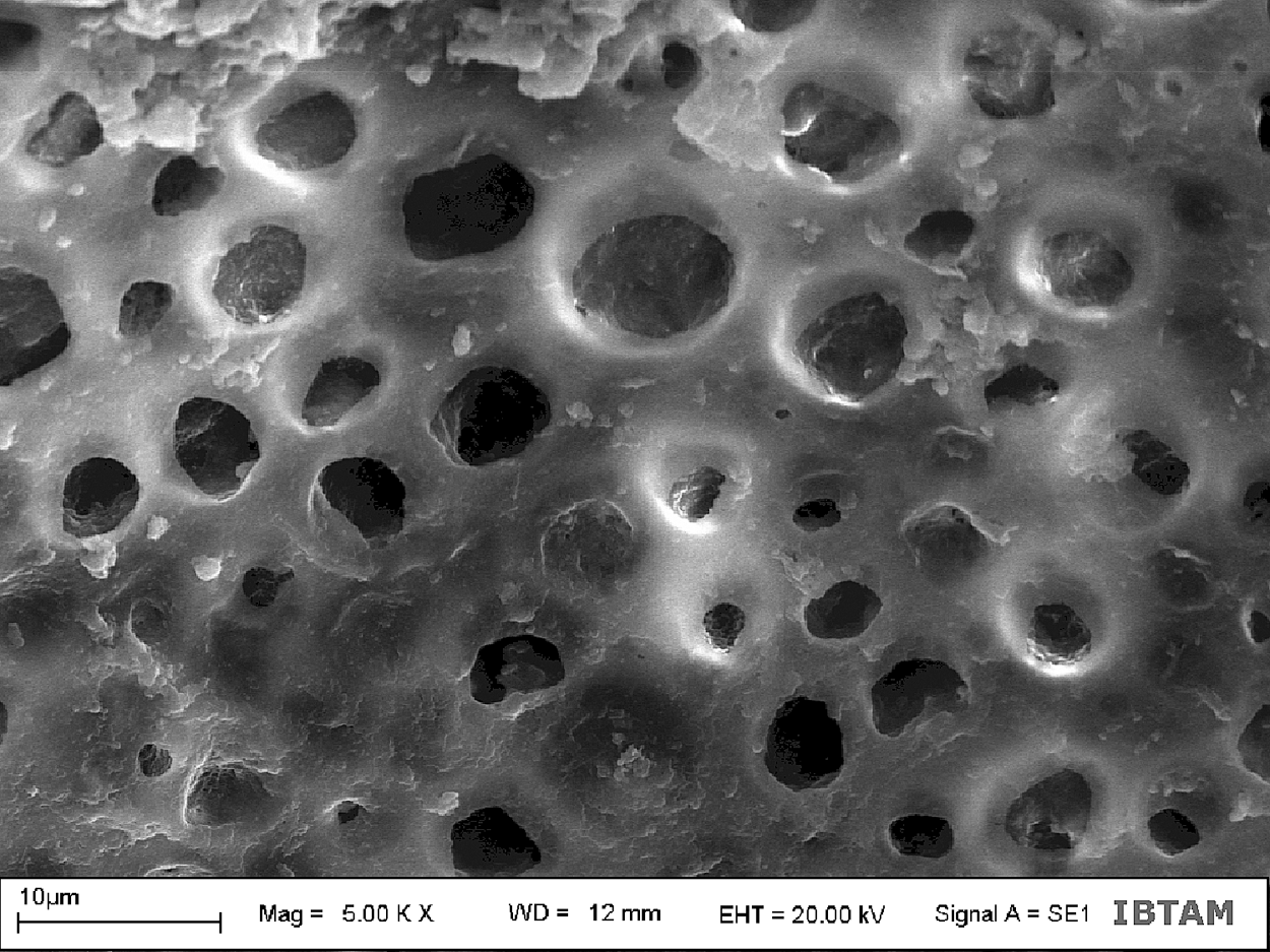
Scanning electron microscope (SEM) photograph of the Er:YAG laser treated dentin surface after microtensile bond strength (μTBS) test (original magnification: ×5000).
Results
Three-way ANOVA tests results for μTBS measurements of the groups are summarized in Tables 2 and 3. Analysis of the data revealed that there were no significant differences between the methods when the cavity preparation methods were compared (p>0.05). In general, the bur-cut dentin displayed similar μTBS to the laser-irradiated dentin. Clearfil S3 Bond presented the highest μTBS to dentin, irrespective of the cavity preparation method used.
For each horizontal row: values with small letters indicate no statistically significant difference (p>0.05).
For each vertical column: values with capital letters indicate no statistically significant difference (p>0.05).
R2=0.339 (Adjusted R2=0.306).
μTBS results were not affected by either cavity preparation method (p=0.112 and F=2.546) or the aging conditions (p=0.149 and F=1.912), whereas, in the interaction of the factors, the cavity preparation method and aging conditions demonstrated a statistically significant difference (p<0.001 and F=14.236). Ultimately, μTBS results were significantly affected by adhesive type (p<0.001 and F=39.33). The interaction of the factors cavity preparation method, adhesive type, and aging conditions showed a statistically significant difference in μTBS (p<0.001 and F=8.319). The bond strength of Clearfil S3 Bond was significantly higher than those of Adper Easy One and AdheSE One (p<0.001). Moreover, there was a significantly different μTBS between Adper Easy One and AdheSE One (p=0.008).
In comparison to the C group, TC and WS did not decrease the mean μTBS of one-step self-etching adhesives (p=0.894 and p=0.148). Similarly, no significant differences were found among the μTBS results for TC and WS (p=0.331). Modes of failure are presented in Table 4. The analysis of failure after the microtensile test revealed that the adhesive failure mode was observed in both C and aged groups for both laser-irradiated and bur-cut cavity methods.
a,adhesive; c, cohesive in composite; d, cohesive in dentin; m, mix.
At 5000× magnification, bur-cut dentin was characterized by a quite irregular topography (Fig. 1) and Er:YAG laser-treated surfaces (Figs. 2 and 3) had a similarly distinctive, irregular, and scaly appearance, exhibiting open tubules and absence of a smear layer.
Discussion
The results obtained in this study clearly demonstrate that the μTBS of laser-irradiated dentin was similar to that of bur-cut dentin, by which the first hypothesis was accepted. The second hypothesis was rejected, because μTBS results were not affected by the aging conditions. Contradictory results and conclusions on tensile bond strengths after Er:YAG laser treatment may be found in the literature, because many different experimental setups have been used. Lee et al. 16 reported that the bur-cut/acid-etched and laser-ablated/acid-etched groups showed no statistical significance. Furthermore, Souza-Zaroni et al. 12 demonstrated that Er:YAG laser groups (250 and 300 mJ) were not statistically different in μTBS from the bur-cut group. The results of the present study were consistent with Lee et al. 16 and Souza-Zaroni et al. 12
Also, in alignment with the results of the present study, do Amaral et al. 11 found that there was no significant difference in μTBS between bur-cut and laser-irradiated specimens in WS for 24 h. However, contrary to the results of the present study, do Amaral and colleagues also reported that there were significant differences when the cavity preparation methods were compared. Moreover, Er:YAG laser-prepared specimens demonstrated lower μTBS after being stored in water for 6 months with 12,000 TC. In addition, Trajtenberg et al. 15 demonstrated that Er:YAG laser-irradiated and bur-cut dentin had similar μTBS when the surfaces were acid etched followed by an etch-and-rinse adhesive. Bur-cut dentin showed higher bond strengths than did laser-irradiated dentin with the use of an experimental self-etching adhesive.
In contrast, De Munck et al. 17 stated that the two-step self-etch adhesive bonded equally well to lased and to bur-cut enamel, but significantly less effectively to lased than to bur-cut dentin. Furthermore, Botta et al. 14 investigated the influence of the cavity preparation and primer application methods on the μTBS of the self-etching system. They found that Er:YAG laser-irradiated dentin presented significantly lower μTBS than did bur-cut dentin. Moreover, Cardoso et al. 19 reported that μTBS to laser-irradiated dentin was significantly lower than to bur-cut dentin. The results of the present study contradicted those of De Munck et al., 17 Botta et al., 14 and Cardoso et al. 19
The mechanism of the laser's cutting effect generally indicated that water droplets produced violent microexpansion after efficiently absorbing the laser energy, which subsequently formed hydrokinetic forces that could quickly ablate the dental hard tissues. 16 Laser-irradiated dentin reveals a scaly, rough surface, lack of a smear layer, open dentinal tubules, and an ultrastructurally modified intertubular dentin. 12 The peritubular dentin protruding from the surrounding intertubular dentin possibly resulted from the higher mineral content and the lower water content of peritubular dentin. 16
No evidence of thermal changes, such as melting or carbonation, which were found in Er:YAG laser irradiation of higher energy, were observed, because laser irradiation was performed in association with water mist. 16,19 In addition, neither Knoop hardness nor Ca/P ratio evaluations on the cavity floor revealed any significant difference between laser and bur treatment. 20 However, some researchers have asserted that Er:YAG treatment promotes morphological changes in the dentin adhesive interface that may have a relationship to reductions in bond strength. The effects of Er:YAG laser irradiation, such as fusion of the collagen fibrils and restriction of resin diffusion into the subsurface dentin, may result in an adhesive interface with failed areas and gaps in which hydrolytic degradation could be facilitated.
Furthermore, during Er:YAG laser irradiation on dentin, the amount of water is decreased, which later can be partly restored by water uptake. 11 This reduction in water content during Er:YAG laser irradiation probably decreases diffusion of adhesive resin and elimination of the solvent. 11 In addition, irregularities of the dentin produced by the laser ablation may prevent a uniform stress distribution at the adhesive interface 19 and cause a reduction of water. 15 It was reported that decomposed organic substance in dentin, 12 microcracks below the hybrid layer, and subsurface damage (which can also exceed the thickness of the hybrid layer) were found in Er:YAG laser-irradiated dentin. 17 Therefore, the decomposed superficial layer obstructed the infiltration of the self-etching primer, and the primer could not fulfill its function for the superficial layer of Er:YAG laser-irradiated dentin. 12
Researchers thought that all the surface alterations might affect hybridization and bonding effectiveness, even when laser irradiation is followed by acid etching. A more acid-resistant surface could have reduced the etching effectiveness, particularly when using the less acidic self-etch adhesive. 17 Nevertheless, no significant difference was found in μTBS between bur-cut and laser-irradiated dentin after WS and thermocycling in the present study.
Many different experimental setups have been used for Er:YAG laser application and described in the literature. Treatments with very long pulses of up to 1000 μs resulted in a dentin surface with chemical and morphological characteristics very similar to that obtained with conventional methods; however, with very short pulses (VSP), a strong modification of collagen aliphatic chains was observed. 20 Therefore, long pulse duration was used in this study. Different levels of Er:YAG laser energy have also been used by researchers. 11 –15,17,24,25 do Amaral et al. 24 used the Er:YAG laser set at 300 mJ and 4 Hz to prepare cavities in the enamel. In another study, do Amaral et al. 11 performed Er:YAG laser cavity preparation on a dentin surface with output energy at 260 mJ and repetition rate of 4 Hz. According to De Munck et al. 17 and Firat et al., 25 Er:YAG laser settings for dentin ablation were 80 mJ and 10 Hz.
Trajtenberg et al. 15 used Er:YAG laser energy settings of 160 mJ and 10 Hz for dentin. Souza-Zaroni et al. 12 and Botta et al. 14 used the settings at 250 mJ and 4 Hz. Souza-Zaroni et al. 12 also used 300 mJ and 4 Hz for preparing cavities on dentin. Korkmaz et al. 13 prepared cavities using an Er:YAG laser set at 200 mJ and 20 Hz for dentin substrate. Hence, to determine the laser parameters used in this study, a pilot study was performed. Based on those results, 200 mJ, 10 Hz, and 2W were selected. For Er:YAG laser application, many different experimental setups have been used and described in the literature, and application of lasers in different parameters such as energy, output power, and pulse duration can affect the results of the studies. Future investigations could focus on which parameters are more suitable for dentin bond strength.
The distance of the laser application was selected based on the previous studies. 13,26 –28 Furthermore, application time was not standardized during cavity preparations. Because it was important to prepare occlusal cavities with specific dimensions of 4×4×2 mm for each tooth in the present study, and each tooth's hardness or structure varied, cavity preparation time changed. Therefore, the sizes of the cavities were standardized in the present study instead of laser application time.
Reports in the literature show that researchers have evaluated adhesion in Er:YAG laser-irradiated tooth structure by performing bond strength tests after 24 h of WS without thermocycling. However, only one study evaluated adhesive resin bonding durability in Er:YAG laser cavity preparations after WS and thermocycling. Therefore, aging conditions could not exactly be compared to the literature.
Abdalla et al. 23 found that after 4 years of indirect WS, the bond strength of each adhesive decreased, but this reduction was not significant. The residual hydroxyapatite around the exposed collagen fibrils remained available for additional chemical interaction with the functional monomers. This bonding mechanism seems to be able to tolerate indirect WS for at least 4 years. However, Frankenberger et al. 29 reported that after 90 days, bond strengths were stable, whereas, after 2,190 days of WS, a significant loss of bond strength was evident in adhesive systems. Similar to the results of the Frankenberger et al., 29 Foxton et al. 30 revealed that bond strengths of the adhesive systems to dentin significantly decreased after 1 year of WS. Ülker et al. 31 and Asaka et al. 32 also found that the aging condition of 10.000 thermal cycles was not effective on the μTBS of the one-step self-etch adhesive systems.
Cardoso et al. 19 revealed that mix failures were frequently observed in the laser-irradiated groups because of the prominent irregularities of laser-irradiated dentin. De Munck et al. 17 found cohesive fractures in dentin for Er:YAG laser groups; subsurface damage produced by the laser can cause thickness in the hybrid layer. In the present study, an adhesive failure mode was observed.
Future studies should concentrate on the effects of Er:YAG laser irradiation on the bond strength of other restorative materials, with different experimental setups.
Conclusions
Within the limitations of this study, adhesion of the Er:YAG laser-irradiated dentin was similar to that of the bur-cut dentin. Water storage and thermocycling were found to be ineffective on the μTBS of one-step self-etch adhesive systems. Clearfil S3 Bond presented the highest μTBS to dentin, regardless of the cavity preparation method used.
Footnotes
Acknowledgment
This investigation was supported in part by the Cumhuriyet University Scientific Research Project.
Author Disclosure Statement
No conflicting financial interests exist.