
Abstract
Introduction
Light-emitting diodes (LEDs) have been shown to be effective in inactivating a wide range of medically important bacterial pathogens, including MRSA. 10 Diode laser has a positive effect on the healing of wounds infected with S. aureus and chronic osteomyelitis induced by MRSA. 11,12 Although many workers have used diode laser to kill S. aureus in vivo and in vitro, the use of this approach has not been reported in in vivo animal models of discitis infected with S. aureus. Therefore, this rabbit model was designed to investigate whether a diode laser light wavelength of 810 nm combined with intradiscal VA could be effective in the treatment of ID induced by S. aureus.
Methods
Animal material
Sixty male New Zealand white rabbits (average weight 2.46±0.3 kg) were used. The rabbits were randomly classified into 6 groups, n=10 per group: a model group (group M), a non-treatment group (group N), an operation group (group O), a VA group (group V), a laser group (group L), and a laser combined with intradiscal VA group (group C). All procedures were performed in accordance with the approval of Institutional Animal Ethics Committee. The care of the animals complied with that stipulated by the Guidelines for Ethical Conduct in the Care and Use of Animals produced by the National Institute for the Control of Pharmaceutical and Biological Products. Rabbits were placed individually in cages in a room with a 12 h daylight/darkness cycle and an ambient temperature between 24°C and 28°C. Rabbit food and water were provided ad libitum.
Bacteria
Standard S. aureus ATCC 6538, sensitive to VA, and obtained from Academy of Military Medical Science, was used. The bacteria were inoculated into 10 mL of nutrient broth medium 1 day before the experiment, incubated at 37°C overnight, and then diluted with normal saline to prepare a homogenous suspension. The approximate bacterial concentration of the suspension was 4 bacteria/μL, as measured by a urine flow cytometer (UF-1000I, SYSMEX, Japan).
Induction surgery
Pre-surgery, all animals were screened radiologically to exclude skeletal abnormalities. The surgical procedures were performed under aseptic conditions using general anesthesia. In a right-lateral position, the thoracolumbar fascia was longitudinally cut on the left side, and the psoas major muscle dissected bluntly. The IVDs were exposed laterally. The target IVDs were oriented using sterile forceps and a sterile 26-gauge needle tip attached to a 1 mL syringe was inserted through the annular fibrosus anterolaterally into the NP of the IVDs (5 mm depth), parallel with the end plate. A volume of 20 μL of bacterial suspension was slowly injected into the NP of the IVDs. After marking the operative discs with suture, the incision was closed in a layered fashion and the animals were allowed to move freely around their cages. No antibiotics were administered postoperatively. Group M rabbits had an MRI scan and radiographs 1 week post-injection. MRI scans were acquired using a 1.5-T MR scanner (GE, USA). T1-weighted and T2-weighted sections in the sagittal plane were obtained as a series of multiple two-dimensional slices. These rabbits were then killed and biopsies of the IVDs were used for bacterial culture.
Treatment groups
Once the bacterial suspension had been inoculated in the target IVDs of all rabbits, Group N received no treatment and was established as the control. The other groups received treatment 1 week post-inoculation. The target IVDs were exposed using the previously described surgical procedure. In Group O, an incision was made using a no. 11 scalpel blade through the anterolateral annulus into the NP. The NP and the adjacent annulus were curetted and the disc space was laved thoroughly with sterile solution (0.1% Benzalkonim Bromide Solution and diluted povidone iodine). The incision was then closed. In Group V, a volume of 20 μL of VA solution was slowly injected into the NP of the target IVDs. The VA powder (Eli Lilly Japan K.K) was dissolved in distilled water (500 mg/10 mL). In Group L, the target IVDs were punctured with an 18-gauge needle and a 600 μm optical fiber was inserted into the discs after removal of the needle stylet. The NP and adjacent annulus were irradiated with an 810 nm wavelength continuous-wave GaAlAs diode laser (Diamed, England). The laser energy was delivered at 10 W/sec pulses for 10 exposures, with 10 sec pauses between pulses to prevent overheating. Total energy was 100 J. In Group C, the target IVDs were irradiated with laser and then a volume of 20 μL of VA solution (500 mg/10 mL) was slowly injected into the NP of the IVDs. Rabbits were allowed to move freely around their cages and no antibiotics were given postoperatively.
Animal euthanasia and histopathological analysis
For ethical reasons, the rabbits were killed when paraplegia occurred. The target IVDs and adjacent upper IVDs were isolated for histopathological examination. The survivors were killed for histopathological examination 6 weeks post-bacterial injection. Segments of the lumbar spine were fixed in neutral-buffered 10% formalin, decalcified, and split midsagittally. Pictures were taken of the gross specimen before it was embedded in paraffin, and the specimen was cut into 5 μm sections stained with collagen van Gieson (VG) method for light microscopy examination. A skilled pathologist analyzed both the pictures and the sections in a double-blind manner. A rabbit ID scoring system (Table 1) based on micrographic and microscopic findings was used to assess the degree of tissue destruction. A grade score ranging from 1 to 3 was assigned to the destruction of the IVD and the bony end plate, the height loss of the disc space, the percentage stenosis of the spinal canal compressed by epidural abscess, and the pathological instability of the spinal segment. The scores were summed to obtain an overall score that reflected the degree of specimen destruction. The overall score ranged from 5 to 15, and higher scores represented severe destruction.
IVD, intervertebral disc.
Statistical analysis
All statistical tests were performed using SPSS (version 13.0, SPSS Inc, Chicago, IL). Histopathological differences among groups were analyzed using the Mann–Whitney U test. A p value <0.05 was accepted as statistically significant.
Results
Rabbit model of ID
In Group M, plain radiographic changes were not apparent by 1 week post- inoculation, apart from slight disc space narrowing. MRI findings revealed a narrowed disc space, decreased signal on T1-weighted images, and decreased signal or mixed signal on the T2-weighted images. The rate of positive bacterial culture (S. aureus) was 80% (8/10).
Incidence of complicated paraplegia
In Group N, four rabbits had complicated paraplegia caused by an epidural abscess-induced spinal cord compression 22, 28, 35, and 38 days post-bacterial injection, respectively, an incidence of paraplegia of 40% (4/10). In Group V, two rabbits developed paraplegia 30 and 36 days post-injection, giving an incidence of 20% (2/10). Three rabbits in Group L developed paraplegia 26, 30, and 34 days post-injection, an incidence of 30% (3/10). No paraplegia occurred in Group O and C.
Macroscopic view
As shown in Fig. 1, adjacent normal rabbit IVDs had a gelatinous NP located in the center with the annulus surrounding. Top and bottom end plates were structurally intact (Fig. 1A). In Group N, the structure of the IVDs was completely destroyed by inflammatory tissue, and the disc space had narrowed to 50% (compared with adjacent IVDs), with internal pus accumulation and involvement of adjacent vertebral bodies. A spinal epidural abscess accumulated in the epidural space, and compressed the spinal cord resulting in a degree of thecal sac compression >50 % of the normal sagittal diameter of the spinal canal (Fig. 1B). In group O, the structure of the IVDs was replaced by fibrous tissue. Bone bridged the anterior portion of the disc space, which had narrowed by <25%. No spinal epidural abscess was found (Fig. 1C). In Group V, the disc space narrowed >25% but <50%. Focal hyperostosis of bony end plates was found, and small epidural abscesses were observed (Fig. 1D). In Group L, cavitation from laser irradiation formed in the region of the nucleus, with disc space narrowed >25% but <50% (Fig. 1E). In Group C, cavitation was less and the structure of the IVDs was better preserved than in Group L. The disc space narrowed by <25% in Group C (Fig. 1F).
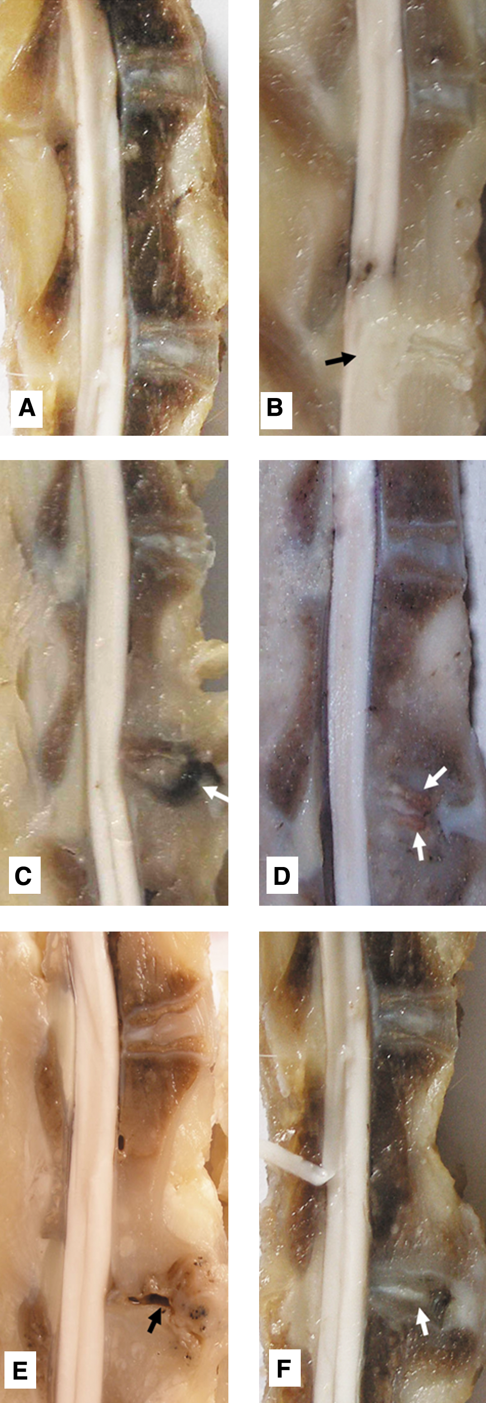
Macroscopic view of intervertebral discs (IVDs) from experimental rabbits. The lower IVDs were the target IVDs.
Micrographic results
As shown in Fig. 2, adjacent normal rabbit IVDs had a gelatinous NP located in the center, with the laminate annulus surrounding. Thin cartilage end plate, bony end plate and epiphyseal cartilage were structurally intact (Fig. 2A). In Group N, a narrowed disc space, inflammatory destruction of disc tissue, extensive erosion and hyperostosis of bony end plates, and epidural abscesses compressing the spinal cord were found. Pus invaded the adjacent vertebral bodies with the formation of osteomyelitis (Fig. 2B). In Group O, the structure of the IVDs was replaced by fibrosis tissue and small amounts of granulation tissue, and narrowed disc space and bone fusion in the anterior portion of the disk space were observed (Fig. 2C). In Group V, narrowed disc space, focal inflammatory changes of disc tissue, focal erosion, and hyperostosis of bony end plates were found (Fig. 2D). In Group L (Fig. 2E) and Group C (Fig. 2F), cavitation formation from laser irradiation, focal inflammatory lesions, focal erosion, and hyperostosis of bony end plates were observed. However, in Group C, fibrosis was found in the region of the nucleus, and the area of cavitation was less and the structure of the IVDs better preserved than in Group L.
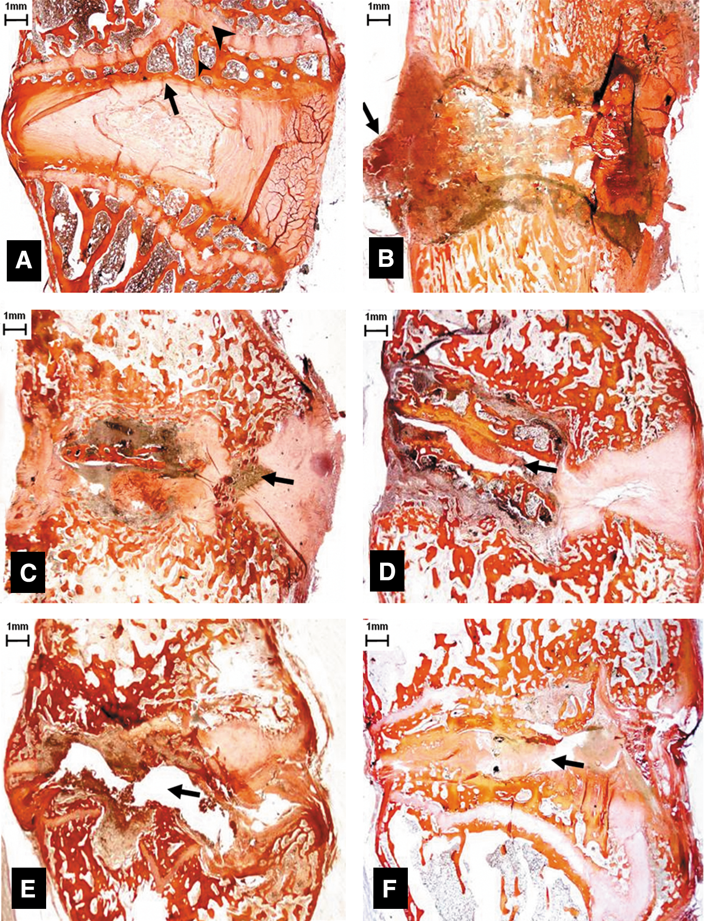
Midsagittal micrograph of intervertebral discs (IVDs) (collagen van Gieson [VG], original magnification×30).
The ID scoring system
The Group O ID score of 9.7±0.95 was significantly less than that of the other groups (p<0.01). The ID scores of Groups V and L were 12.2±1.32 and 12.6±0.97, respectively, both significantly less than that of Group N (p<0.05). The Group C ID score of 10.9±0.99 was significantly less than that of Groups N, V, and L (p<0.05) (Table 2).
ID, iatrogenic discitis.
p<0.05, significantly different from other groups.
p<0.05, significantly different from groups N, O, and C.
Discussion
The structure and composition of mature rabbit discs is remarkably similar to that of adult humans. 13 Therefore, this study used the rabbit model to demonstrate intervertebral disc and vertebral ID pathology. We used S. aureus, the most common pathogen causing IVD infection, and an established ID model of bacterial injection into the rabbit NP. The Group M results were similar to those of Liang et al., 14 with both studies demonstrating changed signal on MRI images and positive bacterial culture. Liang et al. 14 demonstrated that pathological evidence of discitis occurred 1 week post-bacterial injection, and that the pathology observed was similar that in humans.
The avascular nature of the disc renders it vulnerable to the iatrogenic introduction of bacteria during procedures. Treatment of ID is complicated by a poor vascular supply to the infected area and the complexity of the surgical procedure. Conservative medical management is usually accompanied by a long period of immobilization as well as incomplete bony fusion. If bone destruction exists, the rate of pseudarthrosis and instability has been reported as high as 50%. 15 Group N results showed that ID with S. aureus can cause serious inflammation including osteomyelitis and epidural abscess, suggesting that ID should be treated quickly and effectively in order to avoid complications.
Radical surgery has several advantages, including complete debridement, correction of the deformed spine, complete decompression of spinal compression, and immediate spine stabilization. Our study observed slight inflammatory changes and bone fusion in the Group O, the group with the lowest ID score. This result suggests that early debridement is the optimal ID treatment. However, perioperative complications limit its application, particularly in the elderly and those with comorbidities.
Intradiscal antibiotics appear advantageous, as drugs can be delivered locally, avoiding inefficient intravenous therapy. In addition, intradiscal antibiotics are less likely to contribute to antimicrobial resistance. Osti et al. 16 reported that intradiscal administration of antibiotics can prevent the development of discitis even when bacteria are directly introduced into the IVD of sheep. In addition, it has been demonstrated that the application of antibiotics in contrast agents can prevent post-discography discitis. 17 Experimental studies have also shown that intradiscal injection with VA-PLGA sustained-release microspheres is superior to that of intravenous injection, although they failed to completely remove the infection. 18 The Group V results in our study also demonstrated that intradiscal VA injection could partially contain the pathological ID process. However, the therapy appeared to be ineffective at preventing bony endplate destruction in this rabbit model. Intradiscal administration of antibiotics alone into the locally infected region cannot simultaneously achieve debridement. Bacteria replicate very rapidly and a mutation that helps a microbe survive in the presence of an antibiotic will quickly dominate the microbial population. Moreover, the small capacity in the IVD may prevent the application of higher concentrations of antibiotics locally.
High Level Laser Therapy (HLLT) could provide an adjunct or an alternative to surgical debridement and local antimicrobials for infections that are difficult to treat. HLLT can decrease infection levels and bacteria counts in osteomyelitis induced by S. aureus or MRSA, with a positive effect on wound healing infected with S. aureus. 11,12 Compared with some previous studies, this study is mainly using thermal mechanism to sterilize infected wounds as well as ablate inflammatory tissue in IVDs. Some studies using HLLT showed that its results in volatilization of the lesion and sterilization are higher with the use of cautery. 19,20 The Group L results in our study showed that 100 J diode laser irradiation could volatilize infected NP and induce cavitation formation. This technique could effectively achieve “debridement,” by eliminating the focus of infection as well as the abscess. However, focal inflammatory lesions and pathological instability of the spine induced by large cavitations in the disc space still occurred in Group L. A reason for this may be that one time “sterilization” by diode laser irradiation could not kill all bacteria, and bacterial regrowth caused delay of the healing process.
Group C results clearly demonstrate the ability of diode laser combined with intradiscal VA to successfully treat ID in the rabbit model. The cavitation from irradiation was replaced by fibrous tissue and the structure of the IVDs was less damaged. Laser energy can debride the focus of infection and volatilize the abscess. The cavitation contained more local VA, thus exerting enhanced bactericidal activity. Although the effect of combined treatment was better than that of VA or irradiation alone, the efficacy was not equivalent to that of Group O, implying that the combination could not achieve the results of open surgery.
Conclusions
We demonstrated that high power diode laser delivered to the infected NP region could eliminate the focus of infection, which in turn enhanced the efficacy of intradiscal VA. This suggests that the technique may be a useful addition to conservative medical treatment, especially when infection is limited. It is expected to reduce the incidence of operation and avoid the side effects of systemic application of high dose antibiotics.
Footnotes
Acknowledgments
This study was supported by Civil Aviation General Hospital Scientific Research Projects Fund (Project number: 2009016). We thank Dr. Bing-Huai Lu, Dr. Qiu-Lan Lin, and Dr. Ding-Qiong Peng for their assistance in this study.
Author Disclosure Statement
No competing financial interests exist.