
Abstract
Introduction
Resin bonding between a tooth and the restoration is recommended for improving the retention, marginal adaptation, and fracture resistance of restorations. 8,10 Surface treatments create microporosities on the porcelain surface, increase the surface area, and enhance the potential for mechanical retention of the cement into ceramic micro-roughness. 11,12 Although several authors 8,13 –15 have described various surface treatment procedures for all-ceramic restorations, high-strength ceramic materials, such as yttrium-stabilized tetragonal zirconia (Y-TZP), are not suitable for acid etching because they do not have a glassy phase. 6,16 For this reason, prior to luting, alternative methods, such as air abrasion or laser treatment, can be used to treat the inner surfaces of Y-TZP materials.
Air abrasion with aluminum oxide (Al2O3) particles is a commonly used surface treatment method. The abrasive process of this method removes the contamination layers of the ceramic surface, increases the area that is available for bonding, and improves the flow of the resin cement to the ceramic surface. 8,13
In dentistry, the erbium:yttrium-aluminum-garnet (Er:YAG) laser has been proposed for different clinical applications, including carious dentin removal, cavity preparation, and surface conditioning of indirect restorations. 14,17,18 The principal effect of laser energy is the conversion of light energy into heat. Moreover, the most important interaction between the laser and the substrate is the absorption of laser energy by the substrate. 19,20 The macroscopic and microscopic irregularities produced by Er:YAG laser irradiation reportedly constitute a mechanism of adhesion to enamel, dentin, 21,22 and lithium disilicate ceramics. 15 Although there is plenty of information regarding the effects of Er:YAG laser irradiation on enamel and dentin structures, 19 –22 little is known about the use of this laser as a surface treatment option for high-strength dental ceramics.
The aim of this study was to compare the surface roughness and morphologic changes of untreated and air-abraded surfaces of zirconia with those of surfaces irradiated with different energy intensities of the Er:YAG laser. The null hypothesis was that the conditioning treatments would not modify the ceramic surface roughness and morphology.
Methods
A single type of Y-TZP ceramic material (VITA In-Ceram YZ for inLab®; VITA Zahnfabrik, Bad Säckingen, Germany, batch number 24430) was used in this study. It consisted of 92% ZrO2, 5% Y2O3, <3% HfO2, <3% Al2O3, and <1% SiO2 by weight. Ceramic specimens (15 mm×14 mm×3 mm) were cut from pre-sintered blocks using a low-speed sectioning machine (Isomet 1000 Precision Saw; Buehler, Ltd., Lake Bluff, IL) under water cooling. According to the manufacturer's instructions, they were then sintered in a furnace at 1530°C for 7.5 h to the following dimensions: 12 mm×11.2 mm×2.4 mm (VITA ZYrcomat; VITA Zahnfabrik, Bad Säckingen, Germany). After the sintering process, the specimens were embedded in a self-cure acrylic resin (Meliodent, Heraeus Kulzer, Hanau, Germany), leaving one of the surfaces uncovered. The ceramic surfaces were then polished using a series of silicon carbide (SiC) abrasive papers in sequence (grit 120, 200, 600, 1000, 1500, and 2000; Struers, Ballerup, Denmark) for 15 sec under water irrigation to obtain standardized flat surfaces before applying the surface treatments. After the polishing procedure was completed, all of the specimens were ultrasonically (Biosonic JR; Whaledent, NJ) cleaned in distilled water to remove any surface residues and dried. A 5-mm diameter circular area on the ceramic surface of each specimen was exposed to a surface treatment. The specimens were then randomly divided into five groups (n=10) according to the surface treatments performed: 1. Control: No surface conditioning procedure was applied to this group. 2. Laser treatment: The Er:YAG laser (AT Fidelis Er:YAG; Fotona, Ljubljana, Slovenia) with a wavelength of 2940 nm was applied to the ceramics with a special hand piece (R14-C). The laser's cylindrical sapphire optical fiber (1.3 mm in diameter and 8 mm in length) was placed perpendicularly to the surface at a distance of 1 mm (non-contact mode). In addition, the restricted ceramic area was scanned in a sweeping style with water irrigation (6.75 mL/min) and air cooling for 15 sec. During the laser application, the distance between the specimen and the fiber tip was standardized using a special mold that consisted of two parts: a fixed upper part and a mobile lower part. The sample was fixed in the lower part with screws. The lower part of the mold was adjusted vertically by an adjustable screw. The laser parameters were 10 Hz (pulses per second) and MSP mode (100 μs) (pulse width); however, the energy intensities varied according to the experimental groups (200, 300, and 400 mJ). The Er:YAG laser energy intensities of 200, 300, and 400 mJ levels were 15.08, 22.61, and 30.15 J/cm2, respectively. 3. Air abrasion: The ceramic surfaces were air abraded with 110-μm Al2O3 particles (Korox; Bego, Bremen, Germany) from a distance of ∼10 mm and at a pressure of 3 bars for 15 sec.
After each surface treatment, the samples were ultrasonically cleaned in 99% acetone for 5 min and distilled water for another 5 min. Then, one specimen from each group was selected for microscopic analysis. Atomic force microscope (AFM) and scanning electron microscope (SEM) analyses were utilized to observe the morphologic changes on the zirconia surfaces following different surface treatment applications.
AFM analysis
A specimen from each group was evaluated under an AFM (Multimode Nanoscope 3D, Veeco Metrology Group, Santa Barbara, CA) to perform a qualitative evaluation of the samples. Digital images were taken in air. The intermittent contact mode was conducted using a 0.5–2 Ω cm Si tip (at 50 μm) oscillating at a frequency of 297.354 kHz. Changes in the vertical position in images showed the height of the surfaces as bright and dark regions. The tip sample was kept stable via constant oscillation amplitude (set point amplitude). Images with 256×256 pixels were acquired with a scan size of 20 μm×20 μm and a scan rate of 1 Hz. Afterward, the images were evaluated with Nanoscope IIIa (version 5.31R1).
SEM analysis
The same specimens used for the AFM analysis were also used for the SEM analysis (LEO 440; Zeiss, Cambridge, U.K). Prior to the SEM analysis, the specimens were gold-palladium coated with a sputter coater (Quorum Technologies Polaron SC7620; Newhaven, East Sussex, U.K.) for 15 sec at 45°C. Images from each group were taken at 500×magnification. The microscope was operated at an accelerating voltage of 20 kV.
Surface roughness evaluation
After the surface treatments, surface roughness (Ra in μm) measurements were taken for each specimen using a surface texture measuring instrument (Mitutoyo Surftest 402 Analyzer Series 178; Mitutoyo Corporation, Minatoku, Japan) with a cutoff value of 0.8 mm and a measuring length of 4 mm. The Ra value describes the average surface roughness for a surface that was traced by the surface measuring instrument. Ten measurements at different locations were recorded for each specimen, and the average of these measurements was used to obtain the Ra value of each specimen. Prior to measurement, the instrument was calibrated against a reference block for which the Ra value was 2.792 μm.
Statistical analysis
The Statistical Package for the Social Sciences (SPSS) software (version 16.0) was used for the statistical analysis. The data for Ra (μm) were analyzed using a one-way analysis of variance (ANOVA). Multiple pairwise comparisons were conducted with Tukey's honestly statistical difference (HSD) test at a significance level of p<0.05.
Results
The results of the statistical analyses are summarized in Table 1, including the mean and standard deviation values of Ra (μm) and Tukey's HSD results for each group. Results of the one-way ANOVA indicated that there were significant differences among groups (p<0.001). Results of Tukey's HSD test revealed that the mean Ra value of the air abrasion group was significantly higher than that of the other four groups (the control and Er:YAG laser irradiation at 200, 300, and 400 mJ) (p<0.001). With the exception of the air abrasion group, there were no statistically significant differences within groups (p>0.05).
Means with the same letters were not significantly different (p>0.05, Tukey HSD test).
AFM evaluation
The AFM images of the ceramic specimens are shown in Fig. 1. Based on these images, the surface morphology of the laser groups was similar to that of the control group (Fig. 1a–c). However, a specimen irradiated with 400 mJ showed rare pits (Fig. 1d). On the other hand, more irregular and heterogeneous surfaces showing higher peaks and troughs were found with air abrasion (Fig. 1e).
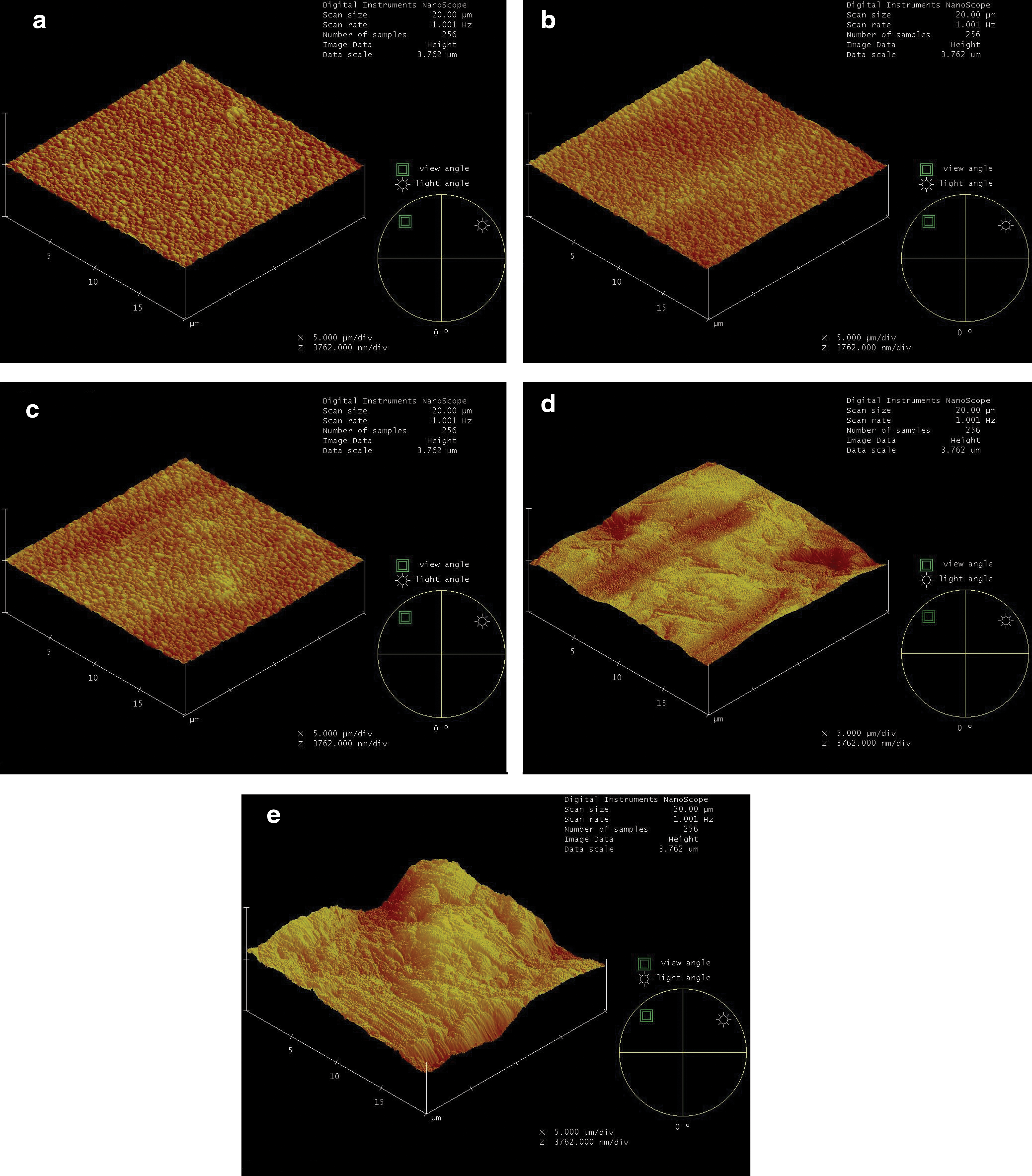
Atomic force microscope (AFM) images of zirconia surfaces (20 μm×20 μm).
SEM evaluation
The SEM images of the ceramic surfaces treated with different surface treatments are presented in Fig. 2. Similar to the AFM images, the SEM images of the ceramic surfaces showed morphologic differences following different surface treatments. The control and laser groups had similar surface topographies (Fig. 2a–d). However, a specimen irradiated at 400 mJ displayed small and rare pits formed by the laser optical fiber tip (Fig. 2d). In addition, the air abrasion group exhibited a change in surface texture with the formation of micro-retentive grooves (Fig. 2e).
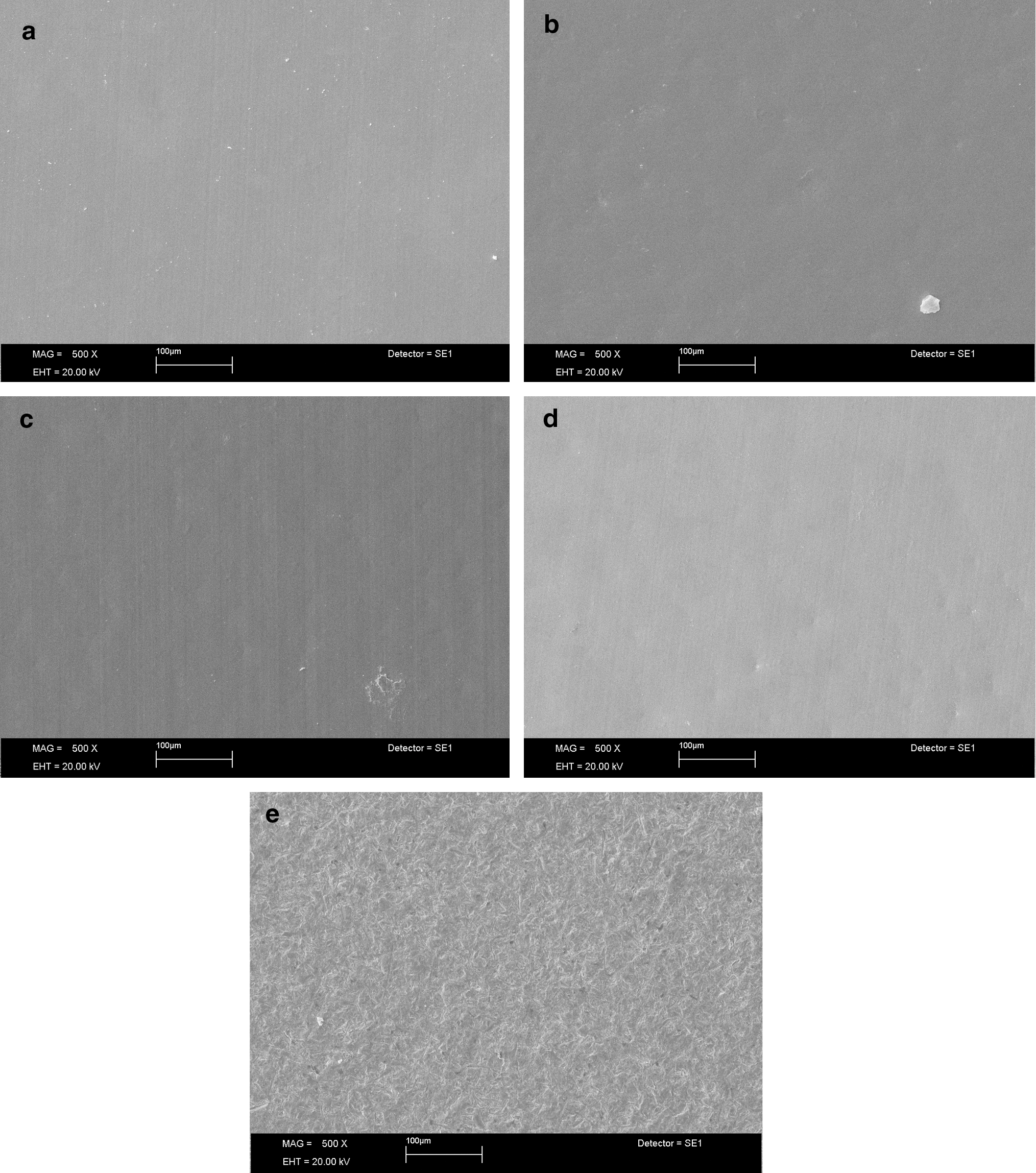
Scanning electron microscope (SEM) images of zirconia surfaces (original magnification×500).
Discussion
The present study aimed to investigate the effect of air abrasion with Al2O3 particles and Er:YAG laser irradiation on the surface roughness and morphology of zirconia. Based on the results of this study, the null hypothesis was rejected, as tested treatments changed the surface roughness and morphology of zirconia. All of these treatments were used to achieve micromechanical retention on ceramic surfaces, although their effect mechanisms differed.
Airborne particle abrasion, an important treatment procedure for promoting strong adhesion of veneering ceramics, works by increasing surface roughness and providing undercuts. 23 In other studies, 24,25 air abrasion influenced crown retention, regardless of the cement used. The increase of retention corresponded to the increase in micro-roughness on the surface of the air-abraded zirconia.
Although surface treatments are used to achieve micromechanical retention on the ceramic surface, they can affect the mechanical properties and long-term performance of ceramics. 26 Curtis et al. 27 evaluated the influence of air abrasion (25, 50, and 110 μm Al2O3 particles) on the biaxial flexural strength of zirconia specimens and found no statistical differences among the groups. Results of other studies 28,29 showed that air abrasion with 110 μm Al2O3 particles might strengthen zirconia. Given the results of these studies, 26 –29 110 μm Al2O3 particles were used for air abrasion in this study.
Laser application leads the deposition of high amounts of radiation energy on the ceramic surface over an extremely short period of time. Therefore, the radiation energy is thermalized, whereas the temperature in a thin superficial layer rises. 19,20,30 The most important interaction between the laser and the substrate is the absorption of laser energy by the substrate. The pigmentation of the surface and its water content, along with other surface characteristics, determines the amount of energy that is absorbed by the irradiated surface. 19,20 Coluzzi 20 reported that the optical penetration depth of the Er:YAG laser was only a few micrometers. This characteristic might be beneficial to the surface treatment of dental ceramics, as structural modifications would be restricted to the outermost surface. Nevertheless, there is limited knowledge regarding the effects of Er:YAG laser irradiation on dental ceramics.
Cavalcanti et al. 31 investigated the effects of different energy intensities of the Er:YAG laser (200, 400, and 600 mJ) and air abrasion with Al2O3 particles on the surface roughness and morphologic characteristics of Y-TZP ceramics (Cercon Smart Ceramics and Procera Zirconia). They coated the surfaces with graphite powder prior to laser irradiation. Finally, they reported that high laser power settings (400 and 600 mJ) caused excessive material deterioration and were unsuitable parameters for roughening zirconia. However, the lower energy intensity tested (200 mJ) might be a potential surface treatment method for Y-TZP ceramics.
Shiu et al. 14 reported that 500 mJ of Er:YAG laser energy could not cause adequate roughness of the feldspathic ceramic surface, probably because of the different composition and reflectance of this ceramic material. In contrast to Cavalcanti et al., 31 they 14 did not apply graphite to the surface prior to laser application. Given the results of these studies, 14,31 200, 300, and 400 mJ of Er:YAG laser energy were used in the present study. Further, ceramic surfaces were not coated with graphite powder prior to application.
In this study, AFM and SEM analyses were used to observe morphologic changes after different surface treatments and to interpret the morphologic changes with roughness analyses. Results revealed that, with the exception of air abrasion and 400 mJ of Er:YAG laser energy, other surface treatments (200 and 300 mJ) did not change the surface morphology of zirconia compared with the control group. Irradiating at 400 mJ changed the morphology of zirconia by forming rare pits, whereas air abrasion formed micro-retentive grooves.
Although the surface roughness of the 400 mJ irradiated specimen was not significantly different than that of the control and other laser groups, rare pits were observed in the AFM and SEM images of this specimen. In addition, other laser irradiations (200 and 300 mJ) did not change the surface morphology of zirconia. Similar to the 400 mJ irradiated specimen, they increased the roughness of zirconia when compared with the control group. However, this difference was not statistically significant. Zirconia is a water-free material with an opaque, white color. Probably because of the differences in the composition of the ceramic, different thermal expansion coefficients can occur during laser application. Therefore, in some regions, superficial ceramic loss can take place. Based on the results of the roughness and morphologic analyses, it seems that all laser irradiation parameters increased the surface roughness, probably resulting from the debris formed by the micro-explosions that might adhere to the melted ceramic surfaces as occurs in tooth substrates. 22 Nevertheless, the application of all laser parameters was not as effective as the air abrasion method.
A reliable bond between zirconium oxide ceramic and resin cement is a crucial factor to ensure clinical success and longevity. 32 It was reported that surface treatments are necessary to obtain reliable bond strength to some resin-based cements (Bis-GMA) because of the lack of a chemical interaction between the resin cement and zirconia. 33 Based on the results of the statistical and microscope analyses, it appears that air abrasion or 400 mJ Er:YAG laser can ease the flow of these resin cements into these surfaces and may improve the bond strength of resin cements to zirconia.
In this study, only one type of ceramic material (Y-TZP) was used. The effects of surface treatments on the mechanical properties and phase transformations of zirconia on the bonding of luting cements and the extent to which this bond will endure under clinical conditions should also be evaluated in future studies.
Conclusions
Results of microscope analyses showed that 400 mJ Er:YAG laser energy changed the surface morphology of zirconia with the formation of pits, whereas air abrasion changed it with the formation of micro-retentive grooves. Other irradiation parameters (200 and 300 mJ) did not change the morphology of zirconia. In addition, except for the air abrasion group, none of the laser irradiation parameters significantly increased zirconia's surface roughness. Based on the results of the statistical and microscopic analyses conducted in this in vitro investigation, 400 mJ Er:YAG laser energy or air abrasion can be used to obtain micromechanical retention prior to luting; however, air abrasion is the most effective surface treatment method.
Footnotes
Acknowledgments
The authors thank Prof. Aslıhan Usumez for supplying the Er:YAG laser unit, Dr. Altinay Boyraz for conducting the AFM and SEM analyses, and Professor Bora Ozturk for completing the statistical analyses.
Author Disclosure Statement
No conflicting financial interests exist.