
Abstract
Introduction
Materials and Methods
Animals
All animal studies were conducted in compliance with the Guidelines for the Treatment of Experimental Animals at the Tokyo Dental College (Approval Number 223206). Sixty-five male Sprague–Dawley rats weighing ∼200 g each were used in this study. Animals were distributed into three groups: a control group without any CO2 laser irradiation, a group in which the animals were killed immediately after the CO2 irradiation, and a group in which the animals were killed 5 days after the CO2 laser irradiation.
The animals were fed a standard laboratory diet during the experimental time periods. The animals were anesthetized with sodium thiopental (Ravonal®; Tanabe, Japan).
Laser irradiation protocol
A CO2 laser (Panalas CO5 Sigma; Panasonic Dental Co., Osaka, Japan) was used to irradiate the occlusal surface of the upper first molar dentin where there was no covering enamel due to because of the natural physical attrition phenomenon in rats. A Type 1A tip (defocus, diameter; 0.15 cm) was used in direct contact with the tooth surface and the exposure time was 8.8 sec. The parameters for the CO2 laser were: wavelength of 10.6 μm, 2 W in the super pulse mode, pulse 0.6 ms, and total laser energy of 4 J (density; 203.84 J/cm2). A peak power of 5 W was used in this study. An air flow system was provided at the tip of the hand piece. These laser conditions were based on clinical irradiation standards 7,8 and on preliminary experiments that established a slightly strong condition in which there were differences between the experimental group and the control group.
Temperature measurement after the CO2 laser irradiation
After euthanasia of the rats, five upper first molars were extracted to measure the temperature changes in the pulp after CO2 laser irradiation. A thermocouple thermometer 0.1 mm diameter was used in this study for the measurement of temperature changes in the pulp. Teeth were sliced transversely at the enamel cementum junction, and pulp was removed mechanically from the crown portion of the teeth. This 0.1 mm thick thermocouple thermometer was too big for temperature measurement to insert directly into the pulp chamber. The thickness of the dentin on the five teeth was ∼0.8 mm on the top to the pulpal side. A super-fine mineral insulated thermocouple, type K (Okazaki Manufacturing Co., Kobe, Japan), 0.10 mm in outside diameter of the sheath, was in direct contact without any intermediate material on the pulp horn, and the CO2 laser was used exactly as described previously. The thermo electromotive force change was recorded using a thermocouple thermometer SR6513 (Graphitic Corp., Yokohama, Japan). The recorded value was converted to temperature (°C) using a conversion chart (mV to °C=X(mV)×25).
Quantitative reverse transcription polymerase chain reaction (RT-PCR) analysis
For quantitative RT-PCR, five animals were sacrificed killed with an overdose of sodium thiopental at 6, 12, and 24 h after CO2 laser irradiation. The upper first molar was extracted and washed in alpha-minimal essential medium (α-MEM, GIBCO, Carlsbad, CA) containing 10% gentamycin and 1.2% fungizone for 5 min. The pulp was then removed mechanically. Total RNA was extracted from each specimen using the acid guanidium thiocyanate/phenol-chloroform method as follows. The cells were homogenized in Trisol® Reagent (Invitrogen, Carlsbad, CA) after rinsing each sample with phosphate-buffered saline (PBS). Each solution was transferred to a tube containing chloroform and was mixed. The solutions were centrifuged at 14,000 rpm at 4°C for 20 min, after which they were incubated in 70% isopropanol at −80°C for 1 h. After centrifugation, the mRNA pellets were washed with 70% cold ethanol, and were then dissolved in RNAase-free (diethylpyrocarbonate [DEPC]-treated) water. Total RNAs were reverse transcribed and amplified using an RT-PCR kit (Takara Biomedicals, Shiga, Japan). RT-PCR products were analyzed by quantitative real-time RT-PCR using TaqMan® Gene Expression Assays for three target genes, TNF-α (Rn 99999017-ml) and IL-1-α (Rn00566700-ml), to confirm the appearance at the mRNA level of inflammatory cytokines, and β-actin (4352340E) as an endogenous control (Applied Biosystems, Foster City, CA), to determine variations in the amount of each RNA. All PCR reactions were performed using the real time PCR 7500 Fast System (Applied Biosystems). Gene expression quantitation using TaqMan® Gene Expression Assays was performed as the second step in a two-step RT-PCR. Assays were performed in singleplex reactions containing TaqMan® Fast Universal PCR Master Mix, TaqMan® Gene Expression Assays, distilled water and cDNA according to the manufacturer's instructions (Applied Biosystems). Reaction conditions consisted of primary denaturation at 95°C for 20 sec, and cycling for 40 cycles at 95°C for 3 sec and at 62°C for 30 sec. The experiments were conducted in triplicate, and data were analyzed using one-way ANOVA (p<0.05) and using Scheffé's test for multiple comparison.
Morphological observations
For morphological observations, the animals were killed immediately and at 5 days after the CO2 laser irradiation with an overdose of sodium thiopental. The upper molar with the maxillary bone of each rat was removed and fixed in 10% buffered formalin and dehydrated and then was embedded in paraffin. Paraffin sections were cut and observed with hematoxylin and eosin (HE) staining.
Immunohistochemical observations
For immunohistochemistry, the streptavidin-biotin immuno-peroxidase method was employed using a Histofine SAB-PO (MULTI) kit (Nichirei Co., Ltd., Tokyo, Japan). Paraffin sections were deparaffinized with xylene, then were washed with 100% alcohol, and then were washed with distilled water. Endogenous peroxidase activity was blocked by incubating the sections with 3% H2O2 in methanol for 30 min. They were then microwaved for 30 min at 65°C in 0.01 M citrate buffer (pH 6.0), cooled to room temperature and then washed in PBS three times for 5 min each. To prevent nonspecific reactions, sections were incubated with 10% serum for 10 min. The HSP-70 antibody, which was used to confirm the expression of HSP-70 heat shock protein (at a dilution of 1:200, Abcam, Nihonbashi, Tokyo, Japan), the VEGF antibody, which was used to confirm the change of tissue restoration (at a dilution of 1:200, Abcam), the nestin antibody, which was used to confirm the appearance of odontoblast function (at a dilution of 1:100, Santa Cruz Biotechnology, Inc., Santa Cruz, CA), and the NFP antibody, which was used to confirm the existence of neural fibers (at a dilution of 1:200, DAKO, Glostrup, Denmark) were used as primary antibodies. They were reacted at 4°C overnight. As a negative control, PBS was used instead of the primary antibody. After the primary antibody reaction, the sections were rinsed in PBS three times for 5 min each. The secondary antibody (biotinylated anti-mouse immunoglobulin G [IgG] or anti-rabbit IgG) was reacted at room temperature for 30 min. After washing in PBS three times for 5 min each, 3,3’-diaminobenzidine-tetra-hydrochloride in Tris-HCl buffer (pH 7.6) was used to visualize the reaction. Finally, sections were counter-stained with Mayer's hematoxylin. Specimens were examined using a light microscope (BX41, Olympus, Shinjuku, Japan) and were photographed.
Results
Temperature change caused by the CO2 laser irradiation
The temperature change at the pulp side caused by the CO2 laser irradiation increased ∼0.9 mV, which was ∼22.5°C as an average (Fig. 1). The temperature increases for each of the five samples were 20°C, 22.5°C, 25°C, 22.5°C, and 22.5°C.
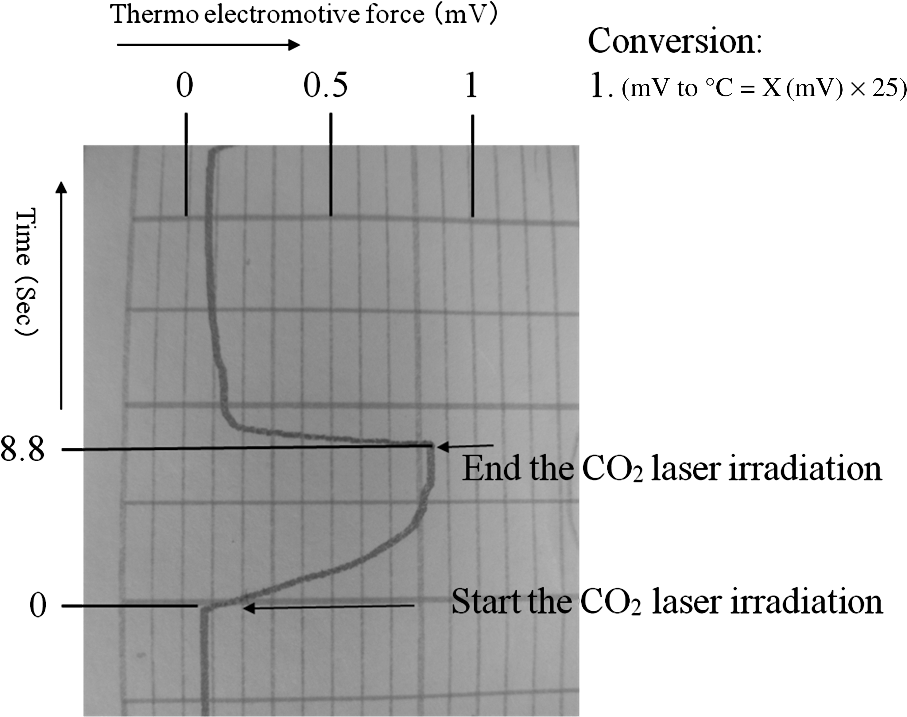
Measurement record of the thermocouple thermometer The electromotive force rises upon exposure to the irradiating CO2 laser and returns to the initial value when the irradiation stops. Recorded values were converted to temperature (°C) using the conversion chart (mV to°C=X(mV)×25).
Quantitative RT-PCR analysis
The expression of TNF-α and IL-1-α mRNAs was significantly higher at 6, 12, and 24 h after the CO2 laser irradiation compared with the control group (p<0.05), as shown in Figs. 2 and 3. The expression of those mRNAs increased in a time-dependent manner and was highest 24 h after the CO2 laser irradiation. However, there were no significant differences in the expression of TNF-α or IL-1-α mRNA at 48 h after the irradiation.
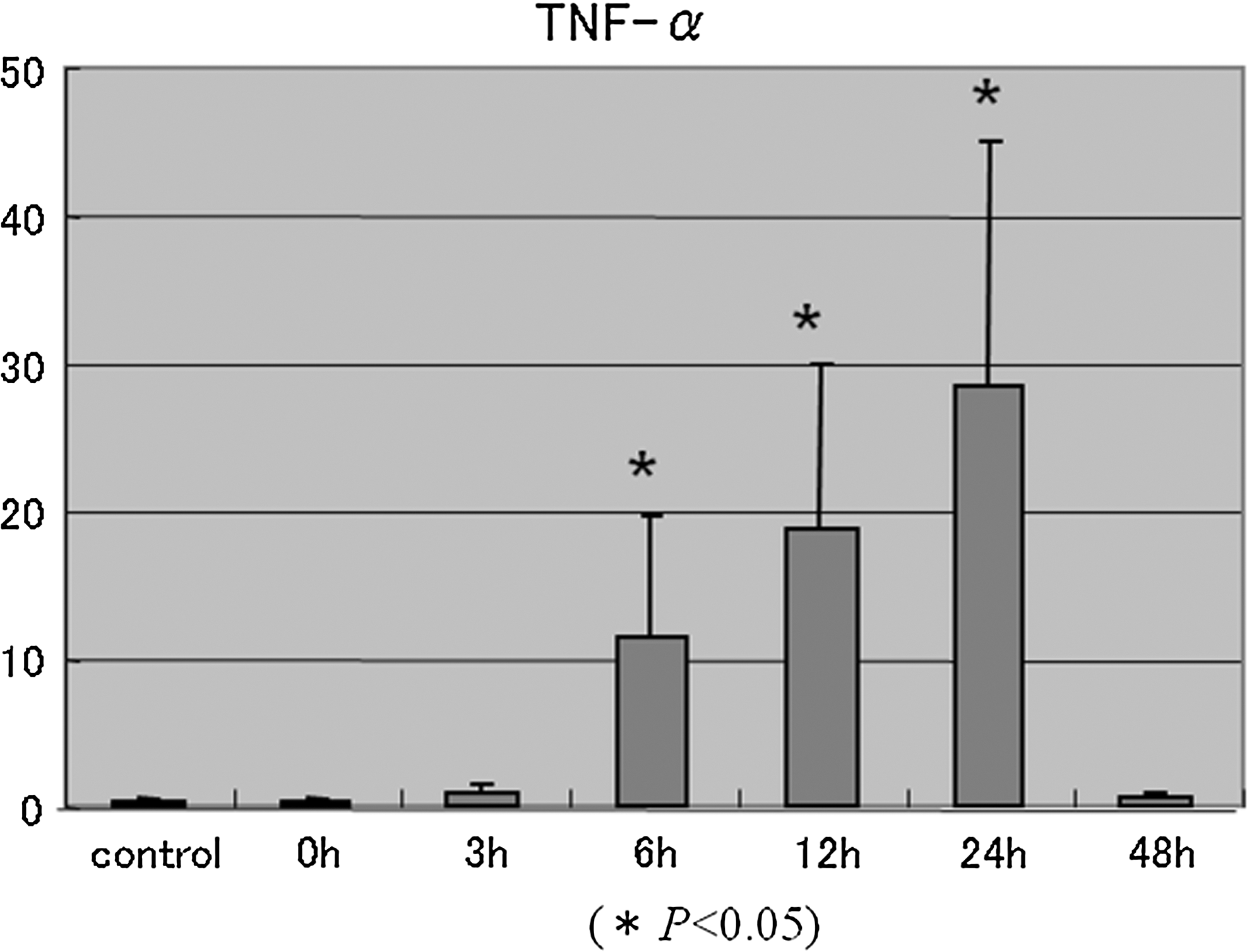
Expression of tumor necrosis factor-alpha (TNF-α) mRNA after CO2 laser irradiation. The expression of TNF-α mRNA was significantly higher at 6, 12, and 24 h after the CO2 laser irradiation compared with the control group (p<0.05). The expression of TNF-α mRNA increased in a time-dependent manner and was highest 24 h after irradiation. However, there are no significant differences in the expression of TNF-α mRNA at 48 h after irradiation.
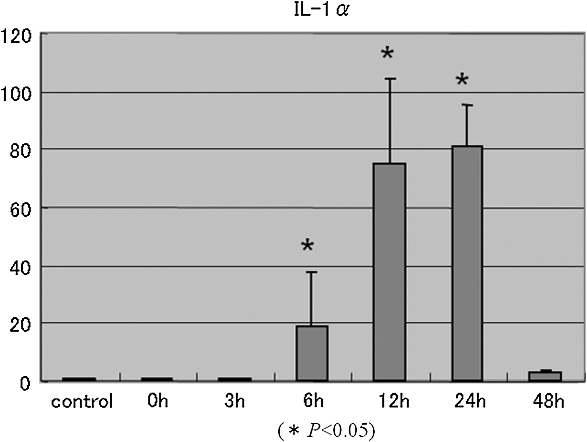
Expression of interleukin (IL)-1-α mRNA after CO2 laser irradiation. The expression of IL-1-α mRNA was significantly higher at 6, 12, and 24 h after the CO2 laser irradiation compared with the control group (p<0.05). The expression of IL-1-α mRNA increased in a time-dependent manner and was highest 24 h after irradiation. However, there are no significant differences in the expression of IL-1 mRNA at 48 h after irradiation.
Morphological observations
Histologically, small vacuoles were observed in the cells immediately after the CO2 laser irradiation (Fig. 4a). In another specimen, a slight hyaline degeneration in the pulp horn was observed underneath the odontoblast layer (Fig. 4b).
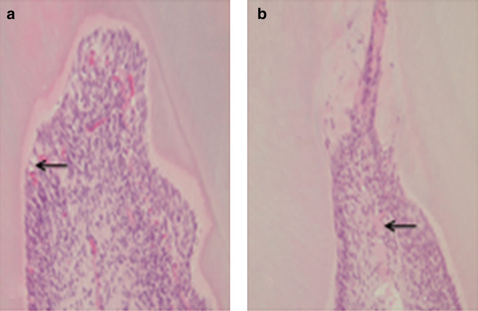
Hematoxylin-eosin staining.
Immunohistochemical observations
Positive reactions for HSP-70 were observed both in odontoblasts and in pulp cells just beneath the odontoblast layer immediately after the CO2 laser irradiation (Fig. 5b). Five days after the irradiation, HSP-70-positive reactions became weaker (Fig. 5c), but were still stronger than the control (Fig. 5a).
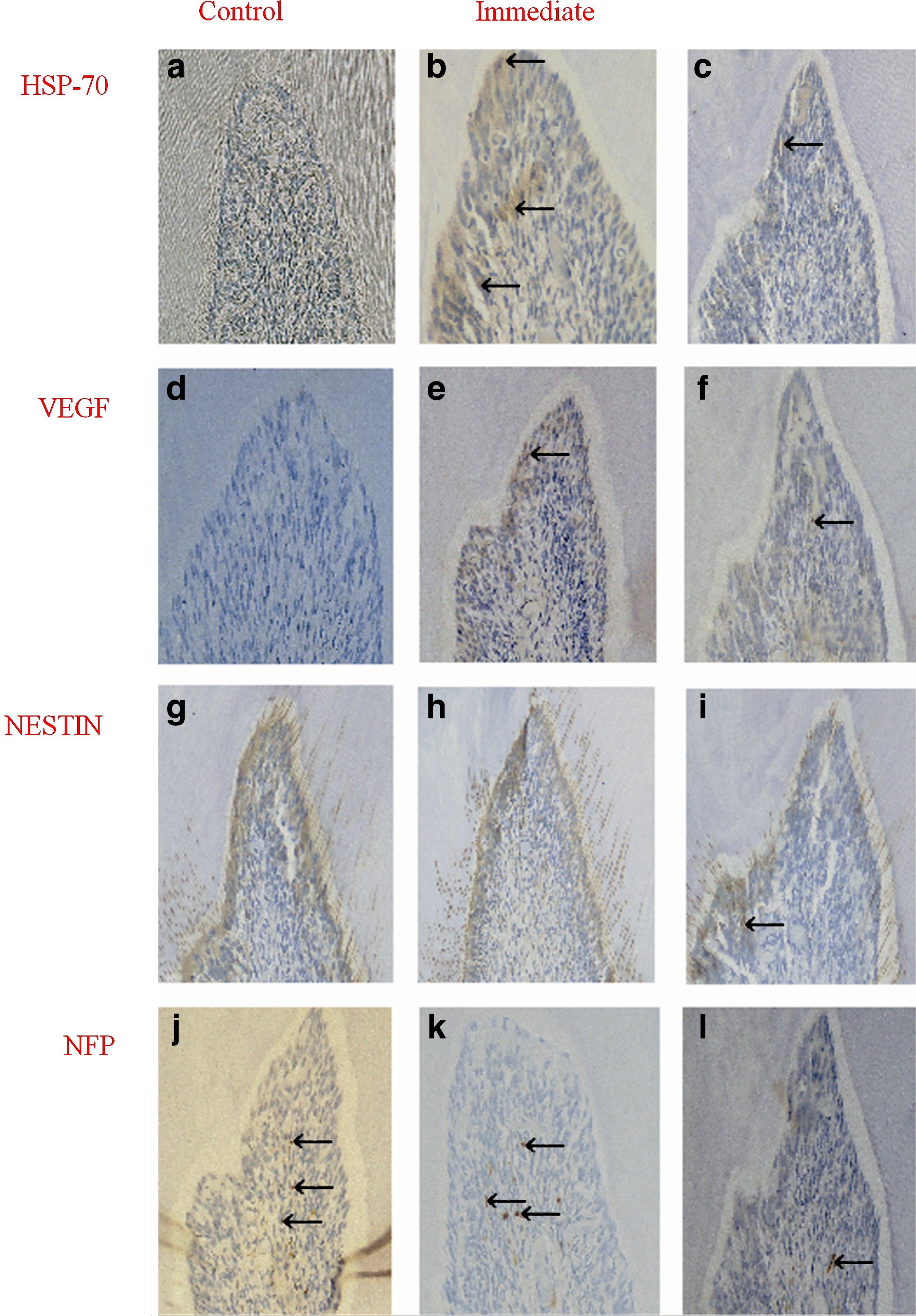
Immunohistochemical staining.
Positive reactions for VEGF were recognized in dental pulp cells just beneath the odontoblast layer and in odontoblasts and pulp cells just beneath the odontoblast layer immediately after the irradiation (Fig. 5e). The positive reaction for VEGF became weaker at 5 days (Fig. 5f).
Positive reactions for nestin were observed in pulp cells just beneath the odontoblast layer, in odontoblasts, and in the dentinal canaliculi immediately after the laser irradiation (Fig. 5h) and in the control pulp (Fig. 5g). At 5 days after irradiation, cells positive for nestin were not observed in the odontoblast layer, but were observed in deeper areas of the dental pulp (Fig. 5i).
Positive reactions for NFP were observed in the pulp horn area immediately after the laser irradiation (Fig. 5k) and in the control pulp (Fig. 5j), and were observed in the deeper pulp cells 5 days after the laser irradiation (Fig. 5l).
Discussion
In our morphological observations, attrition was observed on the occlusal surface of rat molar teeth where the dentin was exposed without covering enamel. As has been reported previously, commercial food pellets for laboratory animals are abrasive and increase tooth wear in rats compared with a powdered diet, 9 and the attrition is not only caused by the abrasive hard food but because the animals also spend more time chewing it. 10,11 That is why we consider that the molar teeth of rats are suitable experimental models for dentin hypersensitivity caused by natural physical attrition.
Many fundamental studies about the treatment of dentinal hypersensitivity with various types of laser irradiation, such as the Nd: or Er:YAG laser, the diode laser (8.5 J/cm2 for 60 sec) 12 and the CO2 laser (irradiation condition: energy: 0.3 J, 0.5 J, 0.9 J, 1.5 J, 2.5 J; application: one exposure/mm, five times within 5 mm of gingiva 13,14 ) has been reported to have extremely good clinical success. Zhang et al. 15 reported that after CO2 laser treatment of dentinal hypersensitivity, all patients were immediately free from sensitive pain. In another study, all patients showed absolutely identical perfusion indices immediately before and after the CO2 laser treatment as well as 1 week after treatment. 16 Furthermore, the CO2 laser treatment reduced dentinal hypersensitivity to an air stimulus, and all teeth remained vital with no adverse effects at 3 months after the irradiation. 15
However, the thermal effects of laser irradiation on oral tissues have been of concern. 17 –19 Irradiation that causes rises in temperature exceeding the threshold of pulpal tolerance will cause thermal injury to the dental pulp. Previous studies have demonstrated that healthy pulp tissue is not injured thermally if the laser equipment is used within appropriate parameters so that any temperature rise within the dental pulp remains <5°C. 20 Using a CO2 laser, no damage was reported after pulpal exposure to 3 W of power for 2 sec in the continuous wave (CW) mode in monkeys and dogs. In our studies using a CO2 laser, 2 W of power for 8.8 sec induced a slight pulpal degeneration, and this power of the CO2 laser increased the temperature ∼22.5°C, causing pulpal degeneration. This is because in our study the dentine thickness of the rat molar teeth was less than the average depth of the cavities used for the pulpal wall radiations in the monkey and dog study.
HSP-70 has been shown to play a significant role in rescuing stressed cells by helping damaged proteins refold or by participating in the synthesis of new proteins to replace damaged proteins. 21,22 Amemiya et al. 23 reported that HSP-70 is expressed in dental pulp cells under the stress condition of hypoxia. Our immunohistochemical investigations showed that HSP-70-positive cells were observed strongly in pulp cells compared with the control immediately after irradiation with the CO2 laser. This suggests that the increased temperature stresses the pulp cells causing slight morphological damage immediately after the laser irradiation within 5 days. This suggests that the remaining cells in the pulp tissue may react, and the expression of TNF-α and IL-1-α mRNAs may increases.
In this study, a weakly positive reaction for VEGF was detected at the depth of dental pulp cells at 5 days after the CO2 laser irradiation. VEGF is a signal protein produced by cells which stimulates the growth of new blood vessels, and is part of the system that restores the oxygen supply to tissues when the blood circulation is inadequate. VEGF function is essential to endothelial cell proliferation and migration. This phenomenon may represent the start of tissue repair after the CO2 laser irradiation, at least up to day 5.
The expression of nestin at 5 days after the CO2 laser irradiation decreased compared with the control in this study. Nestin is an intermediate filament protein most related to neurofilaments of stem cells. In injured teeth, nestin expression is upregulated in a selective manner in odontoblasts surrounding the injury site, showing a link between tissue repair and competence. Nestin is also distributed in the processes of mature odontoblasts and takes the place of degenerated odontoblasts in the case of pulpal damage. 24 In our study, the expression of nestin was observed in deeper pulpal cells, but only weakly in odontoblasts at 5 days after the CO2 laser irradiation. This suggests that pulpal stem cells in deeper areas probably begin migrating toward the degenerated pulp horn area.
It is known that neurofilament protein NFP is a special protein required for nerve fibers. In this study, the expression of NFP was seen in the irradiated pulp compared with the control 5 days after the CO2 laser irradiation. This must be is another reason for the reduction of dentinal sensitivity in hypersensitive patients. In conclusion, these results suggest that 203.84 J/cm2 of CO2 laser irradiation, such as is used in the clinic, to the pulp tissue through the dentin, such as is used in the clinic, induces the inflammatory and pathological cytokine pathways to repair the damaged rat pulp tissue.
Conclusions
These results suggest that 203.84 J/cm2 of CO2 laser irradiation to the pulp tissue through the dentin, such as is used in the clinic, induces the inflammatory and pathological cytokine pathways to repair the damaged rat pulp tissue.
Footnotes
Author Disclosure Statement
No competing financial interests exist.