
Abstract
Introduction
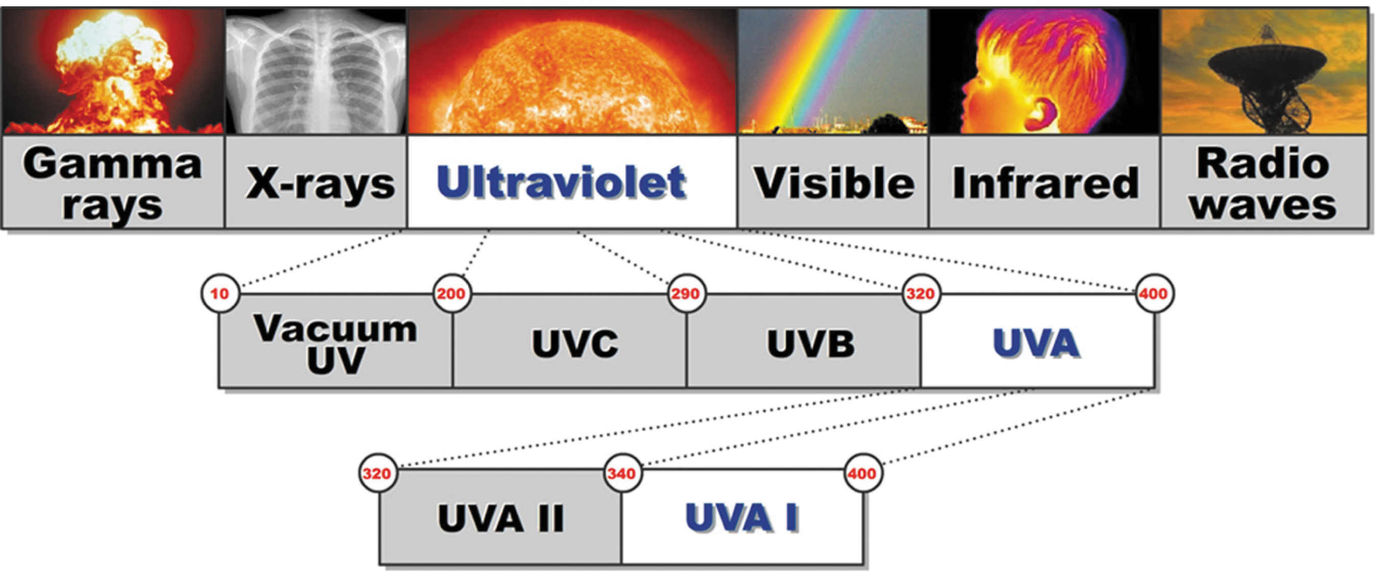
Electromagnetic spectrum.
From a therapeutic perspective, UVA1 irradiation has been categorized into low (10–40 J/cm2), medium (40–80 J/cm2), and high (80–120 J/cm2) dose regimens. Low doses are administered with fluorescent lamps, with low-cost instrumentation and maintenance costs. High energy fluences (>100 J/cm2) can be delivered only with metal-halide lamps that are associated with high costs and time-limited fluorescence. 6,7
UVA1 and UVAB radiation produce photoisomerization of trans-urocanic acid (UCA). Furthermore, exposure of human skin to UVA1 and UVB light results in an increase in percentage of cis-UCA in suction-blister fluid. 8 However, UVA1 therapy presents peculiar characteristics. For example, in contrast with UVB and psolaren UVA (PUVA) therapy, UVA1 radiation produces an immediate apoptotic rather than a delayed apoptotic state by means of constitutive intracellular proteins, instead of requiring accumulation of newly synthesized proteins such as p53. 9 Furthermore, whereas UVB light produces an increase in tumor necrosis factor-alpha (TNF-α) in suction-blister fluid in human skin, an opposite effect is observed following UVA1 therapy. 8 Interleukin (IL)-10 is also significantly increased in suction-blister fluid in human skin following UVB, but not UVA1, irradiation. 8
UV lasers have been extensively evaluated in the field of dermatology. For example, Gomez and colleagues 10 showed that UV radiation (355 nm), emitted by a Nd:YAG laser, required a lower energy for the ablation of the stratum corneum, inducing a greater impact on the lipid structures without any risk of producing lesions to the epidermis, if compared with infrared (IR; 1064 nm) radiation. Sato and colleagues 11 evaluated UV radiation-mediated ablation (355 nm) in porcine myocardium tissue samples using 1064, 532, and 266 nm radiations, showing that the ablation depth was maximized at 355 and 1064 nm through a photo-dermal process. This study emphasised how this laser could be potentially relevant for achieving transmyocardial revascularization for treatment of ischemic heart disease.
Excimer light devices (ELD) have also been widely used for treatment of psoriasis. For example, a study using a 308 nm monochromatic excimer light, showed a complete remission in >50% of patients (average of 12 sessions) with psoriasis involved in this investigation (152 patients with stable and localized plaque psoriasis, 47 with palmoplantar psoriasis). 12 This evidence was also confirmed by Wollina and coworkers 13 who showed an improvement in Psoriasis Area and Severity Index (PASI) score in patients with moderate plaque-type psoriasis treated with a 307 nm ELD. This improvement was equivalent to topical dithranol twice daily, but it was achieved in a significantly shorter time. The literature, regarding the use of excimer light, has also been reviewed by Gattu and coworkers showing that 18 clinical trials report positive results concerning the use of 308 nm excimer devices in psoriasis vulgaris, scalp psoriasis, and palmoplantar psoriasis, 14 further supporting the use of this device for treatment of different types of psoriasis.
UVA1 therapy has also been used for treatment of atopic dermatitis, localized scleroderma, systemic lupus erythematosus, polymorphic light eruption, cutaneous T cell lymphoma, lichen sclerosus, keloids, systemic sclerosis, and hand dermatitis. 5
In the context of psoriasis, UVA1 therapy has been used in combination with tacrolimus 15 and calcipotriol, 16 showing negative and positive results respectively. A study has also used UVA1 light to treat three patients affected by psoriasis of the palms, reporting no improvement in a patient and a 25–50% improvement in two subjects. 17
Objective
The aim of the present study was to investigate the therapeutic effectiveness of UVA1 355 nm laser for treatment of mild, moderate, and severe psoriasis.
Materials and Methods
Patients
A total of 14 patients (n [men]=10; n [women]=4; age=25–50 years [37.7±2.3; mean±SEM]), affected by psoriasis, were involved in this study. Two men and a woman were classified as affected by mild psoriasis (basal PASI score=7.4, 9.6, 8.1). A woman was classified as affected by moderate psoriasis (basal PASI score=19.6) and eight men and two women were classified as affected by severe psoriasis (basal PASI score=20.5, 30.7, 28.9, 22.9, 21.7, 29.1, 26.5, 43, 37.8, 38). All patients signed the informed consent to participate in this study. The study was conducted in accordance with the Declaration of Helsinki and the local Institutional Review Board (IRB). All the patients were asked to suspend previous pharmacological and physical therapies 30 days before the beginning of the study. Exclusion criteria were absence of concomitant cutaneous pathologies, such as cutaneous epithelioma and HIV-associated psoriasis. The PASI was used to measure the severity of the psoriatic lesions following UVA1 laser therapy. 18 PASI assessed four body regions: head, trunk, upper extremities, and lower extremities. For each region, the surface area involved was graded from 0 to 6 and each of the three parameters, erythema, thickness, and scaling of the plaques, was graded from 0 to 4. The scores from the regions were summed to give a PASI score ranging from 0 to 72. Psoriatic patients were classified into mild (PASI<10), moderate (PASI≥10 but ≤20), and severe (PASI>20) according to the working definitions of disease severity in psoriasis adapted from the European Medicines Agency (see EMEA Committee for Proprietary Medical Products. Note for guidance on clinical investigation of medical products indicated for the treatment of psoriasis. CPMP/EWP 2454/02).
Patients underwent a preliminary hematochemical screening and an evaluation of lesions from a morphologic and photographic perspective. The therapeutic protocol consisted of the administration of moderate-to-high frequencies (80–140 J/cm2), according to the lesion phototype and morphology. Lesions were treated four times a week for up to 3 weeks (13±1.21; mean±SEM). The patients rated their satisfaction with the outcome of the procedure as 1 not satisfied, 2 quite satisfied, 3 very satisfied. Each session lasted up to 1 h and 40 min. Every lesion was treated for 25 min, which consisted of the laser beam moving back and forth on top of the area that required treatment. During the therapeutic sessions, the patients were wearing protective glasses, whereas the cutis did not need any protection, as psoriatic plaques were selectively treated.
Light source
The energy administered in the UVA1 spectrum was produced using a new laser technology, laser Alba 355 (Elettronica
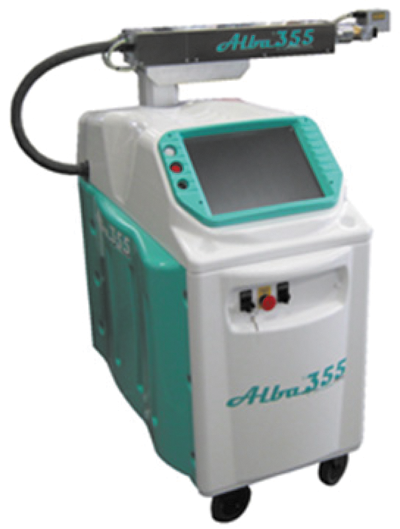
Laser Alba 355 (Elettronica Valseriana, Casnigo, Italy).
Statistical analysis
Statistical analyses were performed using GraphPad® (v5.04, USA). Data were first checked for normality using the Anderson-Darling test. A one-way ANOVA followed by Bonferroni post-hoc test was used to compare each time point to baseline. Data are presented as mean±SEM. A value of p<0.05 was considered significant.
Results
The overall laser treatment of psoriatic plaques produced a significant improvement in the PASI score (F [3, 55]=57.86; p<0.0001) (Fig. 3). The mean PASI score decreased from a baseline value of 24.5±2.9 to a value of 15.6±1.9 at 1 week (p<0.001), 9.1±1.2 at 2 weeks (p<0.001), and 5.8±1.2 at 3 weeks (p<0.001). A reduction of 76.7±10.9% in the PASI score was observed at 3 weeks in the four patients classified as affected by mild and moderate psoriasis after 9.2±0.3 sessions (Fig. 3). In the 10 patients classified as affected by severe psoriasis, a reduction of 89.4±2.4% was observed after 11.4±0.2 sessions (Fig. 3). Therefore a 75% reduction in the PASI score (PASI 75), which is considered a benchmark of primary endpoints for many clinical trials of psoriasis, 19 –21 was observed in 12 out of 14 patients who participated in this investigation. All the patients were very satisfied with the outcome of the procedure. No adverse reactions were observed during the study. Examples of patients affected by psoriasis and after laser treatment are shown in Figs. 4 –7.
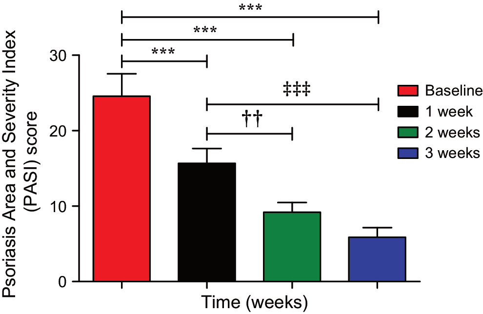
Psoriasis Area and Severity Index (PASI) score at baseline and at 1, 2, and 3 weeks of treatment by means of laser Alba 355. ***p<0.001 versus baseline, †† p<0.01 versus 1 week, ‡‡‡ p<0.001 versus 1 week.
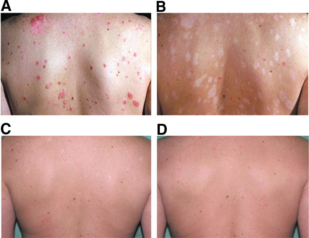
Patient affected by mild psoriasis
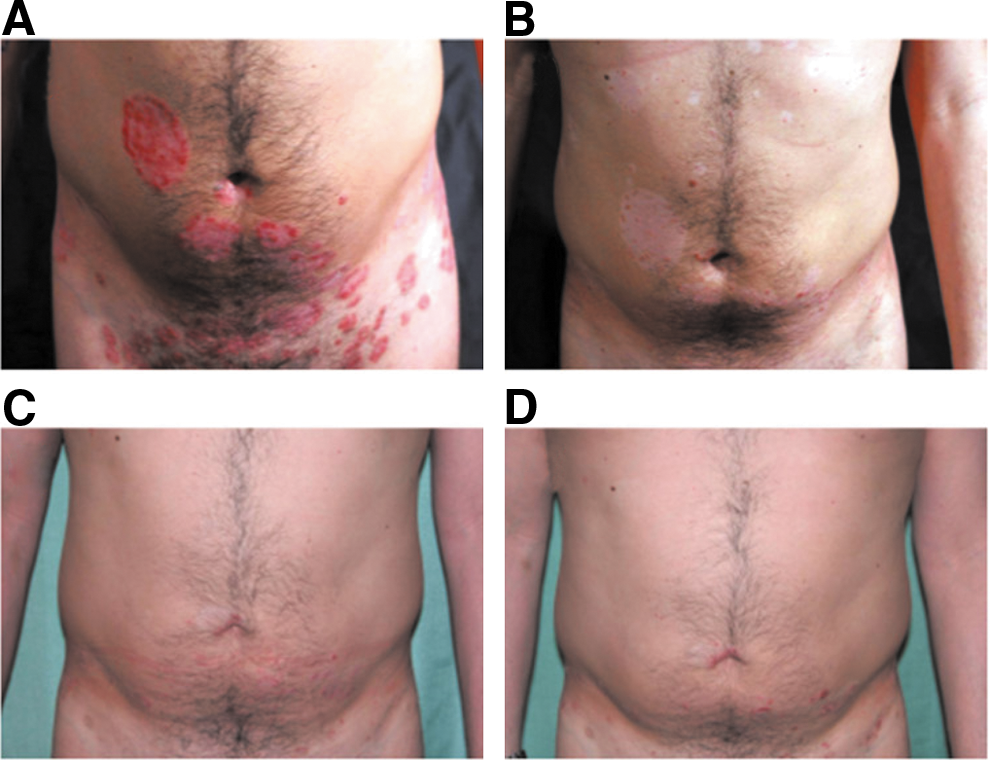
Patient affected by severe psoriasis
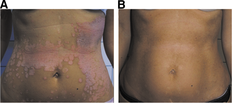
Patient affected by severe psoriasis
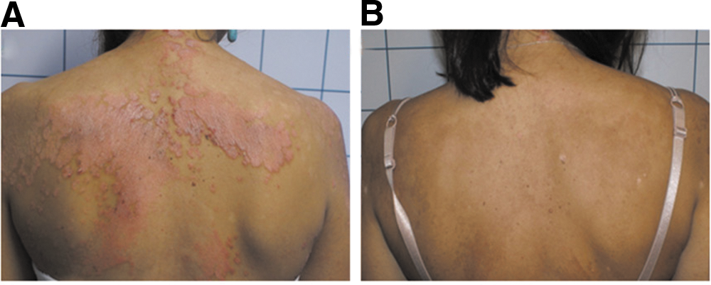
Patient affected by severe psoriasis
Discussion
The present study shows an overall significant improvement in the PASI score in patients treated for up to 3 weeks with four sessions a week of UVA1 laser therapy. These patients were affected by mild, moderate, and severe psoriasis and were treated with moderate-to-high frequencies (80–140 J/cm2). Our findings support the clinical use of a monochromatic coherent and coordinate UVA1 light laser therapy alone for treatment of this condition. These results are not in agreement with a previous study where treatment with UVA1 irradiation of three patients, affected by psoriasis of the palms, produced no improvement in a patient and only a 25–50% improvement in the other two. 17 With regard to UVA1 irradiation and pharmacological treatment, contrasting results have been previously reported. For example, a previous study of medium-dose UVA1 (50 J/cm2) and tacrolimus ointment showed no dramatic changes in plaque thickness or scaling in five patients affected by palmar plantar psoriasis. 15 On the other hand, another study, comparing calcipotriol in combination with UVA1 to calcipotriol with narrow-band UVB phototherapy in 45 patients with plaque psoriasis, concluded that UVA1 phototherapy with calcipotriol is effective and could be an alternative to narrow-band UVB phototherapy with calcipotriol. 16
The UVA1 355 nm laser (Alba 355) used in this study presents many advantages. First, it is cheaper if compared with metal-halide lamps that also require constant maintenance to maintain high tube brightness. Second, it is able to maintain a stable brightness for a number of hours consisting of 20,000 deliveries and with the possibility to deliver up to 100 J/cm2 in 20 sec. This feature allows a selective treatment of 100 J/10 cm2 psoriatic plaques in 4 min.
In the present investigation, we did not observe any side effects. However, side effects following UVA1 irradiation have been reported. Acute side effects include hyperpigmentation, redness, dryness and pruritus, herpes simplex virus reactivation, and polymorphic light eruption induction. 7 Chronic side effects include photoaging and possible photocarcinogenesis. 7 It is important to underline that previous studies reporting these complications could not confirm their association with UVA1 radiation, as the patients had also received other treatments. 22,23 Furthermore, side effects associated with UVA1 radiation are fewer if compared with those from other types of phototherapy. 7
Conclusions
In conclusion, our preliminary study of UVA1 therapy alone for treatment of mild, moderate, and severe psoriasis produced positive results in our cohort of patients. Larger clinical trials are needed to definitely support the role of this medical device not only for treatment of psoriasis, but also for other skin-related diseases. We speculate that, in the near future, the use of this laser will grow in the dermatology clinic.
Footnotes
Author Disclosure Statement
No competing financial interests exist.