
Abstract
Introduction
Er:YAG laser has a wavelength of 2.94 μm, which coincides with the absorption peak of water, collagen, and hydroxyapatite. 5 This laser acts on hard dental tissues by thermomechanical ablation, causing vaporization of water and expansion within a mineral substrate with ejection of both organic and inorganic particles by micro-explosions. 6,7 After irradiation, Er:YAG lasers leave a microscopically rough, clean surface with no smear layer and with open dentinal tubuli, 8 without causing major thermal damage. 9,10 However, effects on hard dental tissues caused by laser irradiation depend upon many factors: the fluence applied on the surface, the focal distance, beam spot size, repetition rate, structural properties of target tissue, amount of water during irradiation, and pulse duration. 11 Studies that analyzed the bond strength of self-etching systems after Er:YAG irradiation in dentin found lower bond strength of the lased surfaces. 12 –14 Laser irradiation can interfere with bonding, because of collagen thermal degradation after laser irradiation, 15,16 or because of chemically modified dentin surface with loss of carbonate, formation of new hydroxyapatite-like crystals, and, consequently, more acid-resistant surfaces. 15,17,18
Introduction of the Er:YAG laser with variable square pulse technology (VSPt) provided very short, square-shaped pulses of adjustable duration. The pulse profile is controlled and ensures that the power within the pulse is approximately constant with all pulse energy being used up for ablation. 19 Nishimoto et al. 20 found that increasing the pulse duration resulted in increased depth of ablated dentin, but decreased diameter, with no difference in ablated volume of dentin. However, these observations were recorded for pulse durations between 100 and 500 μs. 20 Majaron et al. 21 showed that if the laser pulse is <100 μs, heat diffusion effects during the laser pulse can be neglected, enabling more efficient ablation in comparison with longer pulse durations where the ablation threshold is expected to increase because of the conductive loss of heat from the interaction layer.
Considering the fact that Er:YAG laser does not produce a smear layer and removes the already-created one, which will be otherwise incorporated into hybrid layer when bur and self-etch adhesives are used, it is necessary to clarify the effect of Er:YAG laser and lack of smear layer on adhesion properties of one step self-etch adhesive system to dental hard tissues.
The aim of this in vitro study was to evaluate the microtensile bond strength of a composite resin to dentin modified with three different pulse durations of the VSPt Er:YAG laser and with one self-etch adhesive.
Materials and Methods
Forty-eight human molar teeth, extracted for periodontal or orthodontic reasons, were obtained for the experiment under approval of the Ethical Committee of the School of Dental Medicine, University of Zagreb, Croatia. After extraction, the teeth were thoroughly cleaned using brushes and curettes and stored in 1% chloramine solution until use. The teeth were randomly divided into four experimental groups (n=12 per group), according to the pretreatment of dentin: (1) control group; (2) super short pulse (SSP) (50 μsec), 10 Hz, 80 mJ with Er:YAG laser; (3) medium short pulse (MSP) (100 μsec), 10 Hz, 80 mJ with Er:YAG laser; and (4) short pulse (SP) (300 μsec), 10 Hz, 80 mJ with Er:YAG laser. The average power for laser groups was 0.8 W and the peak power for the SSP, MSP, and SP groups was 1.3, 0.85, and 0.45 kW, respectively.
Preparation of specimens
The entire occlusal enamel was cut off with a diamond blade of the Isomet 1000 saw (Buehler, Düsseldorf, Germany), with a speed of 150–200 rpm under continuous water cooling to obtain a flat dentin surface. The dentin surface was polished with sandpapers of different grit size, from coarser to finer (400, 600, 1000 grit), 22 in order to form smear layer on the bonding surface of dentin. The bonding surface was washed with distilled water and gently dried with a dental unit air syringe (Kavo Primus, 1058 S/TM/C/G, Biberach/Riss, Germany) prior to the pretreatment. The dentin surface of teeth in the control group was not modified by any treatment. For the groups treated with laser, the second generation Fidelis Plus II Er:YAG (Fotona, Ljubljana, Slovenia) dental laser with SP (300 μsec, 10 Hz, 80 mJ), MSP (100 μsec, 10 Hz, 80 mJ), and SSP (50 μsec, 10 Hz, 80 mJ) was used for 15 sec over the entire surface of each specimen. The Er:YAG laser energy was delivered by a RO2-C handpiece with the spot size of 0.9 mm in diameter, in a noncontact mode under continuous water spray (15 mL/min) at a focus distance of 7 mm from the target point. Ten teeth from each experimental group were selected for bonding procedure for microtensile testing. Following the application of the self-etch one bottle adhesive system (G-bond, GC, Tokyo, Japan) according to the manufacturer's instructions (Table 1), a composite resin block (Gradia Direct A2, GC, Tokyo, Japan) 5 mm high was built up on the bonding surface, with the application of layers of the material not thicker than 2 mm, each one cured with a Bluephase LED light (Ivoclar Vivadent, Schaan, Liechtenstein, 1200 mW/cm2, soft start) for 20 sec. The bonded specimens were stored in distilled water at 37°C for 24 h. The teeth were embedded into acrylic resin (Orthocryl, Dentaurum, Ispringen, Germany). Afterwards, the teeth were multiply cross and longitudinally sectioned with a diamond blade of an Isomet 1000 precision saw (Buehler, Düsseldorf, Germany), with a speed of 150–200 rpm under continuous water cooling, to obtain beam-shaped sticks, with a cross-sectional area of ∼1 mm2. Before testing the bond strength, each beam was checked under a stereomicroscope (Olympus SZX-12, Optical Co, Europe, GMBH, Hamburg, Germany) to verify that the adhesive interface was perpendicular to its long axis. Only the beams with the adhesive interface perpendicular to its long axis were used for the experiment.
Testing microtensile bond strength
The microtensile bond strength was tested with a universal testing machine (Triax Digital 50, Controls, Milan, Italy). Ends of each beam were glued with cyanoacrylate adhesive (Loctite gel, Henkel, Düsseldorf, Germany) to specially designed metal plates. Each beam was mounted in the machine and a tensile load was applied at a crosshead speed of 0.5 mm/min, until it fractured. At this point, the load at failure was recorded in newtons (N). Beams were observed under a stereomicroscope to verify the failure mode (adhesive, cohesive, or mixed). Adhesive failure was failure at the dentin/adhesive interface; failure was considered cohesive if it occurred in the composite resin material or dentin, and failure was considered mixed if it involved the dentin/adhesive interface and the composite resin material or dentin at the same time. The cross-sectional area at the site of fracture was measured to the nearest 0.01 with a digital caliper (Roc International Industry Co., Ltd., Guangdong, China) so the bond strength at failure in MPa could be calculated.
Scanning electron microscopic (SEM) evaluation
Two specimens from each experimental group were selected randomly and subjected to SEM examination, to observe the irradiated surface. For the SEM analysis, samples were cleaned in an ultrasonic bath (Elmasonic E60H, Tovatech, South Orange NJ) for 5 min. The interface was brought into relief by etching with 32% silica-free phosphoric acid (Bisco, Schaumburg, IL) for 30 sec, washed with de-ionized water, and air dried. Samples were dehydrated in an ascending ethyl alcohol series (25%, 50%, 70%, 80%, 90%, and absolute alcohol) and dried using hexamethyldisilazane (HMDS, Carlo Erba, Rodano, Italy). Specimens were then mounted on aluminum stubs, coated with a 15–20 nm thick layer of gold by means of the SC7620 Sputter Coater device (Polaron Range, Quorum Technologies, England) and inspected by a scanning electron microscope (JSM-6060LV, JEOL, Tokyo, Japan) at 500×, 1500×, 2000×, 3000×, and 4000×magnifications.
Data analysis
For the statistical analysis of the microtensile bond strength, the data were analyzed by a one way ANOVA, after confirming normal distribution of the results with Kolmogorov–Smirnov statistical test. Level of significance was set at 5%. Statistical analysis was performed using Statistica 7.0 (StatSoft, Tulsa, OK).
Results
Microtensile bond strength
Means and standard deviations of microtensile bond strength expressed in MPa are shown in Table 2. Dentin surface treated with SSP showed significantly lower microtensile bond strength values in comparison with the control group and SP (p<0.05), (Table 2). The MSP and SP groups did not show any statistically significant difference in microtensile bond strength in comparison with the control group (p>0.05), (Table 2). There was also no statistically significant difference in microtensile bond strength between the MSP and SP groups (p>0.05) (Table 2). In all groups, fractures were observed mostly between resin and dentin (adhesive failure); mixed failures were not observed (Table 2).
Different superscript letters indicate groups that are significantly different (p<0.05). Groups identified with the same superscript letters are not significantly different (p>0.05).
SSP, super short pulse; MSP, medium short pulse; SP, short pulse; A, adhesive; C, cohesive.
SEM observation of dentin surfaces
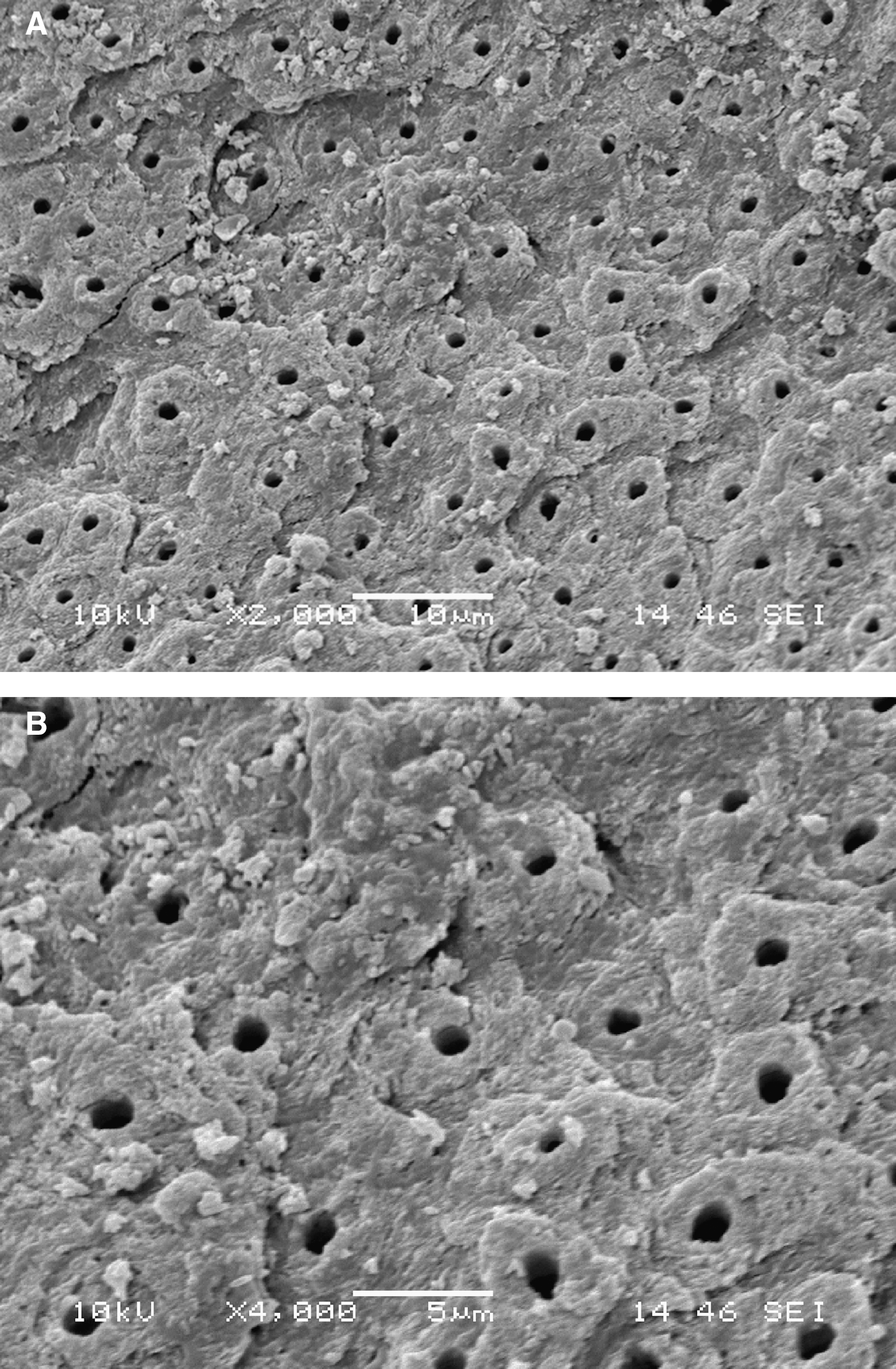
The control group revealed a dentin surface with a small number of exposed dentinal tubules and intact peritubular and intertubular dentin (Fig. 1a and b). It was also possible to verify an intact smear layer (Fig. 1a and b).

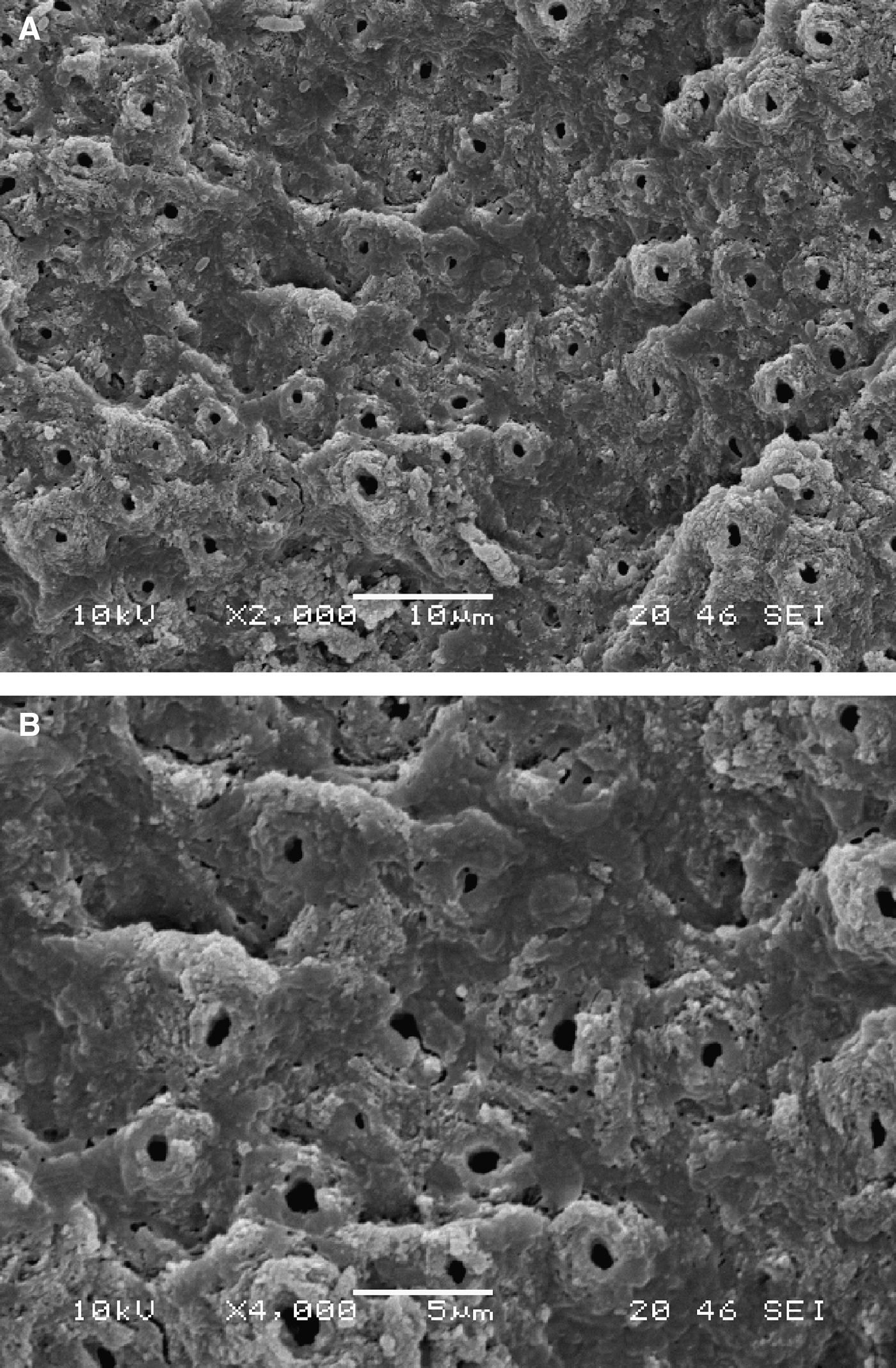
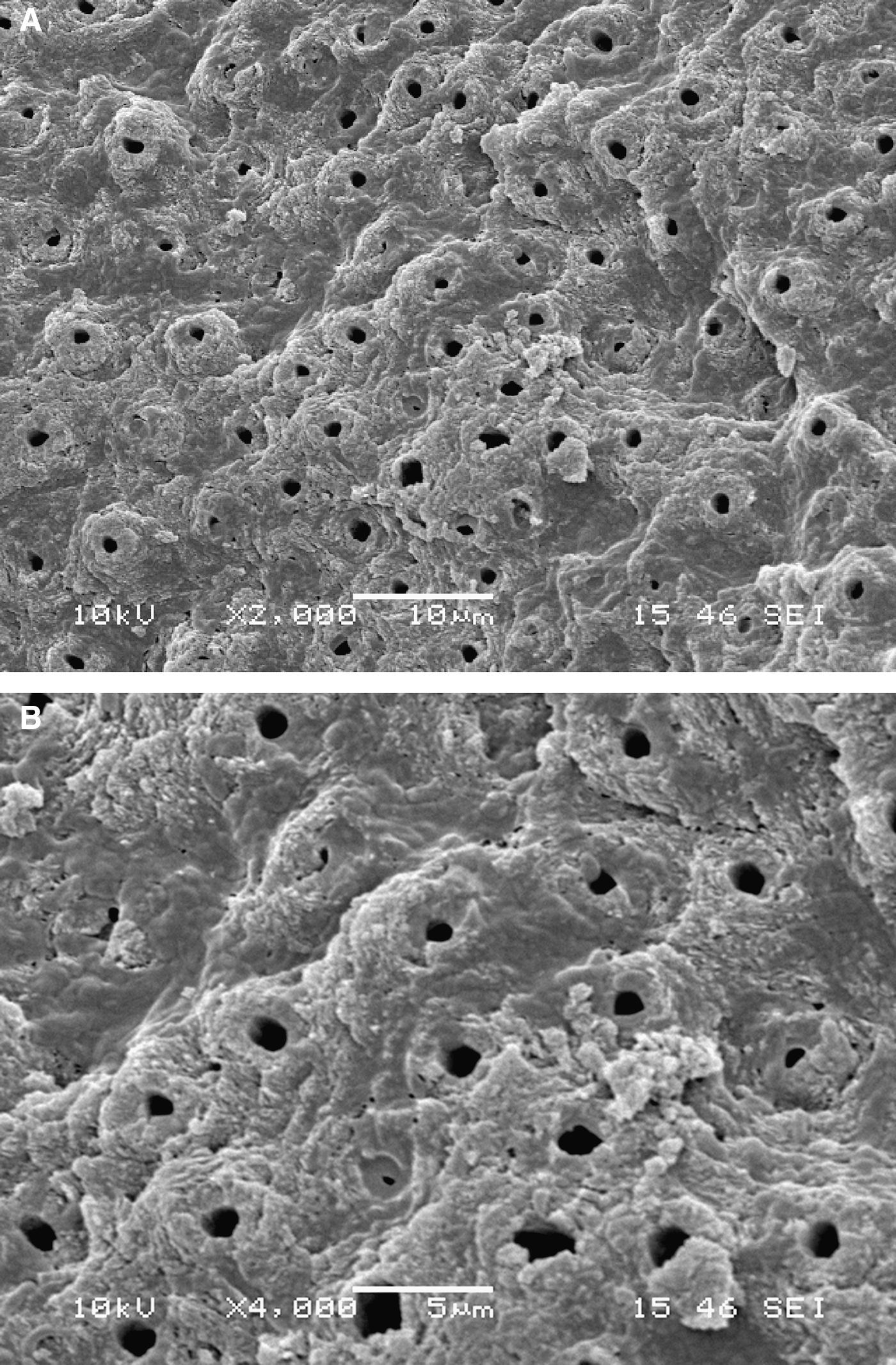
Laser treatment of dentin (SSP, MSP, and SP groups), resulted in an irregular, crater-like surface (Fig. 2a and b, Fig. 3a and b, Fig. 4a and b). In all laser groups, dentin surfaces were clean of the smear layer, exposing the orifices of the dentinal tubules (Fig. 2a and b, Fig. 3a and b, Fig. 4a and b). Intertubular dentin was selectivly ablated more than the peritubular dentin, making the tubules protrude (cuff-like appearance) for all three laser groups (Fig. 2a and b, Fig. 3a and b, Fig. 4a and b). These characteristics of lased dentin were especially noted in the SSP group (Fig. 2a and b).
Discussion
After irradiation with Er:YAG laser, dentin surface was clean, roughened, with little smear layer, and with open dentinal tubules. The irregularities of the dentin surface of all three laser groups resulted from the microexplosions through rapid water evaporation. 6,7 SEM analysis also showed that the intertubular dentin in laser groups was ablated more than the peritubular dentin because of the greater amount of water and collagen and lower mineral content of the intertubular dentin. As a result, dentinal tubules were protruded with a cuff-like appearence, confirming the results of other studies. 23 –25 The cleanest and the most irregular dentin surface was found in the SSP group. Such shorter pulses are more efficient in hard dental tissue ablation, 19 and, therefore, in ablation of the smear layer which consists of water, inorganic and organic compound, in which the energy of Er:YAG laser is absorbed.
Although the absence of smear layer, microretentive pattern, and open dentinal tubules observed in this study are expected to promote good bonding, SSP decreased the dentin bond strength. Irregularities produced on the dentin surface in this group may result in a non-uniform thickness of the adhesive layer 26 and lower the bonding strength. With SSP, the energy is used completely for ablation, unlike with longer pulse durations when the ablation efficency is reduced because of conductive loss of heat. 27 Furthermore, shorter pulses, like SSP, can generate strong elastic waves in the hard dental tissues being ablated because of laser heating, thermal expansion, and recoil of ablation products. 24 Because of these stress waves, cracks and fractures can occur during irradiation of hard dental tissue and negatively influence bond strength. 28 Kataumi et al. 25 also found microcraks below the hybrid layer because of subsurface damage caused by Er:YAG irradiation, whereas De Munck et al. 13 found significantly more microcracks at the laser-irradiated dentin surface using field emission SEM.
The longer pulses employed in the present study, MSP and SP, did not decrease the bond strength, which confirms the results of Ramos et al. 29 who examined the effects of 150 and 250 μs pulses of the Er:YAG laser on the bond strength of self-etch adhesives in dentin, and found no difference in bonding strength in comparison with control. However, according to the results of Ramos et al., 30 irradiation of dentin with Er:YAG laser adversely affected the interaction pattern of self-etch adhesive systems with the lased substrate. In the study by Ramos et al., 30 the dentin surface was irradiated for a longer period of time and tensile test, not a microtensile test, was used to assess the bonding strength. Although Belikov et al. 31 showed with SEM that laser irradiation produces a surface that could increase restorative material retention, MSP and SP did not improve bonding strength in the present study.
Omae et al. 32 investigated dentin surface irradiated by Er:YAG laser with lower energy settings, similar to ones the present study, using X-ray photoelectron spectroscopy, and found reduced ratio of calcium (Ca) and phosphate (P) ions. This reduction in the Ca/P ratio indicates a decline in the concentration of Ca. 32 Mild self-etching adhesives, such as the one used in the present study, do not completely expose collagen for micromechanical retention, but there is an additional mechanisam of ionic bonding of acidic monomers and calcium in hydroxyapatite. 33 4-methacryloxy-ethyl trimellitate anhydride (4-META), a demineralizing monomer with carboxylic groups, also found in the adhesive used in the present study, has been reported to improve adhesion to both enamel and dentin by establishing that ionic bond to calcium in hydroxyapatite. 34 If there is a deficiency of Ca in dentin, the chemical bonding of the mild adhesive is probably decreased, and this could impair the bond strength. Camerlingo et al. 35 showed that chemical changes in dentin after Er:YAG irradiation depend upon pulse length, with very short laser pulses producing a strong modification of the dentin components, especially the organic part. As functional monomers in self-etching adhesives have also been shown to bond chemically to both dentin apatite and collagen, 33,36 chemical changes of both the organic and inorganic parts of dentin, induced by shorter pulses, such as SSP in this study, could explain the lower bond strength of SSP, but an unchanged bond strength when longer pulses, MSP and SP, were used.
Furher studies should examine the effect of different energy and frequency settings of Er:YAG laser based on VSPt on microtensile bond strength of different adhesive systems.
Conclusions
The use of SP and MSP of the Er:YAG laser with one step self-etch adhesive does not impair or improve the microtensile bond strength in dentin, whereas SSP may not be suitable for dentin surface treatment prior to bonding procedures.
On the basis of our results, there is no beneficial effect of laser pretreatment of dentin surface prior to adhesive procedure with one step self-etch adhesive.
Footnotes
Acknowledgments
This study was performed and financed within the research project “Experimental and Clinical Endodontology” No. 065-0650444-0418, approved by the Ministry of Science and Technology of the Republic of Croatia.
Author Disclosure Statement
No competing financial interests exist.