




Abstract
Introduction
Biopure MTAD has been used widely as a final irrigation solution for removing smear layer and disinfecting the root canal, because of its antibacterial potential. 13 –15 Some authors have questioned the effect of MTAD on the bond strength of MTA. 11,12 The acidic pH of MTAD, because of citric acid and doxycycline, was claimed to have an adverse effect on calcium silicate-based cement materials. 16,17
Furthermore, in the field of laser assisted endodontics, various laser systems have been used as final treatments effectively. Laser devices such as Nd:YAG and diode lasers have shown to be effective in root canal disinfection. 18,19 Other laser lights such as Er:YAG, Er:YSGG, and CO2 lasers were used for dental hard tissues with high absorption property and efficient ablation made on either enamel or dentin. 20 –22 Some authors have indicated that Er:YAG yields lower bond strength even after surface acid etch following laser irradiations. 23,24 Also, in previous studies it was acclaimed that CO2 laser might affect the bond strength of composite adhesion adversely. 25 The effect of diode laser on the apical sealing ability of MTA in the retrofilling treatments was questioned by some authors. 26
Accordingly, the hypothesis tested was whether diode laser or MTAD might have an effect on the push-out bond strength of MTA-dentin.
Materials and Methods
The research protocol was approved by the Research Ethics Committee of Kamal Asgar Research Center (protocol no. KARC/42E2011-32-7). Forty extracted single-rooted human teeth were used for this study. Samples were decoronated and sectioned horizontally at the midroot parts into 1.5 mm dentin slices. The canal spaces of the root slices were instrumented by #2 through #5 Gates-Glidden burs (Mani, Tochigi, Japan) to form 1.3-mm diameter standardized cavities. The specimens were then randomly divided into four groups (n=10). In groups A and B the root slices first were irrigated with 5 mL of normal saline (Darou Pakhsh Inc., Tehran, Iran), then dried out with cotton pellet, finally undergoing diode laser irradiation five times for 5 sec each time, with a 15 sec interval between irradiations. Laser treatments were performed with a GaAlAs diode laser (TwiLite™, Biolase Technology, San Clemente, CA), at a wavelength of 810 nm and output power of 2 W with the continuous pulse mode, using a pulse duration of 20 ms and a pulse interval of 40 ms. The laser irradiation was delivered into the canal spaces via a fiber tip 400 μm in diameter. The hand piece was held to form an angle of ∼10 degrees between the fiber and the canal wall. Irradiations were performed with circling movements.
White ProRoot MTA (WMTA) (Dentsply Tulsa Dental, Tulsa, OK) was mixed according to the manufacturer's instructions and placed inside the canal spaces of all root slices. Saline-moistened Gelatamp (Roeko-Colte`ne/Whaledent, Langenau, Germany) was used as a matrix while excess materials were trimmed from the surface of the specimens with a scalpel. These specimens were wrapped in wet gauze and sealed in a plastic bag, which was placed in an incubator and allowed to set for 7 days at 37°C. After incubation, dentin slices in groups B and C were placed in plastic cryo-tubes (Cryo. S,PP Greiner Bio-One GmbH., Frickenhausen, Germany) containing Biopure MTAD (Dentsply Tulsa Dental, Tulsa, OK) for 5 min according to manufacturer's instructions. After immersion, the slices were removed from the solutions and rinsed with distilled water and dried with cotton pellet before testing. In group D, samples had no further treatments and after placing mixed MTA, dentin slices were dried with cotton pellet and served as control group.
Push-Out Test
The push-out bond strengths were measured by using Zwick/Roell Z050 universal testing machine (Ulm, Germany). The WMTA was loaded with a 0.7 mm diameter cylindrical stainless steel plunger at a speed of 1 mm/min. The maximum load applied to the WMTA was recorded in Newton before the occurrence of dislodgement. To express the bond strength in megapascals, the recorded value in Newton was divided by area in mm2 calculated by the following formula: 2 Φr×h, where Φ is the constant 3.14, r is the root canal radius, and h is the thickness of the root slice in millimeters. The slices were then examined under light microscope at×40 magnification to determine the mode of the bond failure. Each sample was placed into one of three failure modes: adhesive failure that occurred at the WMTA and dentin interface, cohesive failure within the WMTA, and mixed failure mode. The data were analyzed by using one-way analysis of variance (ANOVA) and Tukey's post-hoc tests.
One sample from each study group was chosen randomly and underwent scanning electronic microscopy (SEM) examination. After the dislodgment of MTA, the specimens were irrigated with 10 mL of distilled water and vertically grooved on the buccal and lingual surfaces with a diamond disc without entering the canals, and split longitudinally with a chisel. One half of each sample was randomly chosen, placed in 2% glutaraldehyde for 24 h and then rinsed three times with sodium cacodylate buffered solution (0.1 M, pH=7.2). After incubation in osmium tetroxide for 1 h, the samples were dehydrated with ascending concentrations of ethyl alcohol (30–100%), placed in a dessicator for 24 h, and mounted on a metallic stub. After coating the samples with gold, SEM micrographs were taken (XL 30, Philips, Holland) (×2000).
Results
The means±standard deviations of push-out bond strength were 6.74±0.48, 5.95±0.40, 6.86±0.66, and 7.88±0.37 for groups A (laser), B (laser plus MTAD), C (MTAD), and D (control), respectively (Fig. 1). ANOVA test revealed significant differences between experimental groups and control group (p<0.0001).Turkey's test did not show any significant difference between groups A and C (p=0.93). However, these differences were significant between groups A and B (p=0.006) and between groups B and C (p=0.001).
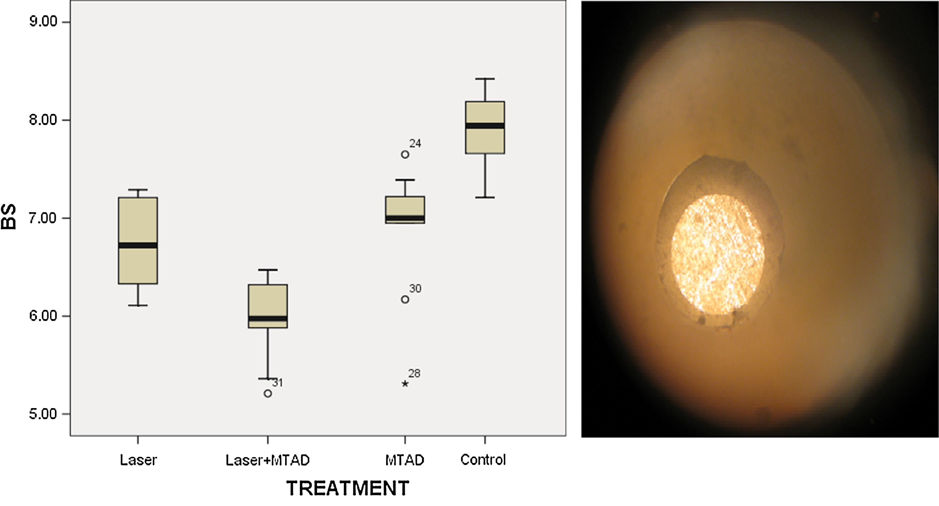
Left: Box plots of push-out bond strength for laser (group A), laser plus MTAD (group B), MTAD (group C), and control (group D). Right: The image of light microscope examination of the bond failure, which demonstrates the surface of the canal wall, which exhibits the debonded surface of the dentinal wall. The adhesive mode of failure occurred at the interface of dentin and set mineral trioxide aggregate (MTA) cement.
The SEM images of samples after push-out bond testing revealed the mode of bond failures in experimental groups (Fig. 2). In laser and MTAD groups, 7 samples out of 10 showed adhesive mode of failure whereas the other three specimens revealed cohesive mode (Fig. 2 A and C). In the laser plus MTAD group, eight dentin slices showed adhesive mode of bond failure whereas other two revealed cohesive type (Fig. 2 B). In the control group, samples that received no treatments, six samples showed adhesive failure whereas the remaining four specimens showed cohesive type (Fig. 2D).
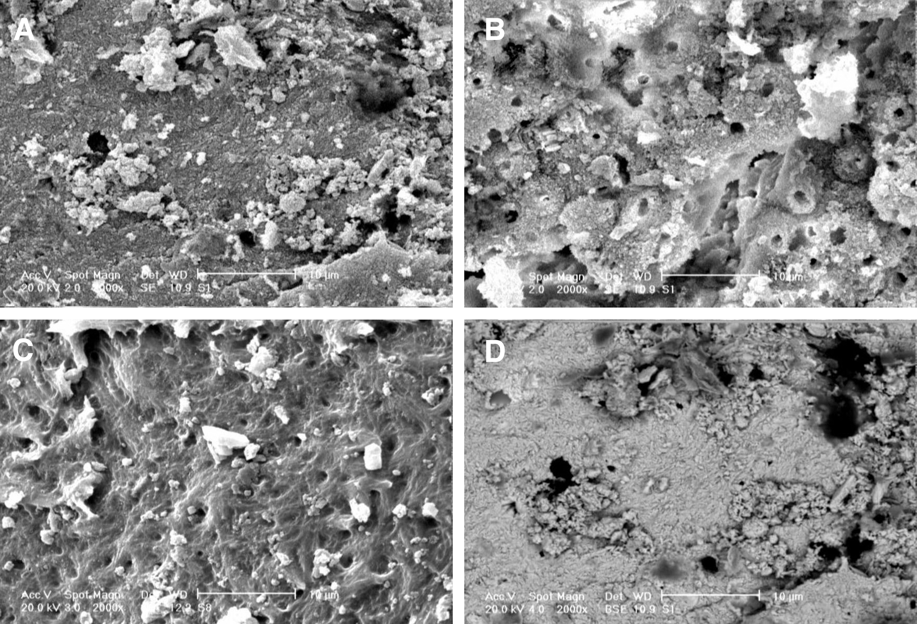
Image of group
Discussion
MTA is a mechanical mixture of calcium silicate cement, bismuth oxide, and gypsum. 27 The sealing ability of MTA relies on chemical bond through diffusion-controlled reaction between MTA surface and dentin walls. 6 In addition, it was mentioned that the hydration phases were responsible for the strength and barrier properties of MTA-based cements. 28 This chemical adhesion is shown to be affected by endodontic irrigants. Smith et al. concluded that the BioPure MTAD-treated dentin surfaces exhibited higher surface roughness and more calcium extracted when compared with those treated with EDTA. 29 Although these authors claimed that EDTA or BioPure MTAD as final rinses were unlikely to result in complete dissolution of the MTA as a repair material, they suggested that BioPure MTAD might cause decomposition of the particle-binding hydration phases in the remaining white MTA after BioPure MTAD irrigation. In the present study, the push-out bond strength of MTA-dentin in samples irrigated with MTAD was decreased, which is consistent with previous studies. These results can be explained by the process of acid corrosion in which calcium hydroxide, calcium silicate hydrate (C-S-H), and the calcium sulfoaluminate phases decompose. These data were supported by other investigators. 17,30 This decomposition can remove MTA and produce porosities that might have an adverse effect on push-out bond strength of MTA dentin surfaces.
The diode laser irradiations in this investigation were performed prior to MTA placement in order to avoid interaction between laser beam and MTA, as the laser light could increase the temperature and create cracks. The power settings used were in accordance with previous thermal analysis. 31 The results of the present study indicated that diode laser irradiations decreased the bond strength of MTA compared with the control group. Souza et al. have questioned the effect of diode laser on apical sealing of MTA and concluded that this laser device did not improve the sealing ability of MTA. 26 This issue has been discussed regarding other laser systems, such as CO2 laser, by other investigators. Hedayatollah Najafi et al. indicated that CO2 laser irradiations can lower the bond strength of composite-dentin after shear-bond test. 25
The effect of laser beam might be explained by the ablation mechanism induced by laser systems, which can cause melting on dentin surfaces in irradiated areas. It seems that this melted part of dentin can interfere with the chemical bond of MTA-dentin surfaces. which results in the decreasing of push-out bond strength. These results were also noticed in samples treated by both diode laser and MTAD as the final rinse. In this group, the push-out bond strength was significantly lower than in specimens treated either with diode laser or MTAD alone. The results of this group indicated that diode laser and MTAD might have an additional effect on decreasing of push-out bond strength of MTA.
Following the push-out test, the light microscope and scanning electron microscope examinations revealed that the failure mode was adhesive in most of the cases. This result is consistent with previous studies that also reported the same failure mode in samples evaluated with the push-out test. 32 The adhesive failure mode might be explained by the short time incubation before push-out testing. Gancedo-Caravia et al. showed that in the presence of humidity, the lengthening of curing time up to 21 days produced a moderate increase in bonding strength compared with the first 3 days. 34 In addition, the particle size of MTA cement used might prevent penetrating more in dentinal tubules, which can cause adhesive failure mode. This issue was also presented by other investigators. 34
Conclusions
According to the results of present study, the authors have concluded that diode laser within the parameters used in this investigation or BioPure MTAD as final irrigation lowered the push-out bond strength of MTA-dentin contact surfaces. Also, it was noted that by using both diode laser and MTAD together, the push-out bond strength was further decreased. Further investigations, especially under clinical conditions, are needed to find an irrigant that may increase or at least not impair the bond strengths of cements such as MTA.
Footnotes
Acknowledgments
We are indebted to Nader Sheibani, Maryam Elyasi, and Neda Bayati for all of their contributions to this research.
Author Disclosure Statement
No conflicting financial interests exist.