
Abstract
Introduction
The light emitting diode (LED) is a semiconductor device that emits noncoherent narrow-spectrum light when electrically biased in the forward direction. LED has become an effective alternative to laser systems for reasons including lower cost, availability in a variety of wavelengths ranging from ultraviolet to near-infrared region of the spectrum, narrow emission band (∼5–10 nm), and a light fluence rate that can achieve hundreds of mW/cm2. In addition, the arrays can be constructed in various sizes to accommodate large areas, and they do not emit any heat, which may cause additional tissue damage. 12
Visible light is used clinically in the treatment of dermatitis, Alzheimer's disease, and muscle analgesia, 13 and is effective at removing bacterial biofilms. 14 Visible light phototherapy seems to be a promising alternative approach to eradicating bacteria with blue light. 15,16 Guffey and Wilborn 17,18 examined the in vitro effects of 405 and 470 nm light on two common aerobes, Staphylococcus aureus and Pseudomonas aeruginosa, and the anaerobe Propionibacterium acnes, and reported different bactericidal effects depending upon wavelength. The killing rate of 405 nm light was greater than 470 nm light (90% for S. aureus and 95.1% for P. aeruginosa). Hamblin et al. 19 showed that high intensity visible light could kill Helicobacter pylori in the stomach of humans. High-intensity broad-spectrum polychromatic light with wavelengths in the range of 400–1000 nm killed bacteria in infected diabetic ulcers. 20
The effects of LED wavelength and optical density on bacteria growth and bactericidal effects are unclear. The bactericidal effects and bacteria growth of LED illumination are unclear, although the bactericidal effects of blue light are well known. 21 Before conducting clinical trials, the wavelength, appropriate dose, and exposure time of the irradiation must be determined.
The acceleration or retardation of bacteria growth might be affected by the type and number of bacteria. The aim of this study was to evaluate the relationship of 625, 525, and 425 nm wavelengths, providing average power output and effects on three common pathogenic bacteria: Porphyromonas gingivalis, Escherichia coli DH5α, and S. aureus in vitro. We hoped to replicate the bactericidal effects of UV using visible light to remove bacterial biofilms without posing risks to human, but with high efficiency and minimal side effects.
Materials and Methods
Reagents
Luria–Bertani agar (LBA), Luria–Bertani broth (LBB), Brain–Heart Infusion Agar (BHIA), Brain–Heart Infusion Broth (BHIB), Baird–Parker Agar (BPA), and Baird–Parker Broth (BPB) were purchased from Difco Laboratories (Detroit, MI).
Bacterial strains and growth media
P. gingivalis (KCTC 5352) is an anaerobic bacterium. S. aureus (KCTC 1916) and E. coli DH5α (ATCC 25922) are aerobic bacteria. E. coli DH5α was grown on LBA and LBB for 24 h at 37°C. P. gingivalis was grown on BHIA and BHIB for 24 h in a 37°C anaerobic chamber. S. aureus was grown on BPA and BPB for 24 h in a 37°C incubator. They were harvested by centrifugation and suspended in 0.85% saline. The final bacterial density in each 0.1 mL aliquot used in the experiments was 2×10⁸ colony-forming units (CFU)/ mL or 1.6×10⁹ CFU/mL.
LED light irradiation devices
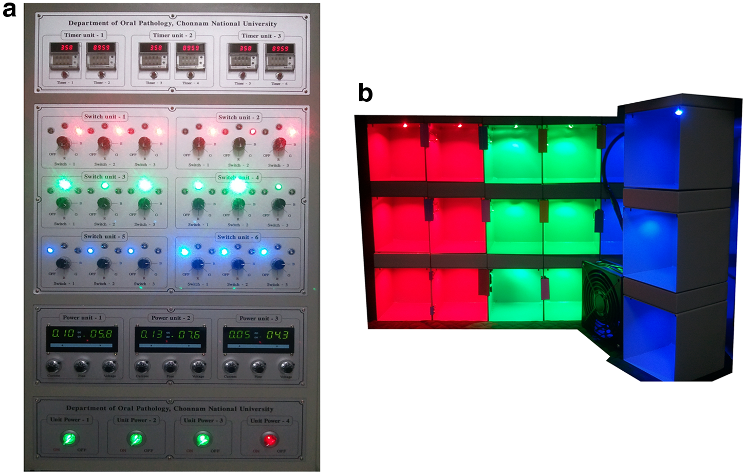
A three-in-one mounted RGB LED package for light irradiation was a continuous wave LED (HELIO Optoelectronics, Taiwan) emitting three wavelengths: red LED peak at 625 nm (spectral width 620∼630 nm), green LED peak at 525 nm (spectral width 520∼530 nm), and blue LED peak at 425 nm (spectral width 420∼430 nm), providing a power density of 10 mW/cm2. An average power output of 6 mW/cm2/h was set, and individual potency was determined using a model AQ215OA handheld optical Multi meter (Ando Electric, Japan). The optical density of microorganisms was analyzed by spectrophotometry at a wavelength of 600 nm in a 0.1 mL cubic cell (GeneQuant Pro OD600; Biochrom, UK). The bacteria were irradiated with 21.6, 43.2, 86.4, and 172.8 J/cm2. To determine the effects of LED irradiation depending upon the cell density of P. gingivalis, 0.1 mL suspensions containing 2×10⁸ or 1.6×10⁹ CFU/mL were dispensed in wells of a 100 mm dish and irradiated with LED for 0, 8, 12, and 24 h. To deal with the generation of heat from the long-term use of each LED source, bacteria were cultured in an incubator attached to a cooling fan (KA1238HA2/H.T.R.130; KAKU, China). Also, LED lamps contained an attached heat sink. In agar plates, 0.1 mL suspensions containing 2×10⁸ CFU/mL of P. gingivalis, E. coli DH5α, or S. aureus were dispensed and LED irradiated for 1, 2, 4, and 8 h. Number of CFU was determined and compared with the untreated group. In broth culture, 0.1 mL of P. gingivalis, E. coli DH5α, or S. aureus (2×10⁸ CFU/mL) were inoculated to 50 mL of the medium and LED irradiated for 1, 2, 4, or 8 h. Bacterial growth was evaluated as the optical density at 600 nm (OD600). Additionally, 0.1 mL of a suspension irradiated for 8 h was added to a well of a 100 mm dish. After 24 h, the number of CFU was measured. The distance between the LED and the Petri dish or Erlenmeyer flask was kept constant, to maintain the proper power density. The LED light was positioned 50 mm directly over the center of the Petri dish and Erlenmeyer flask (Fig. 1). The three-in-one mounted RGB LED package was powered by a DC power control system (0∼9 A and 0∼30 V). The current was set at 2±0.05 A at a voltage of 5±0.1 V, giving an approximate irradiance of 6 mW/cm2 for 1 h at the surface of each plate or dish. Each sample exposure was calculated as E=P × t, where E=energy density (dose) in J/cm2, P=power density (irradiance) in W/cm2, and t=time in seconds. The power supply system obtained from the Department of Oral Pathology, Chonnam National University School of Dentistry, was set in an incubator for the experiment (Fig. 2).
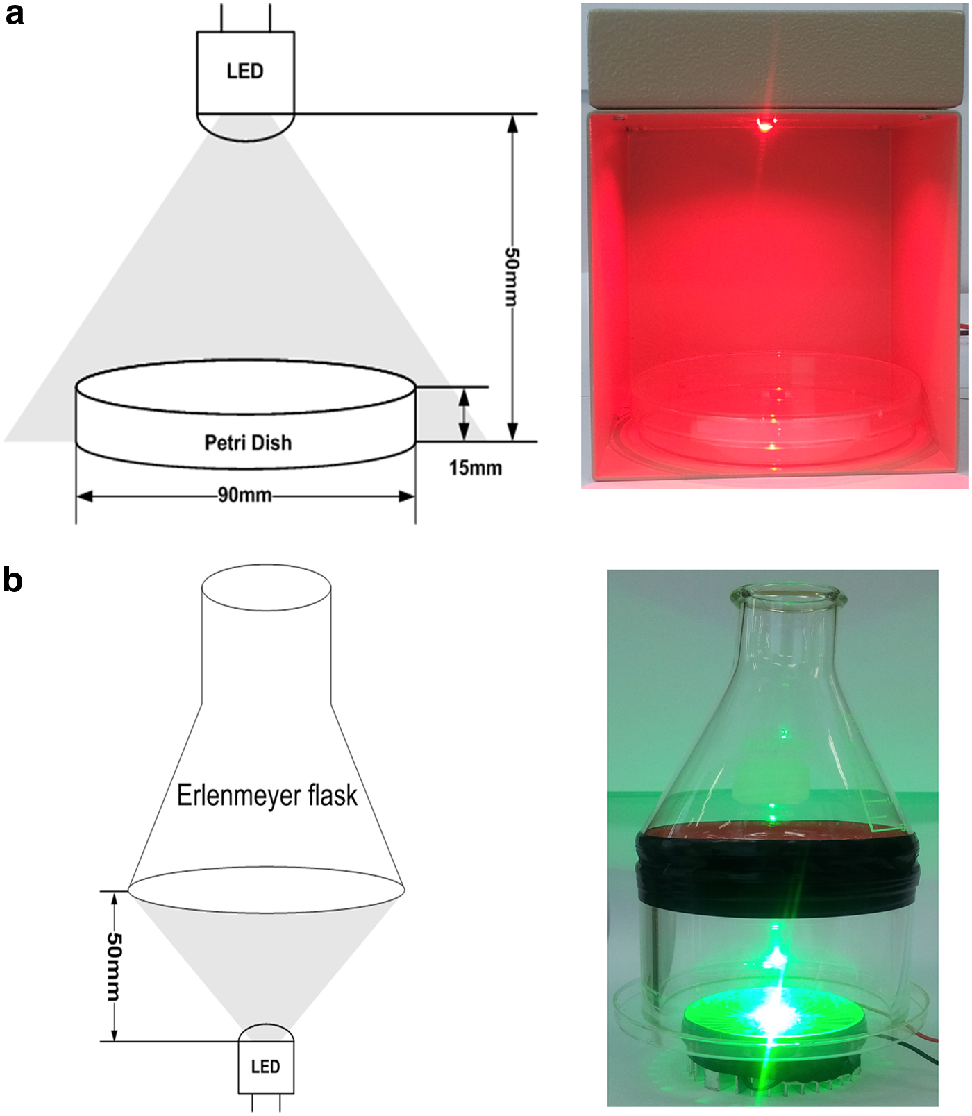
Schematic diagram of setup for the three-in-one approach mounting the RGB light-emitting diode (LED) package
CFU
The results were provided by calculation of 0.1 mL of 2×10⁸ CFU/mL and 0.1 mL of 1.6×10⁹ CFU/mL at 24 h after incubation at 37°C. CFU/mL is the number of colonies multiplied by 101+dilution factor. It takes into account the need to multiply by an extra 10, because of the addition of 0.1 mL to the plate from the tube. The number of CFU/mL was calculated with and without irradiation. The experiment was repeated two times to ensure accurate results.
Statistical analysis
Values obtained in CFU and OD600 were expressed as mean and standard deviation. Statistical calculations were done using Graph Pad Prism version 5.01 (GraphPad Software, San Diego, CA) statistical data analysis software. The data are expressed as the mean±SEM and analyzed with two way analysis of variance (ANOVA). Bonferonni's test was used to perform multiple comparisons among different groups. Null hypotheses of no difference were rejected if p values were <0.05.
Results
LED irradiation of P. gingivalis
To determine the effects of LED irradiation on P. gingivalis, 2×10⁸ and 1.6×10⁹ CFU/mL were irradiated at 625, 525, and 425 nm for 0, 8, 12, and 24 h. For the samples containing 1.6×10⁹ CFU/mL, OD600 was decreased by 425 nm light by 60% at 8 h, but not decreased as precipitously by 525 and 625 nm light (Fig. 3A). However, bacterial growth recovered to the control level within 24 h following irradiation at all three wavelengths. For the suspensions containing 2×10⁸ CFU/mL, the OD600 was decreased at 425 nm by 90∼100% at all time points (Fig. 3B). After 24 h, irradiation at 425 nm was completely bactericidal. In contrast, bacterial growth peaked in samples irradiated at 625 nm for 8 h as much as 2.5-fold over control values.
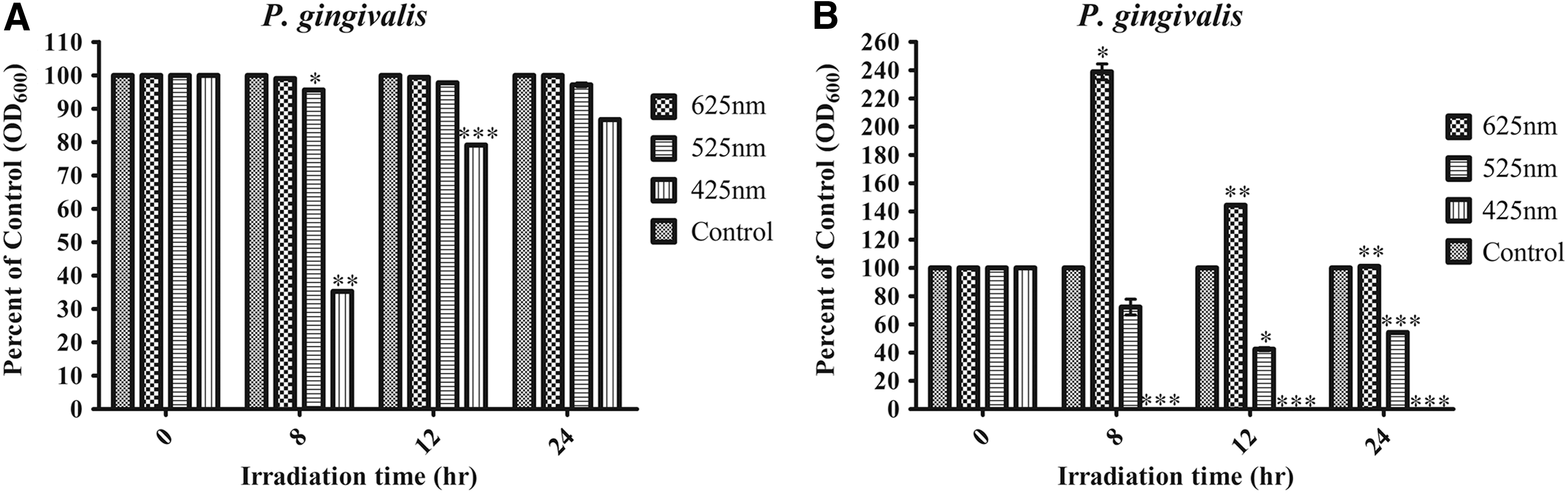
Bacterial survival by LED irradiation on Porphyromonas gingivalis.
Time dependence of LED irradiation on P. gingivalis on agar and in broth
To examine the effects of LED irradiation on P. gingivalis, bacteria (2×10⁸ CFU/mL) were irradiated with LED wavelengths of 625, 525, and 425 nm at 6 mW/cm2/h. P. gingivalis viability was decreased by irradiation at 425 nm (40∼60% reduction) and 525 nm (10∼20% reduction) both in agar (Fig. 4A) and broth (Fig. 4C). In contrast, P. gingivalis viability was only slightly reduced by irradiation at 625 nm for 4 h. To confirm the growth and bactericidal effects on P. gingivalis, bacteria were LED irradiated in broth, and viability was analyzed by OD600 and CFU. Survival of P. gingivalis was markedly reduced by 425 nm (90∼100% reduction) and 525 nm (40∼70% reduction) after 8 h (Fig. 4C and D). In contrast, P. gingivalis viability increased during an 8 h irradiation at 625 nm compared with control.
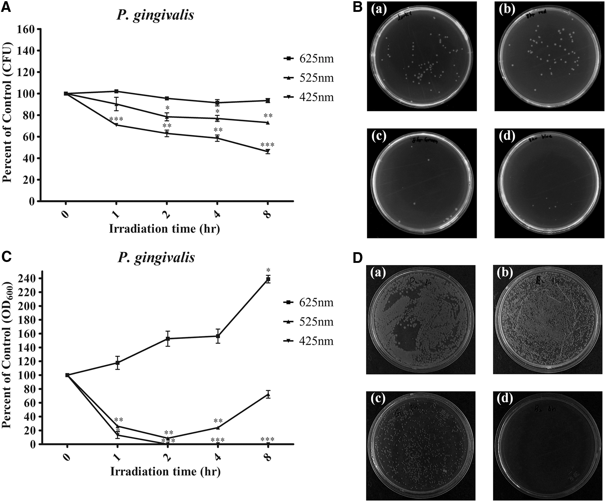
Bacterial survival of light-emitting diode (LED) irradiation by 6 mW/cm2; for 1 h irradiation in Porphyromonas gingivalis.
Time dependence of LED irradiation of E. coli DH5α in solid and liquid media
To examine the effects of LED irradiation on E. coli DH5α, cells (2×10⁸ CFU/mL) were exposed to LED wavelengths of 625, 525, and 425 nm (density of 6 mW/cm2/h) when growing on agar and in broth. Viability was decreased during irradiation at 425 nm (90∼100% reduction) and 525 nm (∼40% reduction) in agar (Fig. 5A). Viability was not affected at 625 nm. Survival was markedly reduced by irradiation at 425 nm (80∼100% reduction) and 525 nm (40∼50% reduction) for 8 h (Fig. 5C and D). In contrast, viability was unaffected by irradiation at 625 nm.
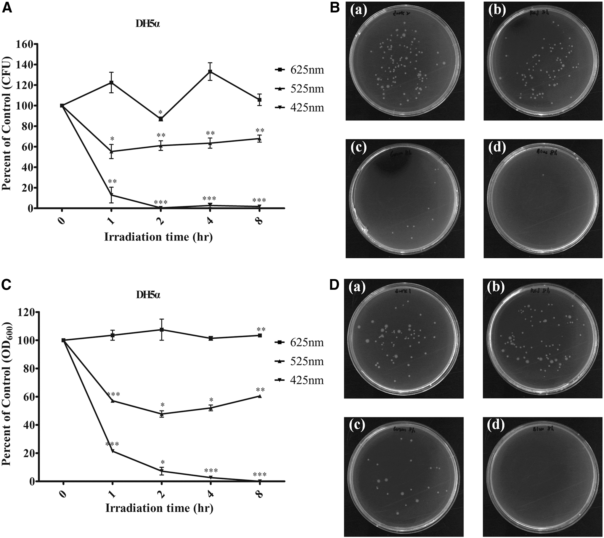
Bacterial survival of light-emitting diode (LED) irradiation at 6 mW/cm2 for 1 h irradiation in DH5α.
Time dependence of LED irradiation of S. aureus in solid and liquid media
To examine the effects of LED irradiation on S. aureus, bacteria (2×10⁸ CFU/mL) were LED irradiated at 625, 525, and 425 nm (density of 6 mW/cm2;) for 1 h during growth on agar and in broth. As shown in Fig. 6A, S. aureus viability decreased during an 8 h irradiation at 525 nm (10∼60% reduction) on agar. The bactericidal effects of S. aureus were increased by 8 h irradiation at 425 nm (40% reduction) and 525 nm (10∼60% reduction) in broth (Fig. 6C). In contrast, S. aureus viability was unaffected at 625 nm (Fig. 6A). To confirm the growth and bactericidal effects on S. aureus, S. aureus exposed to LED irradiation in broth was analyzed by OD600 and CFU. Survival of S. aureus was reduced by 30∼90% at 525 nm irradiation, both for 8 h (Fig. 6C and D). Viability was unaffected by irradiation at 625 nm.
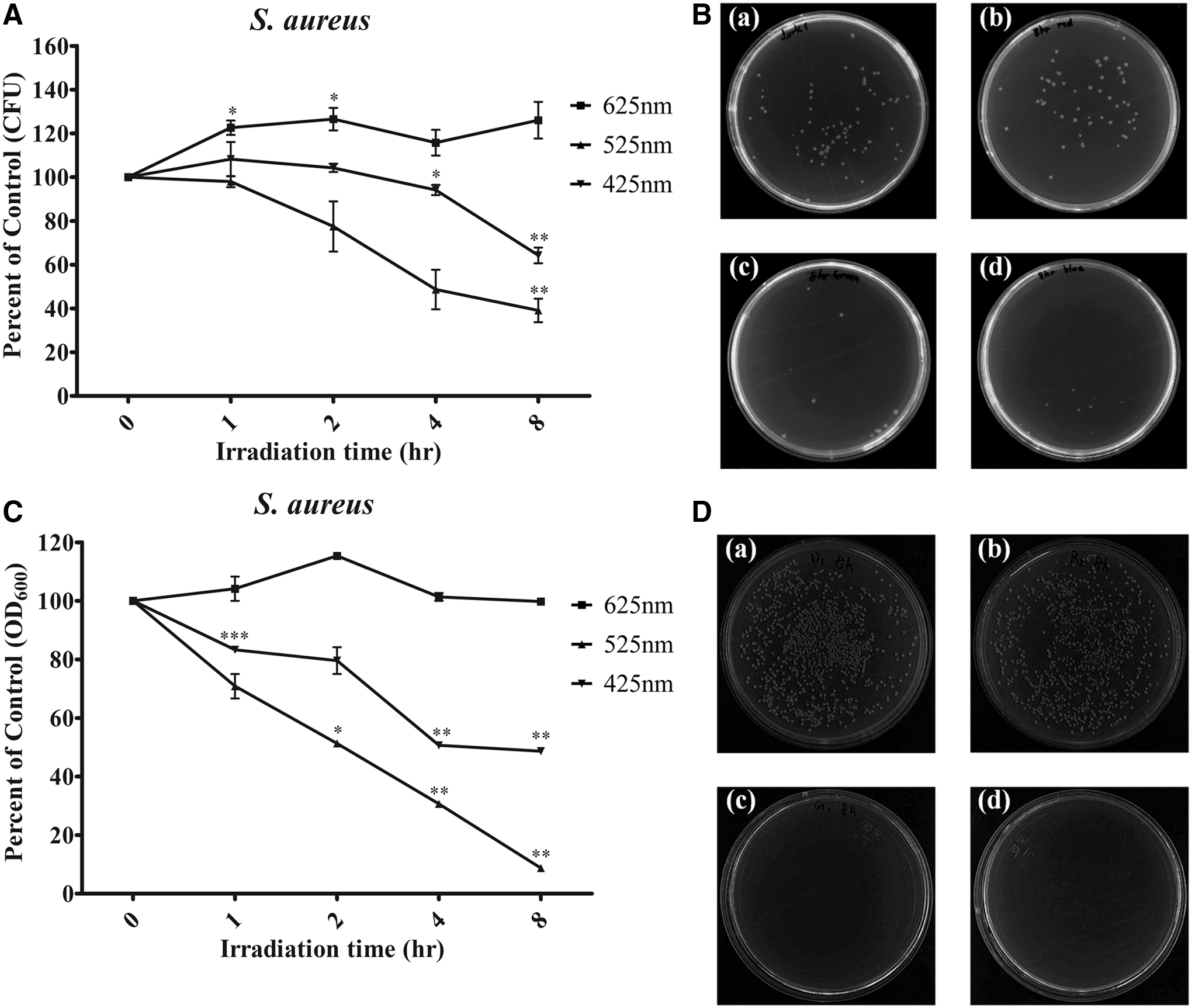
Bacterial survival of LED irradiation at 6 mW/cm2 for 1 h in Staphylococcus aureus.
Discussion
Light therapy has begun to be considered for various treatments. Bactericidal effects of LED irradiation have been reported, with results differing by wavelength. 17,22 Wavelengths of red, green, and blue light are commonly used in dentistry for wound healing, 23 periodontitis, 23 and photopolymerization of restorative materials.
In the present study, two gram-negative bacteria (P. gingivalis and E. coli DH5α) and a gram-positive species (S. aureus) were chosen. E. coli DH5α is not pathogenic and was used as a model organism for the experimental procedures. P. gingivalis is associated with periodontal bone loss. 24 S. aureus is one of the most common bacteria present at infected soft tissue lesions. 25 Increasing evidence suggests that S. aureus may be an important pathogen in the initiation of peri-implantitis. 26
Light irradiation has been used as a bactericidal agent against oral disease. 27 Presently, the lethal exposure dose is dependent upon bacterial species and bacterial density. As is shown in Fig. 3, the growth or inhibition at each wavelength seemed to be dependent upon the inoculum quantity of P. gingivalis. When 1.6×10⁹ CFU/mL was inoculated, bacterial growth recovered by blue light irradiation in 24 h, whereas red and green light had no effect. However, with a smaller number (2×10⁸ CFU/mL) of P. gingivalis as the inoculum, blue light irradiation inhibited bacteria growth over time. Neither red nor green light irradiation was bactericidal. The collective results indicate that bacterial growth was dependent upon the viable numbers of the inoculated bacteria. Light therapy is a proper measure for prevention and/or reduction, not for an absolute treatment and/or elimination of bacteria.
Bactericidal effects were also dependent upon wavelength. Blue light irradiation was bactericidal against P. gingivalis, E. coli DH5α, and S. aureus, whereas red light irradiation was not. However, P. gingivalis was increased only at 8 h irradiation, because of the energy dose. Blue light might induce bactericidal effects by the generation of reactive oxygen species (ROS). 15 Lipovsky et al. 28 reported that visible light (400∼800 nm) at high intensity killed bacteria frequently found in infected wounds, whereas low power white light enhanced bacterial proliferation. ROS production following blue light (400∼500 nm) illumination was higher than that of red light (500∼800 nm). 28 Feuerstein et al. 29 showed that blue light (400∼500 nm) is phototoxic to P. gingivalis and Fusobacterium nucleatum. The minimal inhibitory dose for P. gingivalis and F. nucleatum was reported to be 16∼62 J/cm2. 29 Near infrared diode laser irradiation did not affect any of the bacteria tested. 29 The results of the latter study suggest that visible light sources without exogenous photosensitizers have a phototoxic effect, mainly on gram-negative periodontal pathogens. 29 In contrast to their findings, bacteria exposed to 625 nm light in the present study was not affected (Fig. 4).
Green light was especially effective against S. aureus (Fig. 6). These results suggest that bacteria species is an important factor in the bactericidal effects of light therapy, because of differences of bacteria structure, such as membranes. Dadras et al. 30 proposed that an Ar-ion laser at 514 nm be used to determine the effect of various energy densities of green light on these bacteria. All energy densities of Ar-ion laser had a proliferative effect on P. aeruginosa and an inhibitory effect on S. aureus. Similarly, second harmonic generation (SHG) Nd:YAG (532 nm) and He-Ne (633 nm) lasers with given energy densities increased proliferation of P. aeruginosa, but inhibited S. aureus. 30 Application of 525 nm green light in our study would be expected to have a bactericidal effect on other gram-positive microbes such as S. aureus. Additionally, it can be expected to have bactericidal effects when dental bleaching is treated with green light.
Regarding the power density for bactericidal effects, high light power (6 mW/cm2) was more effective for bactericidal effects than low power (3 mW/cm2) (data not shown). The reason may be a high concentration of light-induced ROS, which are lethal to the cell, although low levels stimulate cell growth. 31
Finally, these results suggest that 625 nm irradiation has potential applications for oral periodontal disease, as P. gingivalis grew after irradiation. This might be expected to have additional merits for dental bleaching and resin curing in dental clinical practice. Irradiation at 625 nm irradiation can reduce inflammatory reactions and induce cell proliferation 32 in periodontal disease, which involves the interaction of bacteria and oral cells. However, the absence of a bactericidal effect at 625 nm and the lack of bacterial proliferation were noted in the present study. We can infer that 625 nm irradiation has potential applications for periodontal disease.
Conclusions
The effects of LED phototherapy are dependent upon wavelength, power density, quantity (or number) of bacteria, and species. At 625 nm, the growth of S. aureus, E. coli, and P. gingivalis increased, whereas 425 and 525 nm were bactericidal. Especially, S. aureus was killed only by 525 nm.
Footnotes
Acknowledgments
This study was supported by the National Research Foundation of Korea (NRF) grants funded by the Korea government (MSIP) (No. 2011-0030759).
Author Disclosure Statement
No competing financial interests exist.