
Abstract
Introduction
This analytical technique is based on the measurement of the intensity (number of photons collected per unit of time) of the characteristic X-rays emitted by elements in the sample when properly excited. 3 The intensity of the characteristic energy components emitted by the sample is directly proportional to the concentration of each element present in the sample. 3
Osteoporosis has been defined as a skeletal disorder characterized by loss of bone mass and deterioration of microarchitectural bone tissue. 4 –8 The main consequence of this disease is the occurrence of low-impact fractures caused by loss of power of resistance of bone tissue. 4,5
It is estimated that 40–50% of women and 25–35% of men will sustain fractures resulting from osteoporosis at some point in their lives. The number of cases of osteoporosis will double worldwide by 2025, an increase that is greater among men. This disease is not detected and treated in men the way it is in women, who have been monitored by women's health programs during menopause. 5
Therefore, osteoporosis is a major public health problem with significant economic consequences because of the high cost of treatment 4,5,9 and high rates of morbidity and mortality associated with osteoporotic fractures. 5,8,9 The disease is known to have multiple etiologies related to gender, age, genetics, and environmental factors. 4,8
Although the implications of these factors in osteoporosis are not yet well determined, a range of hormones and local factors also act in its development. 4,8 Among them are estrogen, testosterone, calcitonin, growth hormone, insulin, thyroid, and parathyroid, as well as local factors such as interleukins IL-1, IL-6, IL-11, prostaglandins, and tumor necrosis factor (TNF). 4,8,10 –13 Although testosterone plays an important role in maintaining bone density in men, estrogen is a dominant steroid in the regulation of bone metabolism in men as well. 8,10
Several treatment protocols have been proposed for osteoporosis, but calcitonin replacement through intramuscular injections or nasal sprays is already widely used. 4,6,14,15 Calcitonin is a polypeptide hormone produced by the thyroid gland that plays an important role in bone reabsorption by inhibiting osteoclast activity. 4,15 The administration of exogenous calcitonin inhibits this process, thereby decreasing the risk of fractures. 6
Low-level laser therapy (LLLT) has also been used in health promotion in several areas of medicine 16 because of its photobiomodulation abilities. 17 Osteoblastic activity can be stimulated when the bone tissue is irradiated by appropriated wavelengths, accelerating the production of organic bone matrix as a result of increased vascularity in the irradiated site and elevated levels of adenosine triphosphate (ATP). Soft tissues also exhibit proliferation of young fibroblasts and increased production of collagen fibers, thus accelerating the tissue repair process and maintaining quality of tissue. 17,18
The use of light photons in the modulation of cellular activity leads to intracellular biochemical responses without the propagation of heat and without causing tissue damage. 19 The biochemical effects are still not completely understood, 17,19 but results indicate that the absorption of light by mitochondria promotes the production of ATP, activating or inactivating enzymes, enhancing the cell permeability of calcium by altering the chemical properties of macromolecules and promoting cell proliferation. 17,19
The types and parameters of diodes used for light emission vary between different devices available and in accordance with the goal of therapy. Gallium-aluminum-arsenide (GaAlAs) diode devices have been used successfully in the medical field. Specifically, LLLT has been effective in the containment of edema, pain control, treatment of dermatitis, relief of musculoskeletal pain, and the treatment of chronic inflammation and autoimmune diseases. 17,18
The high incidence of osteoporosis, the available treatments associated with the high technology of therapeutic techniques, and the search for new noninvasive analytical methods have motivated this study. Considering that the stiffness and strength of bone tissue derive from the deposition of hydroxyapatite crystals (inorganic material) on collagen fibers (organic matrix), this study aimed to evaluate, ex vivo, biochemical changes in the composition of the bone tissue during the repair process in rats with induced osteoporosis treated with calcitonin, with and without LLLT, using a potential analytical measurement tool, μ-EDX.
Methods
This study was approved at Vale do Paraíba University by the Ethics Committee on Animal Research under No. A35/CEP/2008. We used 60 adult male rats (Rathus norvegicus albinus), 60 days old, weighing ∼200 g, fed with chow and water ad libitum. All animals underwent two surgical procedures. The animals were weighed and pre-anesthetized with butorphanol (0.01 mL/kg, i.m.) and acepromazine (0.02 mL/kg, i.m.), and then anesthetized with Zoletil (0.1 mL/kg). After this procedure, the area was treated with an antiseptic solution of polyvinylpyrrolidone iodine (PVP-I).
The testicles were bilaterally removed to induce osteoporosis. Sixty days after the orchietomy, a surgical bone defect 2.8 mm in diameter was produced on the femur using a drill type cast with the help of a low-rotation engine of 1100 rpm under constant irrigation with saline solution at 0.9% throughout the surgery. Figure 1 shows a picture of the surgical defect in the animal femur.
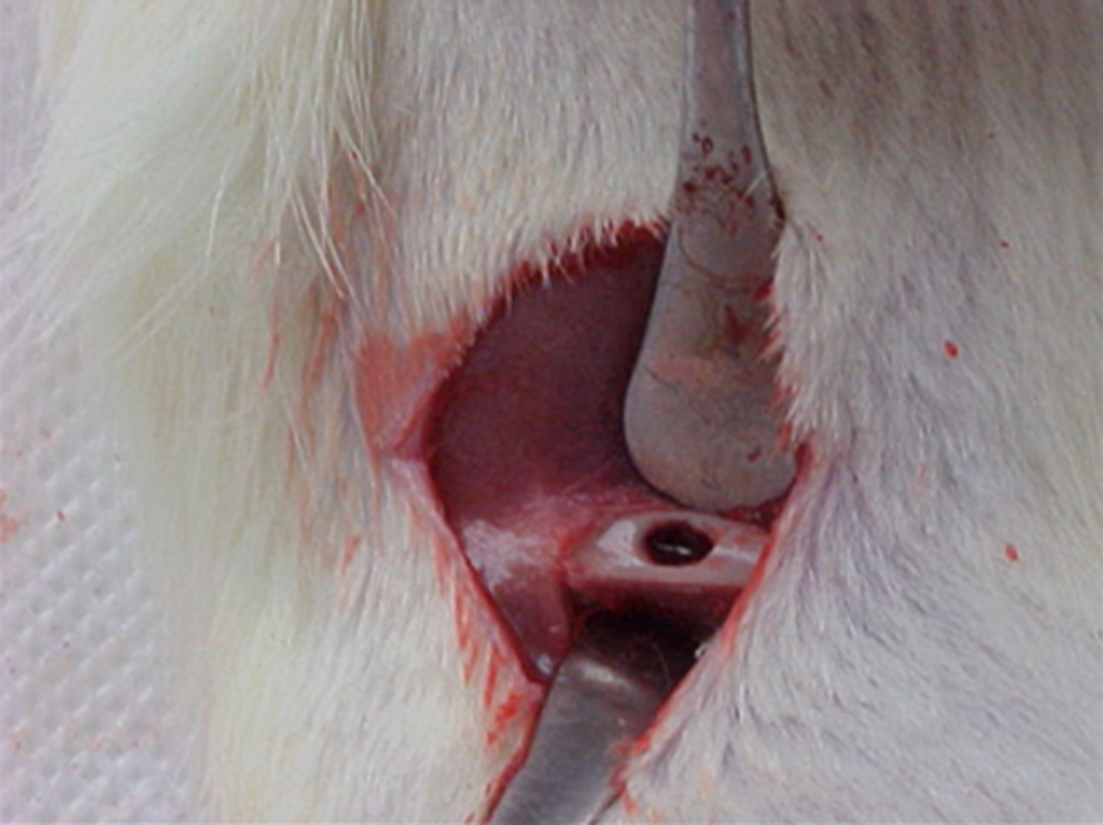
Surgical defect in the animal femur 2.8 mm in diameter.
The animals were randomly divided into four equal groups: control—C, calcitonin—Cal, Laser—La, and calcitonin+Laser—CaLa. The Cal and CaLa groups received salmon calcitonin 2 UI/kg, i.m. (Miacalcic – NOVARTIS, Stein, Switzerland) immediately after the creation of the surgical bone defect, and repeated on alternate days. The La and CaLa groups received LLLT immediately after surgery and on alternate days in a timely manner using the THERA LASER® (D. M. C. Equipamentos Ltda São Carlos – SP) GaAlAs semiconductor diode. Five specimens of each group were euthanized at 7, 14, and 21 days. Table 1 shows the total period including the LLLT and calcitonin application days until the death of animals.
Total of treatment turns in days before euthanasia.
LLLT applications were punctually performed using near infrared light (λ-830 nm) lead by an optical fiber in transcutaneous mode, over the surgical wound extension, as shown in Fig. 2. Table 2 shows the calculated radiation parameters considering the beam area of the optical fiber.

Low-level laser therapy (LLLT) application punctually performed by an optical fiber in transcutaneous mode, over the surgical wound extension.
The femurs were removed and stored in formol 10% until evaluation by X -ray fluorescence. For analysis of mineral components of the samples, calcium (Ca) and phosphorus (P), a fluorescence spectrophotometer X-ray Shimadzu-1300 μ-EDX in LEVB/IP&D/UNIVAP was used.
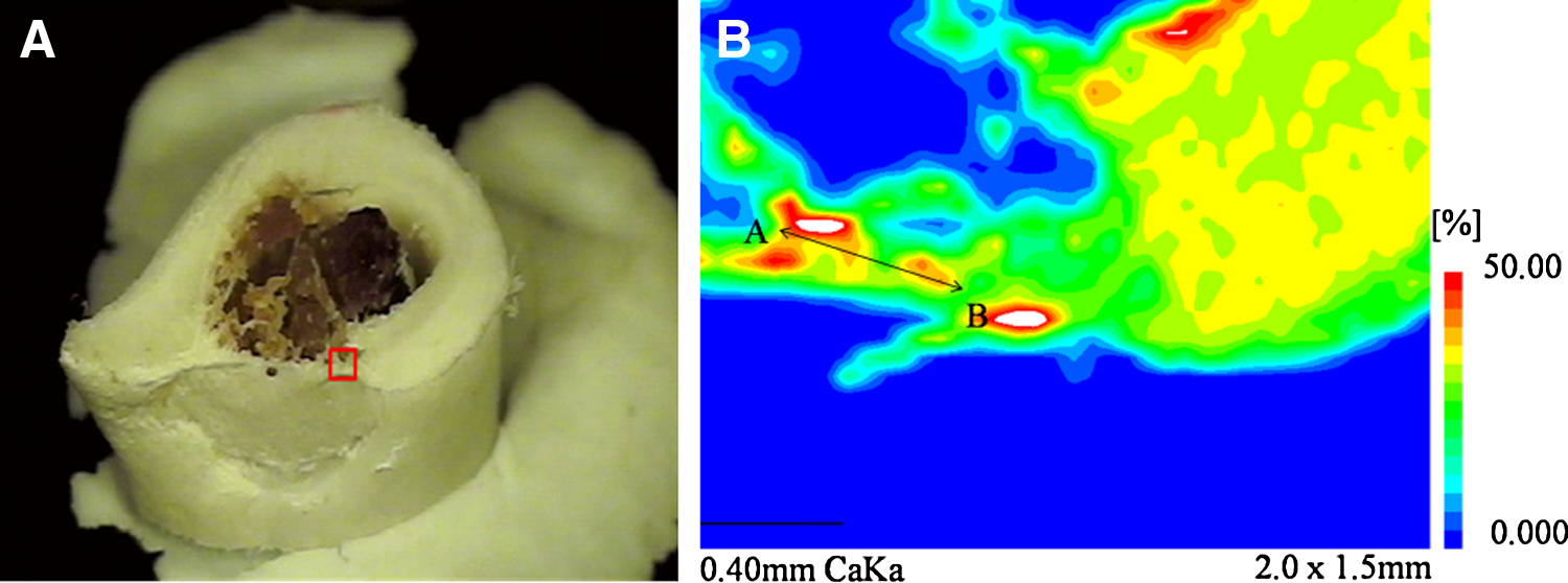
The chemical analysis was done by energy-dispersive X-ray fluorescence using the characteristic radiation from the samples. The surface of the samples was targeted by the radiation from a tube of rhodium (Rh). A semiconductor detector silicon lithium (Si[Li]) cooled by liquid nitrogen was used for counting the sample radiation with voltage set at 15 kV and automatic current. The area of repair tissue was sectioned in half, lengthwise, with hard diamond disc N15 at 500 rpm (Fig. 3A). The scan was made mapping an area with 40×30 points, a 50 μm pitch, and a reading time of 260 sec/area on the edge of the injury.
Sigma-Aldrich brand synthetic stoichiometric hydroxyapatite with a purity of 99.99% Ca10(PO4)6(OH)2 was used as a reagent reference. The basic parameters for calculating the balance of the chemical formula were established for the relative weights of Ca, P, and O.
Descriptive statistical analysis was performed considering the averages, standard pattern, and coefficients of ranges for each of the components and their relationships. Numerical data were obtained from the average of points from a line of 0.5 mm along on the mapped area (Fig. 3B).
Results
Tables 3 –6 show the average of calcium content (Ca mean), in weight%; the standard deviation of the calcium content (SD-Ca); the coefficient of variation, in percentage, of calcium content (% CV), the average of phosphorus content (P mean), in weight%; the standard deviation of the phosphorus content (SD-P), the coefficient of variation, in percentage, of phosphorus content (% CV), the average of the stoichiometric ratio of calcium to phosphorus (average Ca/P) and the standard deviation of the Ca/P (SD-Ca/P).
Ca, calcium; P, phosphorus; CV %, coefficient of variation.
Ca, calcium; P, phosphorus; CV %- coefficient of variation.
Ca, calcium; P, phosphorus; CV %, coefficient of variation.
Ca, calcium; P, phosphorus; CV %, coefficient of variation.
The calcium and phosphorus (Ca/P) relational values of the samples studied in all groups were evaluated and compared, using as a reference the stoichiometric value of hydroxyapatite, 2.16.
Regarding the Ca at 14 and 21 days, considering only the mean values obtained for the samples, experimental groups showed similar values. There was an increased rate of replacement of the mineral content of bone formation in all groups. After 21 days, the group showing the lowest rate of Ca deposition was group C. The rate of deposition of calcium in weight% after 21 days can be seen in Fig. 4. For group C, this value was 28.38%, for group Cal it was 31.91%, for group La it was 31.43%, and for group CaLa it was 32.17%.
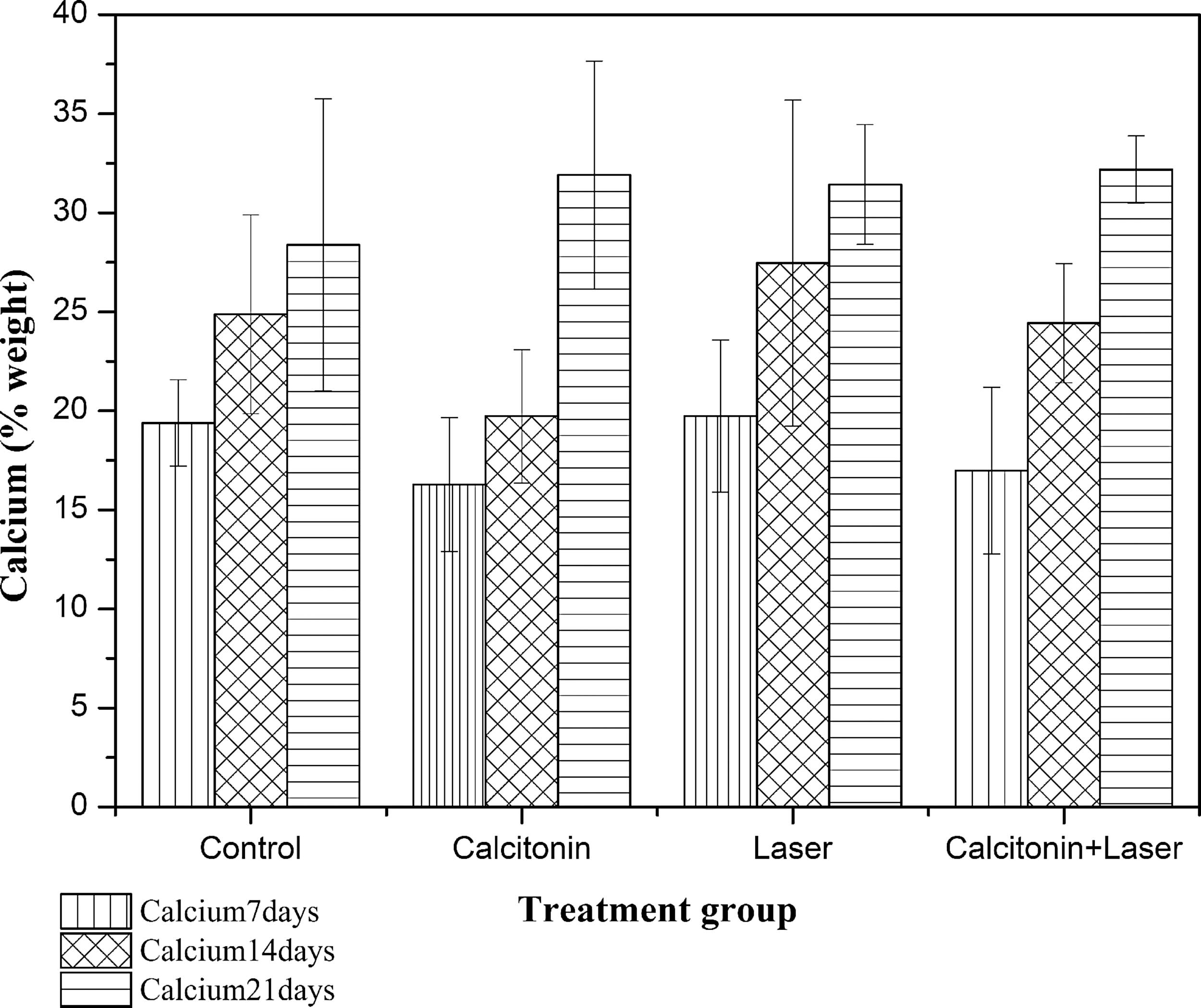
Calcium (wt%) for all groups.
Figure 5 shows that the average values of the P grew significantly at 7, 14, and 21 days. Initially, group C had the highest rate of deposition of this component. However, after 21 days, this is the group that had the lowest replacement. The deposition rate for phosphorus was 13.47% for group C, 15.58% for group Ca, 15.66% for group La, and 14.54% for group CaLa, as shown in Figure 5.
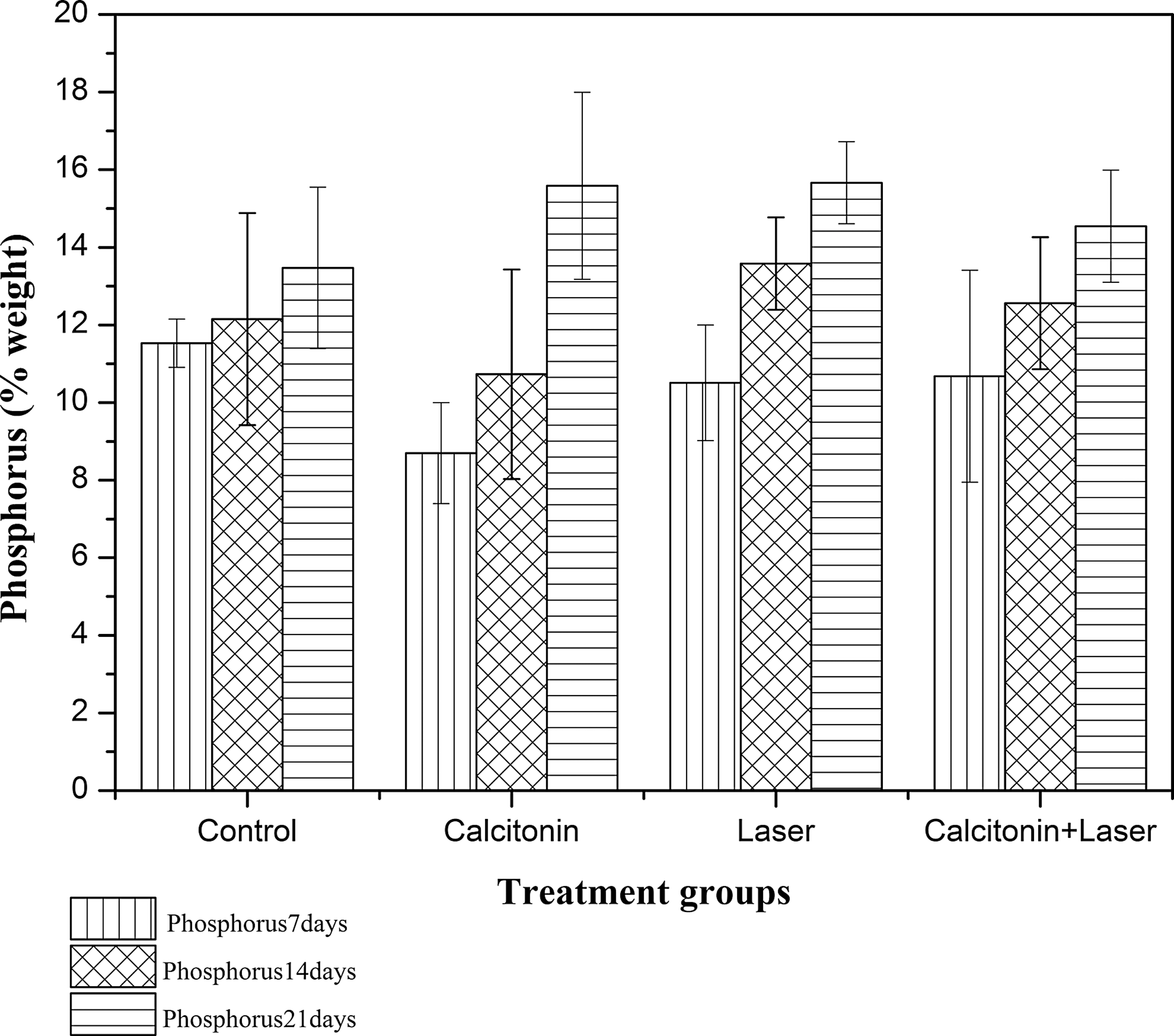
Phosphorus (wt%) for all groups.
The graph in Fig. 6 presents the values obtained in the Ca/P of the experimental groups of animals euthanized at 7, 14, and 21 days. The La group maintained greater homogeneity in the balance of bone mineral replacement at different times. After 21 days, the rate was similar for all groups. It was observed that groups C and CaLa showed similar values close to stoichiometric hydroxyapatite, 2.10 for group C and 2.21 for group CaLa, as shown in Fig. 6. The values for groups Cal and La were 2.04 and 2.00, respectively.
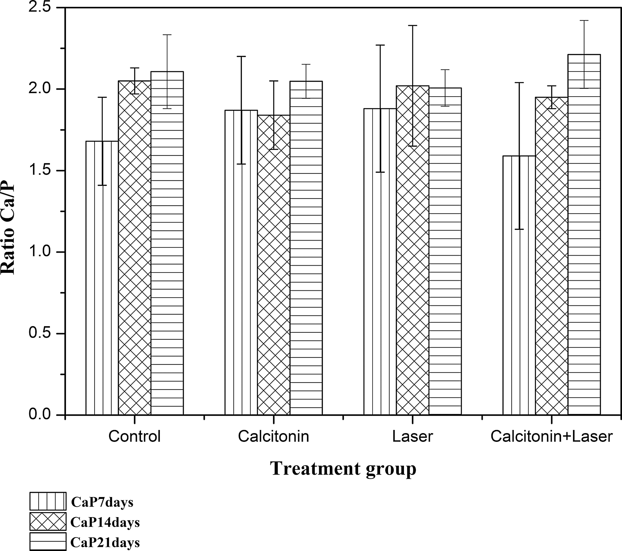
Ratio Ca/P for all groups.
Discussion
The μ-EDX technique that has been used in the analyses of samples of this study was a useful tool in the evaluation of inorganic components present in calcified tissue. 20 The qualitative and semiquantitative analyses of inorganic components of the samples were measured without any destruction of the sample or any change in its structure, showing that it is a nondestructive technique that is safe and effective for biochemical analysis of inorganic compounds in biological tissues. 2,3
The variability within and between groups is related to the intrinsic biological variability of the sample. The homeostasis of calcium and phosphorus in the body depends upon different mechanisms for its regulation. Several treatments based on the pharmacology and laser therapy have been applied to encourage the setting of these minerals in the body. 8,21
The outcome of the healing process with deposition of neo-tissue in the lesion favors the variability of statistical data obtained in the boundary regions of healthy tissue and the margin of the lesion. However, the increase in the number of points analyzed by mapping made the processing of statistical data more efficient by reducing the coefficients of variation. Therefore, it appears that the question of the variability shown in the results is directly related to the heterogeneous chemical and morphological characteristics of biological tissue.
The synthetic calcitonin administered to the animals has been widely used to treat osteoporosis by inhibiting osteoclast activity and promoting bone remodeling, with a 33–36% reduction of bone fractures. 6 Åkesson 4 (2003) point out that calcitonin has a decisive role in the maintenance of calcium in bones, and its absence or reduction will determine the quality of remodeling. 4,11
The results obtained in this study showed that the rate of calcium deposition was greater for animals in groups treated with synthetic calcitonin alone, or with LLLT. The Ca/P indicates a deviation from the stoichiometry of hydroxyapatite, a key component in the resilience of bone tissue. In fact, bone formation process comprehends deposition of organic contents (colagen type I, proteoglycans, and glycoproteins) and mineral content (hydroxiapatite, sodium, and magnesium). These compounds will provide bone quality. Furthermore, the process of modeling and remodeling activity speed plays a fundamental role in the whole process of mineralization. 22
Therefore, the deposition rates for various mineral components analyzed can determine the range of variation in relation to the standard stoichiometric value. Synthetic hydroxyapatite has a value of 2.16. Therefore, the closer the mineral makeup is to this value, the more resilient that tissue will be. LLLT, with or without other treatments, has also been applied in the control of metabolic diseases of bone tissue. 21,23 Kamali 23 and Karu 17 reported that laser light is capable of promoting the following in the area of bone injury: tissue remodeling by stimulating cellular biomodulation, collagen synthesis, improved cellular activity of blood cells, macrophages, fibroblasts, and chondrocytes. The synthesis of RNA, DNA, and ATP is also favored in the application of laser therapy. 21
The La group had the lowest Ca/P of the four groups, indicating that the deposition of phosphorus is greater than the deposition of calcium in this group. These results are supported by the fact that this is a relationship strongly influenced by any change in the process of bone turnover. However, these results did not permit the inference that phosphorus directly affects bone mineral apposition rate, a process that is still not completely elucidated. Our data corroborate those reported in previous studies, which showed that the efficacy of LLLT occurs predominantly in the early stages of the tissue repair process. 13,21
The specimens of the group in which there was a combination of treatments, LLLT and calcitonin, showed the Ca/P closer to the value of stoichiometric hydroxyapatite, agreeing with results obtained by Nascimento et al., which used bone densitometry as an analytic tool for newly formed bone tissue. 14
Biochemical changes are common in the body, and laser is able to induce chemical reorganization. Composition modifications in the Ca/P ratio after laser irradiation (λ-1064 nm) on tissue as hard as enamel were reported. 1 Despite working with a different wavelength, the current study (λ-830 nm) also indicated an increasing of the Ca/P ratio after laser irradiation.
X-ray fluorescence has been successfully employed in biomedical and bioengineering researches on hard tissues and composite biomaterials. Authors reported significant chemical reduction on Ca and P components. 24 The effects of laser irradiation were investigated by the μ-EDX technique on dentin components and enamel, showing significant changes in the amounts of Ca and P. 1,24
Recently, Kourkoumelis et al. 25 reported that the ratio Ca/P is a suitable in vitro biomarker for induced osteoporosis. The authors used two spectroscopic techniques: 1) Auger electron spectroscopy (AES). and 2) microanalysis by energy-dispersive X-ray fluorescence spectroscopy (SEM-EDX). 25 Their findings indicated that there is no significant difference between samples from different genders or among cortical bone sites. On the other hand, the Ca/P ratio of trabecular bone sections is comparable to that of cortical sections with induced osteoporosis. 25 Their results come to us giving an understanding of both the distribution and intrinsic variability of Ca/P ratio values that were verified in our results.
In our study, X-ray fluorescence has been successfully employed as a non-destructive analytical tool to investigate the ratio Ca/P in osteoporotic bones.
Conclusions
The μ-EDX technique used in sample analyses of this study is a potential analytical measurement tool in the evaluation of inorganic Ca and P during the repair of bone defects in animals with osteopenia. It was possible to perform qualitative and semiquantitative analyses of inorganic components of the samples without any destruction of the sample or any change in its structure.
The present study demonstrates that the combination of calcitonin and LLLT is the best treatment protocol, as evidenced by the proximity between the value of the Ca/P and that of synthetic stoichiometric hydroxyapatite.
Footnotes
Acknowledgments
This work was supported by Fundação de Amparo à Pesquisa do Estado de São Paulo - FAPESP - process: 2008/05786-4 and Universidade do Vale do Paraíba.
Author Disclosure Statement
No competing financial interests exist.