
Abstract
Introduction
We recently reported that LLLT application to the bone marrow (BM) of rats post-myocardial infarction (MI) caused a marked reduction in scarring, possibly because of the induction of stem cells in the BM and their migration to the infarcted area. 16,17 The BM hosts a variety of cell types including various progenitor cells of the mesenchymal and hematopoietic system. The possible long-term toxic effects of LLLT application to the BM have not been studied to date, and such study is therefore warranted in light of possible clinical application of the new approach to the induction of stem cells by LLLT.
The present study was designed to investigate the long-term safety of laser application to the BM on the histopathology of the BM, liver, kidneys, and brain of mice that underwent LLLT at several different doses and frequencies, compared with control, sham-treated groups of mice.
Materials and Methods
Experimental procedures
A total of 83 Imprinting Control Region (ICR) mice, weighing 20 g (6 weeks old), were used in this experiment. All the experimental procedures were approved by the Animal Care Committee of Tel-Aviv University. The mice were divided randomly into five groups. Bone marrow was sham treated or laser treated at various power densities as detailed (see “Laser application” section). All mice were kept under optimal conditions (food and water provided ad libitum) at room temperature (22–24°C). Mice were killed 8 months post-laser treatment and mortality was recorded daily to calculate the survival rate over 8 months.
Laser application
In control sham-treated mice (n=35) the laser was placed on the tibia but not turned on. Four groups of mice were laser treated at various power densities at the level of the BM as follows: 4 mW/cm2 (n=8), 10 mW/cm2 (n=16), 18 mW/cm2 (n=6), and 40 mW/cm2 (n=18). All laser treatments were given for a duration of 100 sec. Laser treatment was applied twice weekly for the first 2 weeks at the abovementioned doses, up to a period of 8 month.
Mice were randomly assigned to a laser-treated or control sham-treated group. A diode (Ga-Al-As) laser, wavelength 804 nm with a tunable power output of maximum of 400 mW (Lasotronic Inc., Zug, Switzerland) served for application of LLLT to the BM, as described by us previously. 16 A metal backed glass fiberoptic (30 mm long and 2 mm in diameter) was connected to the power output of the laser device. The distal flat end of this fiberoptic was placed directly on the bony proximal medial part of the tibia bone following a small incision (5 mm) through skin and muscle. The measurements of power density and transmission of the laser through the tibia bone were performed as detailed (see Fig. 1). In a preliminary experiment, three intact mice (same age and weight as the experimental laser-treated mice) were killed by overdose of avertin; the fresh tibia bone was removed, cleaned from attached muscles and tendons, and then carefully cut on its longitudinal axis to yield two sections of bone featuring the medial and lateral parts of the tibia. The medial flat aspect of the bone was cleaned from the BM and used for measurement of laser transmission through the bone. The distal tip of the laser fiberoptic was placed perpendicular to the flat aspect of the tibia, ∼3 mm distal to the in the knee joint. The tunable laser was set to provide various power outputs on its distal tip (see Table 1 for details). The laser meter probe (model PD.300-3W, Ophir Inc. Jerusalem, Israel) was then placed on the opposite side of the bone (inside the bone where the BM is normally located), and the power was measured (Fig. 1). The irradiation was performed using multimode fiberoptics 1.5 mm in diameter. The laser beam profile was measured using the truncated Gaussian beam method according to Urey 18 at the 1/e 2 location on freshly prepared tibia as described. The fiber output Gaussian beam had a numerical aperture of 0.22, that is, a divergence of ∼±13o from its central axis. Measurement of the energy at the output of the fiber appears in Table 1, and the spot size, measured at 1/e 2 of the energy curve, starts at 1.5 mm diameter (see Fig. 1 for further clarification). The diameter of the beam at the BM interface with the bone is ∼1 mm away from the fiber distal end giving a calculated 1/e 2 beam diameter of 1.94 mm, and actually is even larger because of the scattering of light in the bone. The laser transmission through the bone was calculated as the ratio value (in percent) of the power output measured after laser penetration through the bone divided by the power at the tip of the fiberoptic on the external surface of the tibia (see Fig. 1 and Tables 1 and 2 for details).
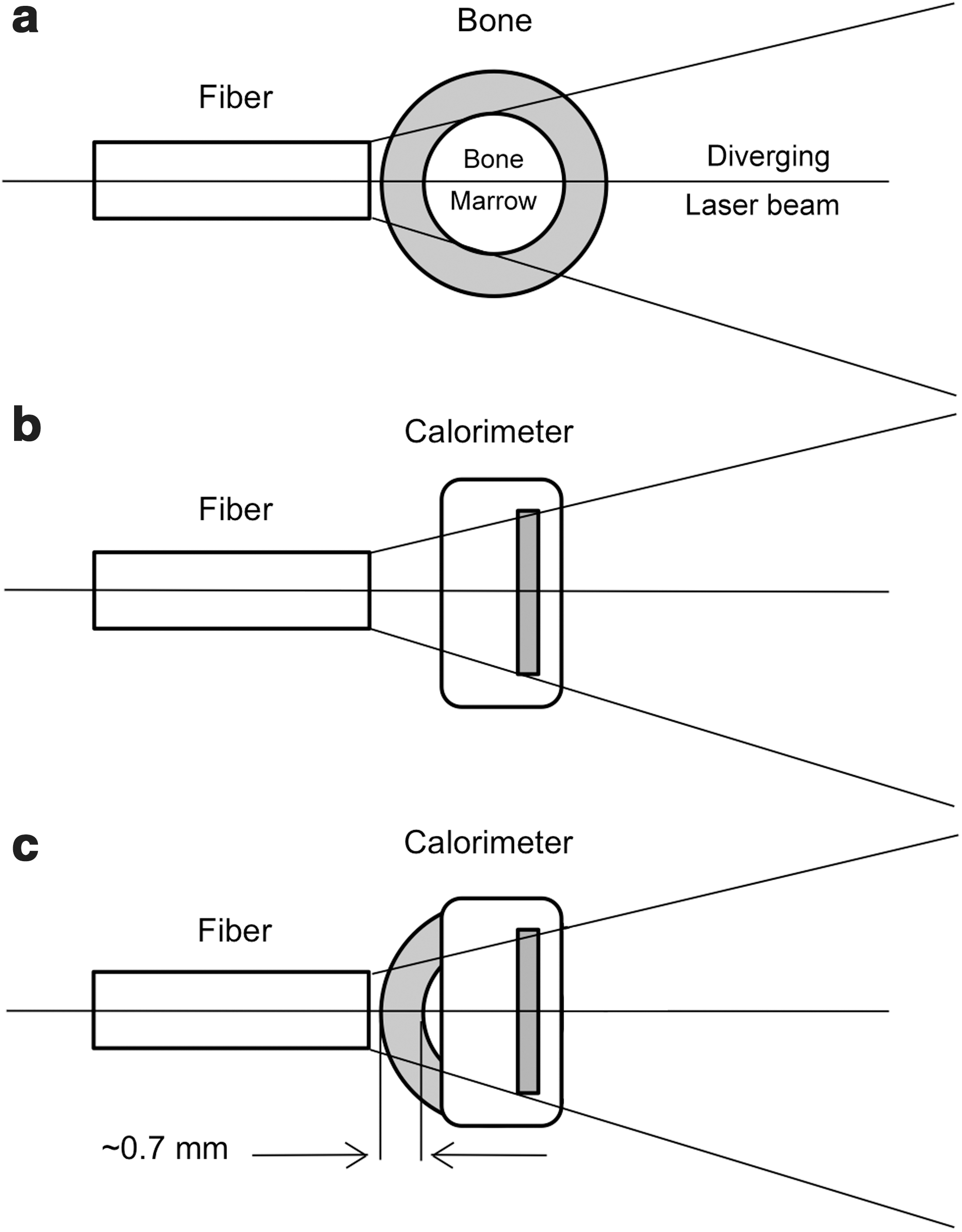
Schematic presentation of
Measured at 1/e2 of energy peak.
The area of the beam where the diameter is measured at 1/e2 of the peak energy.
At the abovementioned setting, the laser beam diameter on the BM was 6 mm, comprising an area of 0.28 cm2, the measured transmission of the laser through the tibial bone was 27%, and the tunable laser power output from the distal tip of the fiberoptic (delivered directly to the external surface of the tibia) was set to provide parameters of 4, 10, and 18 and 40 mW/cm2 power densities on the BM tissue, which comprised 0.4, 1, 1.8, and 4 J/cm2, respectively. Laser application was applied for 100 sec in all rats.
Histology
Mice were killed by overdose of avertin. The tibia bone, liver, kidneys, and brain were excised and washed for 30 sec in extensive saline to remove external blood. The tissue samples (∼0.5×0.5×0.5 cm of tissue randomly cut from each organ) were fixed in 10 mL of 4% neutral buffered formalin solution for 3 days. The formalin solution was replaced with a fresh one after 24 h. The fixative was washed in running tap water for 3 h, and then dehydration was performed with graded 50–100% ethanol solutions. The tissue samples were then embedded in paraffin and 8 μm sections were prepared using a microtome. Defined cross-section samples (2 mm thick) were taken from the abovementioned organs for histology. Eight micron paraffin sections were prepared from the tissue samples of each organ. Sections were stained with hematoxylin-eosin. Two observers, blinded to whether the mice were control or laser treated, reviewed the histological sections.
Isolation and culturing of mesenchymal stem cells (MSCs) from bone marrow
Bone marrow of control, sham-operated and laser-treated mice, at power densities of 4, 18, and 40 mW/cm2 was extracted from the tibias and femurs of each mouse using a steel rod as described by us previously, 16 and pooled for each mouse. Bone marrow was not extracted from the mice treated at a power density of 10 mW/cm2. Mesenchymal stem cell isolation was performed essentially as described by Davani et al. 19 The collected BM was incubated in a shaker with 5 mL Dulbecco Modified Eagle Medium (DMEM) containing type I collagenase (250 U/mL, Sigma Israel) for 45 min at 37°C. Cells were then cultured at a concentration of 1.3×106 cm2 in DMEM supplemented with 10% fetal bovine serum (FBS), 2 mmol/l L-glutamine, 100 U/mL penicillin, and 100 U/mL streptomycin (Biological Industries, Beit Haemek, Israel) in small tissue culture flasks. Forty-eight hours later, the medium was replaced to remove nonadherent cells (MSCs are adherent 18 ), and thereafter replaced every 4 days. C-kit+ staining of these cultures revealed that ∼90% of the cells were positively immunostained for c-kit. The isolated MSCs from the mice were harvested for 7 days (4 days are necessary for cells to adhere, and an additional 3 days are needed to express their proliferative capacity). After 7 days in culture, the cells were harvested and centrifuged, stained with toluidine blue 0.1%, and counted using a hemocytometer.
Statistical analysis
The SigmaStat 2.0 (Sigma, St Louis, MO) software was used for statistical analysis. Tests were performed first for normality distribution, followed by parametric (Student's t test) test.
Results
No histological differences were observed in the liver, kidneys, brain, or BM of the laser-treated mice as compared with the sham-treated control mice (Fig. 2). Moreover, no neoplasmic response in the tissues was observed in the laser-treated groups as compared with the controls.
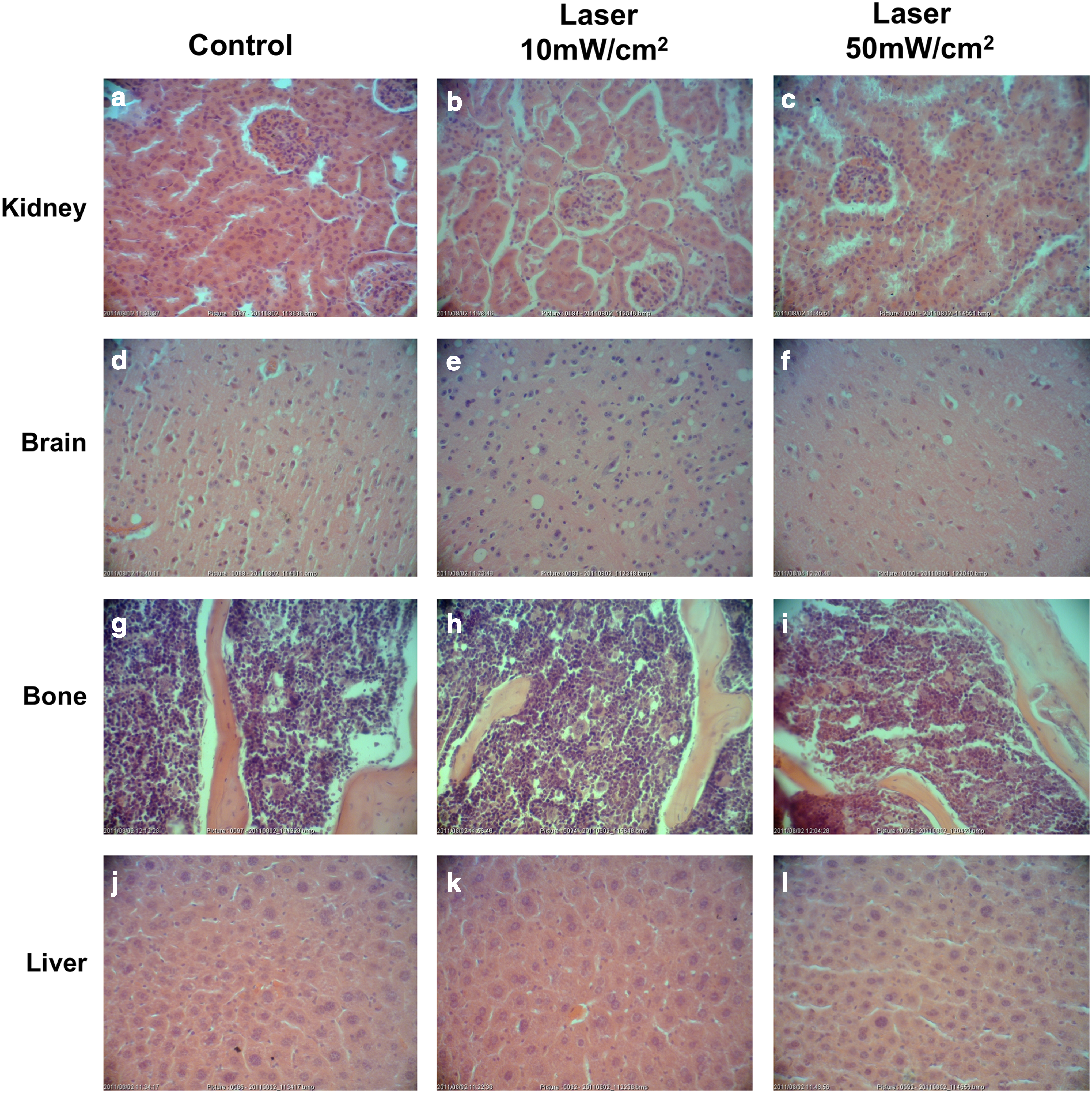
Representative light micrographs of kidney
There was no statistical difference (Fisher's test) in mortality during the 8-month follow-up period between the control group and any of the laser-treated groups.
The mean number of MSCs that were isolated from non-laser-treated mice and grown in culture for 7 days was 9×10⁵±2, not significantly different from the mean number of the MSCs (8.4×10⁵±2, 6.6×10⁵±2, and 6.5×10⁵±2) that were isolated from the BM of laser-treated mice at power densities of 4, 18, and 40 mW/cm2, respectively (Fig. 3).
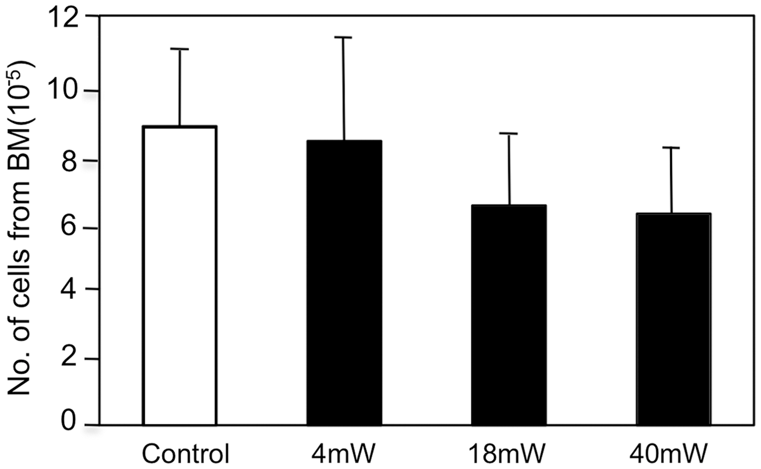
Effect of low-level laser therapy (LLLT) applied to the tibia of mice, on the number of mesenchymal stem cells (MSCs) isolated from the tibia 8 weeks post-LLLT. The entire bone marrow (BM) from control non-laser-treated (open column) or laser-treated (solid column) tibias was collected and pooled together for each experimental group and then grown in six culture wells. The MSCs that were isolated from each group were grown in culture for 7 days and then counted. Results are mean±SEM of six repetitions (wells) in each group.
Discussion
The results of the present study indicate that applying LLLT at a power density dose of up to five times the optimal dose that causes a photobiostimulatory effect on the cells in the BM of rats post-MI, 5 did not induce any pathological effects on the various studied organs in the mice. Histological examination of these organs did not reveal any neoplasmic response over a period of 8 months (almost the entire lifespan of mice) following the application of LLLT to the BM.
The results corroborate a previous long-term safety study on the application of LLLT to the rat brain, which found no toxicological abnormalities in the brain at similar laser power densities to those used in the present study. 9,12 –14 In that study, however, only the brain tissue was analyzed. In the present study, we looked for pathological changes in several organs, as the cells that populate the BM and were induced by means of LLLT might have migrated through the circulation to other organs. The present study is the first to explore possible long-term histopathological changes concomitantly in several organs following multiple LLLT applications to the BM. In another short-term study of LLLT application to the brain, at power densities similar to those in the present study, no side effects were observed. Moreover, when LLLT was applied to the brain post-traumatic brain injury, no changes in the histopathology of the brain were evident. 20,21
Another indication of the safety of LLLT to the BM was found previously when the kinetics of growth of MSCs isolated from laser-treated or non-laser-treated BM were followed in vitro. 16 It was found that MSCs isolated from the BM of infarcted rats 4 weeks post-LLLT had a significant twofold higher rate of proliferation than the non-laser-treated BM. However, this enhanced proliferation was not observed when the MSCs were isolated from the BM 6 weeks post-LLLT. These results indicate that the induction of cell proliferation by LLLT is limited to a relatively short period post-laser application, and that there is no discernible neoplasmic response in the stem cells in the BM that have been induced to proliferate at a faster rate by the LLLT.
In an additional study, we followed the kinetics of the growth factors inducible nitric oxide (iNOS) and vascular endothelial growth factor (VEGF) in rats with post-MI where the laser was applied directly to the heart. 22 The content of these factors was higher in the laser-treated hearts 2–4 days post-MI than in the non-laser-treated hearts, but was identical to the non-laser-treated hearts at 7 days post-MI. The above finding indicates that LLLT does not induce production of these significant growth factors in an uncontrolled manner that differs from their production post-MI without the interference of LLLT.
In the present study, we have also shown that the number of MSCs isolated from the BM of laser-treated and non-laser-treated mice (at 8 months of age and before euthanasia) proliferated in culture to a similar extent. This gives credence to a non-neoplasmic long-term possible effect of LLLT on the BM. This phenomenon, in addition to the histopathology of the BM, which did not indicate neoplasmic response in the laser-treated mice, supports the long-term safety of LLLT application to the BM in mice.
Conclusions
It is concluded that LLLT applied multiple times either at the optimal power and energy density (that induces photobiostimulation of stem cells in the BM), or at higher power densities up to five times the optimal (that yields photobiostimulatory effects), does not cause histopathological changes or neoplasmic response in the various organs of the mice as examined over a period of 8 months.
Footnotes
Author Disclosure Statement
No competing financial interests exist.