
Abstract
Introduction
Some studies of human dentin surfaces have indicated that tubules are larger in size and greater in number in sensitive areas of dentin, when compared with the nonsensitive areas, 4,5 which further substantiates the role of opened dentin tubules in the etiology of DH.
Clinically, DH is often associated with noncarious cervical lesions, which is the loss of tooth structure at the cement–enamel junction, not related to caries. 6 These lesions are of multifactorial origin, 7 in which erosion, abrasion, and abfraction are known to play an important role. 8 Erosion and abrasion are also believed to be implicated in the etiology and development of DH. Corroborating this idea, micromorphological studies have indicated that some acidic beverages 9 –11 and toothbrushing 12,13 can open, and sometimes even enlarge the diameter of, the dentinal tubules.
There are two strategies for the treatment of DH. One is to physically occlude dentin tubules to isolate the tubule contents from the oral environment and prevent the fluid flow movement. Among the agents used to occlude dentin tubules are: adhesives, fluoride solutions or dentifrices containing sodium fluoride, calcium fluoride, or ferric, aluminum, or potassium oxalate. 14 –16 The other strategy is to chemically desensitize the sensory nerves, blocking the transmission of the noxious stimuli from the dentin tubules to the central nervous system. Potassium salts are an example this kind of agent. 17 Both strategies have shown to be effective in reducing DH; however, evidence suggests that the occlusion of dentin tubules is more likely than chemical desensitization to be successful in clinical applications. 18
The desensitizing agents can be applied topically at home by the patients or in the office by a dentist. The at-home methods are known to have an immediate effect and a lower cost, 19 whereas the in-office agents can show a better effect because dental professionals can deliver a wider range of more complex and more potent desensitizing agents. 17 However, repeated desensitization applications are usually necessary to maintain the treatment effect, because exposed dentin continues to sustain erosive and abrasive challenges in the oral cavity. 20
As an alternative to the topical agents, but with the aim of occluding the dentin tubules as well, the use of an Nd:YAG laser has also been suggested. 21 –26 A previous study demonstrated that Nd:YAG laser irradiation has the ability to cause melting and re-solidification of the dentin surface, thus occluding the dentinal tubules. 11
The purpose of this study was to compare the effects of some in-office protocols in promoting the occlusion of dentin tubules with Nd:YAG laser irradiation, and also the short-term efficacy of these agents after erosive and abrasive challenges. This latter is particularly important because until now, little has been known about the long-lasting effects of the agents or forms of treatment of DH described in the literature, whether or not they are able to resist the chemical and mechanical challenges constantly present in the oral environment.
The null hypothesis was that none of the treatments would be able to sustain tubule occlusion after chemical and mechanical challenges.
Materials and Methods
Specimen preparation
The study protocol was approved by the Ethics Committee in Research of the local institution (University of São Paulo, School of Dentistry), protocol 195/2010- CAAE 0021.0.017.000-10. Specimens from human third molars were used. Molar teeth were freshly extracted and they were stored in 0.1% thymol solution at 4°C for <1 month until the beginning of the experiment. From these teeth, dentin slabs (4×4×2 mm) were sectioned from the roots using a microtome (Isomet® 1000- Precision Saw- Büehler, Lake Bluff, IL). The slabs were then embedded in an acrylic resin (Varidur, Buehler, Lake Bluff, IL). The resulting blocks were ground flat and polished with water-cooled abrasive discs (600- and 1200-grit Al2O3 papers; Buehler). Any specimens with cracks or defects were discarded and replaced.
Phase 1: Procedures to open tubules (simulation of hypersensitive dentin)
Three different agents were tested regarding their ability to open dentinal tubules and simulate hypersensitive dentin: 27% ethylenediaminetetraacetic acid (EDTA pH 7.4), 37% phosphoric acid (Villevie®, Joinville, SC, Brazil/ pH 1.28) and an acetate buffer solution (pH 5.5) with equimolar amounts (4.875 mmol/L) of calcium and phosphate, proposed by Shellis and Curtis. 20 Calcium and phosphate were added as CaCl2.2H2O and KH2PO4, respectively.
For this test, nine dentin specimens were randomly divided into three groups (n=3). In the first group, the specimens were immersed in 27% EDTA solution for 2 min. In the second group, specimens were immersed in the buffer acetate solution (pH 5.5) for 2 h and ultrasonicated for 1 min in the same buffer solution. The third group was treated with 37% phosphoric acid for 20 sec. After the treatments, the specimens were washed with water/spray and analyzed by MeV to determine which solution best simulated hypersensitive dentin.
Phase 2: Evaluation of in-office treatments
After simulating hypersensitive dentin by opening the dentinal tubules, 12 dentin specimens were randomly divided into four experimental groups (n=3), according to their respective treatments. Group 1 was the control group in which no treatment was performed. For Group 2, an Nd:YAG laser (Power Laser™ ST6, Lares Research®, Chico, CA) was used in contact mode, focused, and in a perpendicular direction, with the following parameters: 1.0 W power, 10 Hz repetition rate, within the energy parameters of 100 mJ and an energy density of 85 J/cm2 per pulse. Using a quartz fiber of 400 μm, in a pre-established region, scanning movements were made in occlusal-apical and mesio-distal directions, and vice versa. Four 10 sec irradiations were made in each direction, totaling an irradiation time of 40 sec. An interval of 10 sec between irradiations was performed to allow thermal relaxation of the tissue. 11 Before all irradiations, the real power delivered was measured with Power Meter equipment (Coherent, Newport, United States), to ensure no loss of power during any of the irradiations. For Group 3, prophylaxis was performed using a rubber cup mounted in a slow speed handpiece (5000 rpm) and pumice (SS White, Rio de Janeiro, RJ, Brazil) for 20 sec, according to the manufacturer's recommendations. In Group 4, Colgate® Sensitive Pro-Relief™ desensitizing paste (Colgate Palmolive, New York, NY) was applied with a rotary cup at low speed (5000 rpm) for 3 sec, following the manufacturer's instructions.
Erosive/abrasive cycling challenges
For this challenge, samples from all the groups were immersed for 5 min in 1% citric acid (pH adjusted to 3.8 using NaOH), rinsed with distilled water, stored for 60 min in artificial saliva, and then brushed with a slurry of a regular toothpaste, according to the brushing protocol described in the next section. This cycle was repeated twice a day, over 5 days. The specimens were stored overnight in artificial saliva.
Brushing protocol
With the aid of a brushing simulation machine (MSEt - ELQUIP, São Carlos, SP, Brazil), brushing of the specimens was standardized regarding the number of brushing cycles, time of toothpaste injection, temperature, and load. The speed of the equipment and time spent on daily brushing was calculated at 2 min for each brushing episode, at a brushing speed of 258 cycles/min. One cycle was considered to be one complete back and forth movement of the toothbrush. A 200g load was used. Taking into account that people usually brush their teeth for 2 min, twice a day, it would result in 516 cycles per brushing. Therefore, 5 days of erosive challenge and 20 brushing times, corresponding to 5160 cycles, were performed by the end of the study.
Crest Pro-Health (Procter&Gamble, Cincinnati, OH), a medium abrasiveness dentifrice, was chosen to simulate tooth brushing. This dentifrice contains 0.454% of stannous fluoride and 0.16% of fluoride and was used in accordance of the manufacturer's instructions, which corresponds to its use for at least 1 min twice a day. A toothbrush (Sorriso Master, Kolynos, Colgate-Palmolive, Brazil) with round-tipped nylon bristles of soft consistency placed in three rows of tufts, #40, was used. Screws were used to fix the toothbrushes onto the brushing machine arms. Each sample was brushed with a toothbrush designated for the sample and it was not re-used. Also, the equipment has a controllable water-dentifrice suspension injection system, with independently programmed timing and intervals between injections, and is temperature controlled at ∼ 37±1°C. During the brushing abrasion test, a dilution of toothpaste and distilled water was used in a proportion of 1:2 (90 g of toothpaste to 180 mL of distilled water). The dilution was prepared immediately before use with the purpose of preserving its characteristics. The final pH value of this dilution was 5.6. This toothpaste/water slurry was inserted in syringes and injected onto the toothbrush bristles every 1 min. After concluding the brushing simulation, the specimens were carefully removed and washed under running water for 2 min to remove the abrasive toothpaste particles.
Scanning electron microscope (SEM) evaluation
Specimens in Phase 1 were qualitatively evaluated by SEM. In Phase 2, qualitatively and quantitative evaluation was performed at the following experimental times: immediately after treatment and after 4 and 5 days. The response variable was the amount of opened dentin tubules per area. For standardization purposes, the opened tubules with a reduced diameter were considered as opened tubules.
The SEM used for this study was a Hitachi Analytical Table Top Microscope TM3000 (Hitachi, Tokyo, Japan). Representative SEMs were taken at 1000x and 3000x, 15 kVA in the center of the specimen. No special sample preparation, such as coating with metal films, was required.
Qualitative assessment
The micrographs had their surface characteristics evaluated and checked for the patency or occlusion of the dentinal tubules.
Quantitative assessment
A computer program (Windows/PowerPoint, version 7.0) was used to standardize the micrographs for grading at a magnification of 1000x. The amount of opened dentinal tubules were recorded by three different trained examiners and considered as the response variable. The mean number of opened dentinal tubules was calculated for all groups using a representative micrograph for each group. For standardization purposes, opened tubules with a reduced diameter were considered as opened tubules.
Statistical analysis
A descriptive analysis was used in the qualitative assessment. For the quantitative assessment, the statistical analysis of the data was performed using the Sigma Plot software, version 12.0 (Systat Software, Chicago, IL). Initially, Friedman's test was used to check for consistency of the examiners. Then, the homoscedasticity and normal distribution of the data was checked by the Hartley and Shapiro–Wilks tests. Once these assumptions were satisfied, two way repeated measures ANOVA and Tukey tests were performed for comparisons among groups and experimental times. A level of 5% was taken as significant.
Results
Phase 1
The images obtained in Phase 1 are shown in Fig. 1. It can be observed that the 27% EDTA solution (Fig. 1B) was more effective in opening dentinal tubules than the other agents and the experimental solution (Fig. 1A). Therefore, 27% EDTA was the solution used to simulate hypersensitive dentin in this study.

Experimental simulation of hypersensitive dentin.
Phase 2
Qualitative analysis
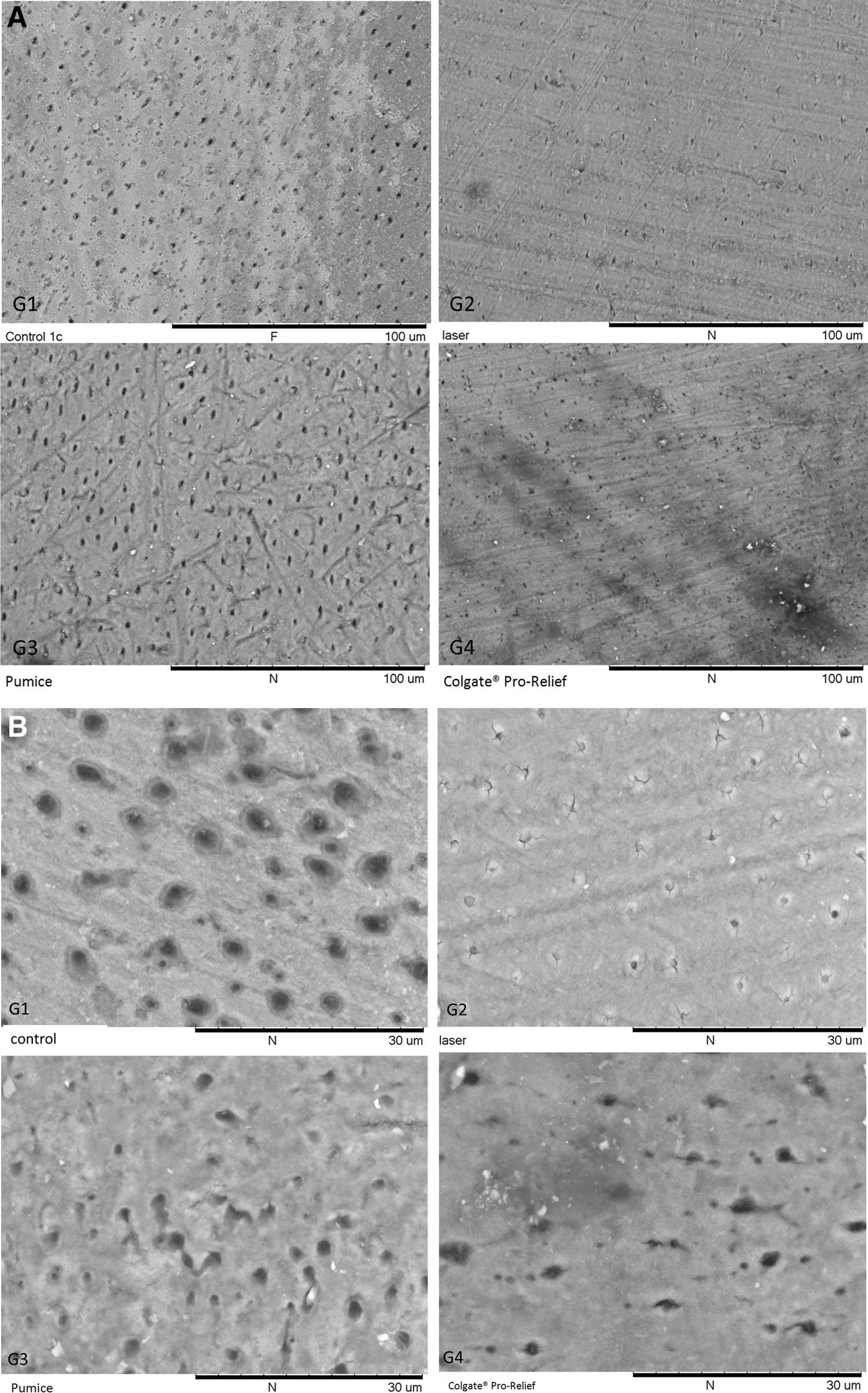
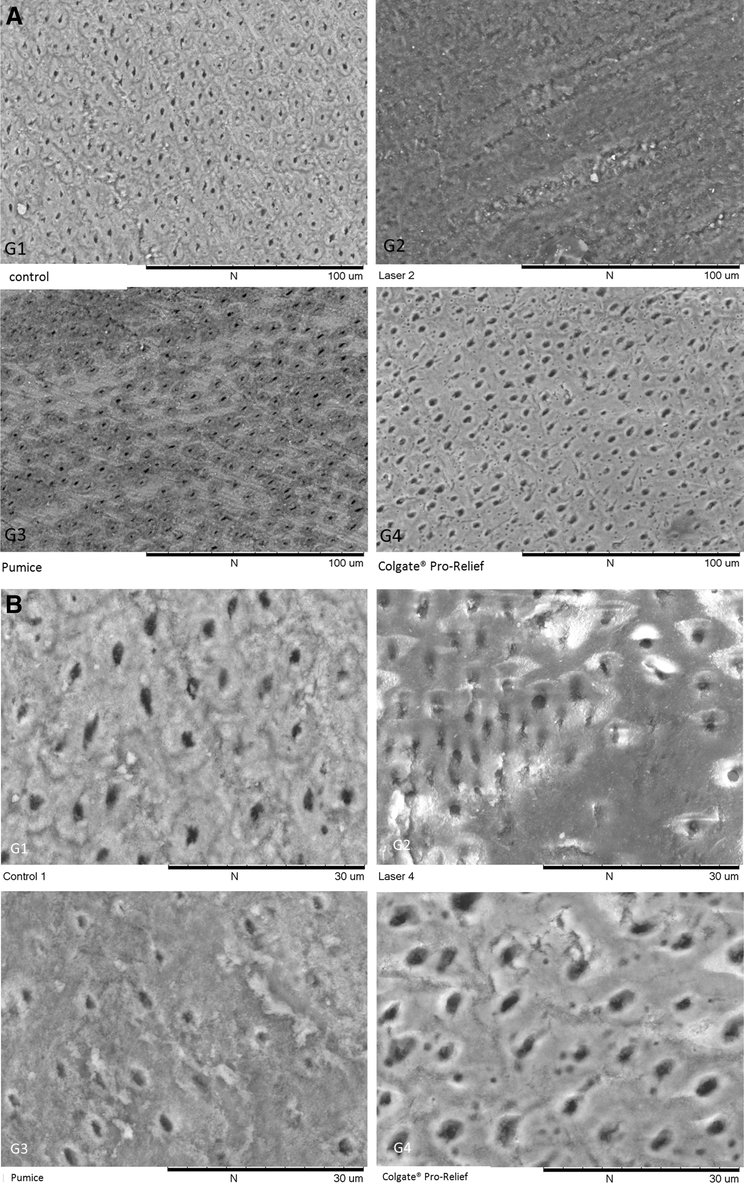
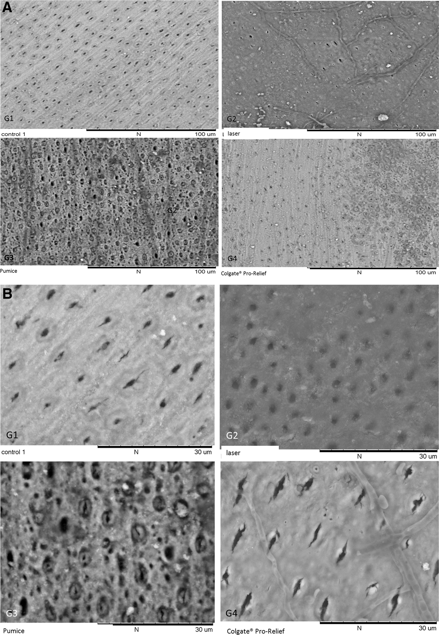
Qualitative assessment of the treated dentin surface micrographs revealed that some tubule occlusion occurred for all groups immediately after treatment (Fig. 2A and B); however, the laser group showed the most satisfactory results (Fig. 2A-G2 and B-G2). A melted and solidified surface was observed. In addition, for this group, the tubules that remained opened presented a smaller diameter than those of the other groups (Fig. 2A-G2 and B-G2). Colgate Pro-Relief sensitive paste showed some degree of tubule occlusion with plugs of the paste observed inside tubules (Fig. 2A-G4 and B-G4). After the erosive/abrasive challenges, the laser group continued with tubule occlusion (Fig. 3A and B). On the 4th day, few opened dentinal tubules could be seen with a smaller diameter; however, opened dentinal tubules were visible for the other groups (Fig. 3A and B). On the 5th day of erosive/abrasive challenges, all groups showed opened dentinal tubules with regular diameter, comparable to the immediate EDTA micrograph (Fig. 4A and B).
(
(
(
Quantitative analysis
Friedman test showed a value of 0.76, which indicates confidence between the examiners. The results of the quantitative analysis are presented in Table 1. The only group that showed a significant reduction in the amount of opened dentin tubules immediately after application of the desensitizing agents in comparison with the control (Group 1) was the laser (Group 2). After 4 days of cycling, there was no significant difference in the number of opened dentin tubules between the groups and the control (p>0.05); however, Group 3 presented significantly lower values than did Group 4. After 5 days of cycling, there was no significant difference in the number of opened dentin tubules among the groups. When comparing the results of each group over the course of time, Groups 1 and 4 presented a significant reduction in the number of opened dentin tubules after 5 days of cycling, but that difference for Groups 2 and 3 was not significant.
In vertical columns, different lower case letters imply significant difference between the groups within time (p<0.05). In horizontal rows, different capital letters imply significant difference among time within a group (p<0.05).
Discussion
The null hypothesis was accepted in this study, as no treatments were able to sustain tubule occlusion after the erosive and abrasive challenges. Similar to previous reports, 27 –32 the Nd:YAG laser irradiated specimens showed a significant occlusion of the dentin tubules immediately after irradiation; however, the number of opened dentin tubules was not significant from the control after the 5th experimental day, implying that, under the conditions of this investigation, the laser was initially effective, but presented no long-term effects after the challenges.
The Nd:YAG laser irradiation has been extensively used for the treatment of DH. 33 –36 The mechanism in which this equipment works is simple and reproducible. The irradiation can melt the hydroxyapatite structure, which, upon cooling, can re-solidify forming hydroxyapatite crystals larger than the initial structure. The result is the formation of a glazed and nonporous surface with occluded dentinal tubule orifices. Dentin has a low value of absorbance at the wavelength of 1064 nm (which is wavelength of the Nd:YAG laser); therefore, the most incident light penetrates into the tooth and may cause an unacceptable increase of the tooth temperature. The use of correct protocols, according to previous studies in the literature, overcomes this difficulty. 11,21 –24,33 –35 Laser protocols may vary depending upon the equipment and diameter of the fiber used, but most of the investigations present protocols in the range of 0.5–1 W in power, 10–15 Hz of repetition rate and 60–150 mJ of energy. Recently, Farmakis et al. showed that the Nd:YAG laser irradiation at 1 W caused more tubule occlusion when compared with the irradiation at 0.5 W, implying that this protocol may lead to a more effective treatment of cervical dentinal hypersensitivity.
In addition to the ability of occluding tubules, Nd:YAG laser irradiation may also have an effect on intradental nerve responses to mechanical stimulation of dentin. This laser is believed to depress intradental nerve responses, similar to low-level laser therapy, by interfering in the sodium pump. 37
The outcomes of this present investigation are in disagreement with the results obtained by Naylor et al. 11 and Gelskey et al. 38 In the study by Naylor et al., dentin tubules occluded by the Nd:YAG laser irradiation remained obliterated even after a subsequent erosive challenge with different beverages. It must be pointed out that the previous investigation did not include an abrasive challenge, as used in the current study; therefore, despite the differences in methodology, it could be speculated that toothbrushing indeed has an impact in the permeability of the dentin tubules and can be considered an important etiological factor of DH. An in vivo study by Gelskey et al. 38 found a significant reduction of DH in 19 patients after Nd:YAG laser irradiation. According to those authors, this effect lasted up to 3 months. Therefore, further in situ and in vivo studies are needed to elucidate the benefits of Nd:YAG laser irradiation on DH.
The Colgate Pro-Relieve® in-office paste formula contains the Pro-Argin™ technology, which is composed of calcium carbonate and arginine, an amino acid naturally found in saliva, which effectively plugs and seals opened dentin tubules based on a natural process of tubule occlusion. 39 The arginine and calcium carbonate are believed to bind to the dentin surface and help attract a calcium-rich layer into the dentin tubules. According to the literature, it provides instant and lasting relief for 4 weeks. 40 –42 However, in the present study, this in-office treatment was not able to significantly reduce the number of opened dentin tubules, in comparison with the control, at all experimental times. A reduction in the number of opened dentin tubules was only observed over time, but this effect was also observed in the control group, and may be a result of the deposition of toothpaste abrasive in the tubules.
The two professionally applied desensitizing treatments used in the present study were selected according to their different mechanisms of action. However, a simple topical prophylaxis, with the use of a rubber cup mounted on a low-speed handpiece and pumice was included, as it is a common procedure performed in the dental office, especially before the application of therapeutic agents. It has been hypothesized that this procedure could reduce dentin permeability with the formation of pumice plugs inside the dentinal tubules and act as a contributing factor for the immediate effect of in-office desensitizing agents. However, in this study, there was no difference between the number of opened dentin tubules in the specimens treated with the pumice in comparison with the control, at all experimental times. According to Al-Saud and Al-Nahedh, 44 the relationship between the surface and intratubular precipitation is not simple. It is important to observe the quality of the deposits, density, and degree of porosity, depth of penetration into tubules, and how the deposits are bound to the dentin surface. Therefore, it may be supposed that the topical agents tested in this in vitro study presented surface deposits that were not firmly bound to the dentinal surface, given the washing procedures performed after the treatments and the dynamic nature of the oral environment also tested throughout the erosive/abrasive challenges.
To simulate the condition of DH with opened dentinal tubules, three different agents were tested: 27% EDTA, 37% phosphoric acid, and a 100 mmol/L acetate buffer solution with calcium and phosphate in equimolar concentrations, proposed by Shellis and Curtis in 2010. 20 According to these authors, the treatments usually used to simulate DH in studies are often aggressive and not only remove the smear layer off the dentin tubules, but also promote a demineralization of the underlying dentin surface, enlarging the tubule diameter by dissolving the peritubular dentin. This would be a matter of concern, especially when testing tubule occluding agents, as the quantification of occlusion would be jeopardized by the lack of peritubular dentin. In view of this fact, the present authors tried to develop a minimally destructive technique to remove the smear layer and open dentin tubules with the use of the acetate buffer solution. However, under the conditions of this in vitro study, it was not possible to observe opened dentin tubules with the use of this solution. As can be observed in the micrographs of Phase 1, the only agent that was able to remove the smear layer or plugs and open the dentin tubules was the 27% EDTA solution (Fig. 1A), which was applied for 2 min, in accordance with a previous study. 11
The SEM equipment used in this investigation presents some advantages over regular equipment, such as the ability to analyze the specimens without previous preparation. It also has a higher magnification range, allowing more detailed observation and better resolution (∼30 nm compared with ∼0.2 μm), and a greater depth of field (>1 mm compared with 2–3 μm), which means that more of the sample can be in focus at the same time, allowing imaging of taller objects. The image also contains compositional and topographical information, if needed, and also permits the load and investigation of larger and taller samples (samples with a diameter of up to 70 mm, and a height of 50 mm).
One of the limitations of this study was the fact that it was not possible to obtain dentin specimens from the same tooth to be distributed among the groups, because of the small size of the roots from human third molars. Although we recognize that this procedure would provide a better standardization of the substrate, efforts were made in order to select dentin specimens with similar amounts of tubules per area. In addition to that, in the quantitative analysis, the dentin tubules with narrow diameter or those that were partially closed had to be considered as open tubules, in order to reduce subjectivity among the examiners. This may have led to underestimation of the protection given by the treatments in some cases. Therefore, caution should be taken to extrapolate the findings of the present investigation to the clinical scenario.
Conclusions
Based on the current results, Nd:YAG laser irradiation was the only treatment capable of occluding dentin tubules immediately after application; however, this effect was not maintained after the erosive/abrasive challenges. It is possible that a combination of in-office techniques or repeated irradiation could show a better effect for dentin tubule occlusion, and should be explored in further studies.
Footnotes
Author Disclosure Statement
No competing financial interests exist.