
Abstract
Introduction
Hard palate wounds can occur directly as a result of various etiologic factors, including tooth extraction, traumatic injuries, and the resection of neoplasms, or indirectly as a result of surgical procedures. 14,15 In particular, during periodontal plastic surgeries and gingival recession treatment procedures, the palatal masticatory mucosa is often injured and widely used as a connective tissue donor source. 16
Based on the information detailed, we aimed to investigate the effects of LLLT on palatal mucoperiosteal wound healing and oxidative stress parameters in experimental diabetic rats.
Materials and Methods
Animals
A total of 42 adult male Wistar rats (250–300 g) from the Department of Medical Science Application and Research Centre of Dicle University were used. In this study, a diabetic state was induced in the rats by an intraperitoneal injection of streptozotocin (STZ, Sigma Chemical Company, St Louis, MO) in 0.1 M citrate buffer, pH 4.5, at a dose of 50 mg/kg. 17 All of the animals were provided with commercial rat chow and water ad libitum, and were maintained on a 12 h light/12 h dark cycle, at a temperature of 22±1°C. The study was performed in accordance with the Helsinki Declaration and with the permission of the Governmental Animal Protection Committee. All of the procedures involved in the experimental protocols were approved by the Animal Ethics Committee of the Dicle University (protocol No: 2010/49). The animals were randomly divided into two groups (n=21) prior to the experiment. Group 1 comprised the control group of diabetic rats that were wounded, and group 2 comprised the experimental group of diabetic rats that were wounded and then treated with laser irradiation. These groups were further divided into three subgroups for euthanasia on the 7th, 14th, and 21st days of wound healing. From each group of seven animals, harvested tissue samples were subjected to histological and biochemical analyses.
Hard palate wound preparation
The surgical procedures were performed with the animals under ketamine HCI (35 mg/kg) and xylazine (3 mg/kg) based anesthesia. On the 7th day after the STZ injection, full-thickness excisional wounds were made on the left side of the mucoperiosteum of the hard palate using a 3 mm biopsy punch. 18 All of the procedures were performed by the same researcher under aseptic conditions. The blood glucose levels of these animals were measured just prior to wounding, using a digital glucometer. The rats exhibiting blood glucose levels >300 mg/dL were considered to be diabetic.
Irradiation protocol
A GaAlAs laser was used for irradiation at a wavelength of 940 nm, an intensity that had been previously calibrated by the manufacturer. The wounds of each experimental group received laser stimulation at doses of 10 J/cm2. The power output was maintained at a constant 0.1 W in the continuous-wave (cw) mode; the spot size was 0.09 cm2. The laser energy was applied via a 400 μm optical fiber. The optical fiber was positioned at a distance of ∼5 mm proximal to the wound, and the irradiation time was 9 sec. Whereas the control group did not receive any irradiation treatment, the experimental group animals received their first dose of irradiation at 2 h after the wounding procedure, and were subsequently irradiated at 2 day intervals following the surgery, for a total of four sessions.
Histological procedure
On the 7th, 14th, and 21st days after the wounding procedures, seven animals from each group were euthanized by cardiac puncture under intraperitoneal anesthesia with ketamine HCI (35 mg/kg) and xylazine (3 mg/kg). Harvested specimens were fixed in 10% formalin for 24 h, decalcified 5% formic acid, dehydrated in graded ethanol baths (100 and 70%), cleared in xylene, embedded in paraffin wax, and serially sectioned at 5 μm. Two sections of each wound were randomly selected and stained with hematoxylin and eosin (H&E) and Masson's trichrome (MT) for evaluation by light microscopy. H&E was used to evaluate the cellular structures (Fig. 1). Fibroblasts and collagen fibers were examined with MT (Fig. 2). The parameters (inflammation, fibroblast proliferation, keratohyalin granules, and vascularization) were scored as follows: 0=absent, 1=mild, 2=moderate, and 3=marked, as previously described by Kirchner et al. 19 The density of collagen fibers was scored according to the following scale: 1=few collagen fibers, 2=few and partially spread collagen fibers, 3=few and fully spread collagen fibers, and 4=dense collagen fibers 20 (Table 1). To avoid observer bias, the histologist was blinded to the study groups, and the data were recorded with respect to the sample codes. The microphotographs of each preparation were acquired using a Nikon Eclipse-400 digital (Coolpix 4500) camera, which was coupled to a standard research microscope.
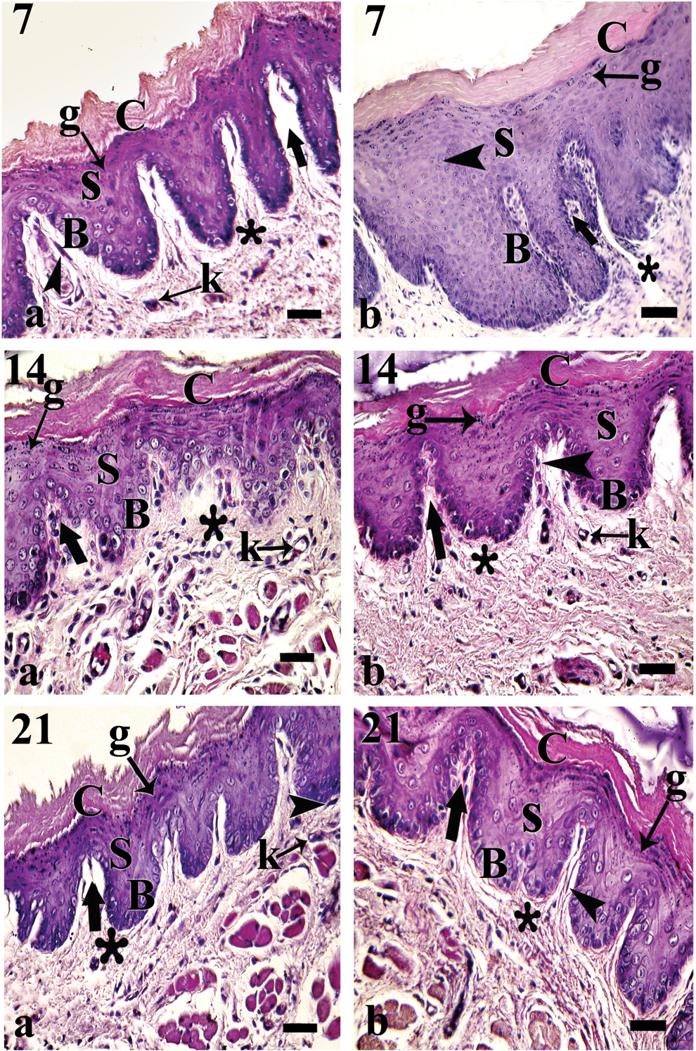
Hematoxylin–eosin (H&E) staining on the 7th, 14th, and 21st day after surgery.
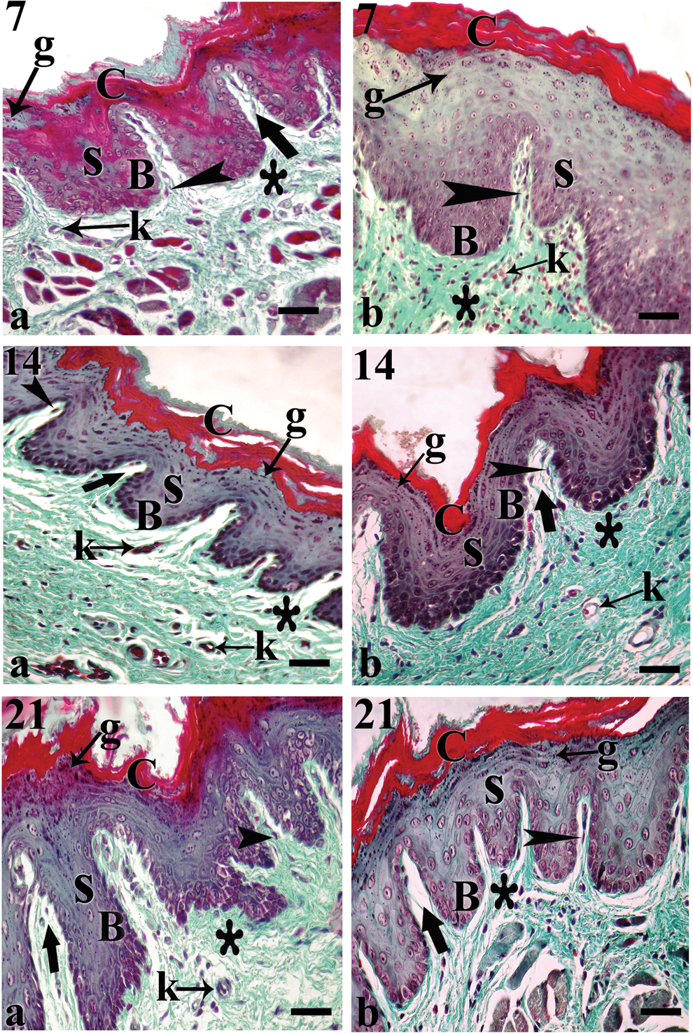
Masson trichrome (MT) staining on the 7th, 14th, and 21st day after surgery.
Scoring: 0=absent; 1=mild; 2=moderate; 3=marked.
Biochemical procedures
The blood samples were collected by cardiac puncture. Each blood sample was immediately centrifuged at 4000 rpm at 4°C for 10 min and analyzed in the central laboratory of the Medical Faculty Hospital. Total antioxidant capacity was measured using Erel's method. 21 This 2,2′-Azino-bis(3-ethylbenzothiazoline-6-sulfonic acid) (ABTSS+)-based method uses colorless, reduced ABTS molecules that are oxidized to a characteristic blue-green ABTSS+ form. When the colored ABTSS+ is mixed with any substance that can be oxidized, it is reduced to its original colorless ABTS form, whereas the reacted substance is oxidized. The total oxidant status was also measured using Erel's method. 22 The oxidants present within the samples oxidized the ferrous ion-o-dianisidine complexes, converting them to ferric ions, a reaction that is increased by glycerol molecules. The conversion to ferric ions yields a colored complex with xylenol orange in an acidic medium.
Statistical analysis
The SPSS 15.0 Windows PC program was used for the statistical analyses. Depending upon the analysis of the data variables, we applied the Friedman test for comparisons within groups, the Wilcoxon test for binary comparisons, and the Mann–Whitney nonparametric statistical test for intergroup comparisons. The data are shown as the mean values±the SD. p Values<0.05 were considered statistically significant. Average semiquantitative evaluation score was presented as mean score±SEM and compared with the nonparametric Kruskal–Wallis test.
Results
Histological results
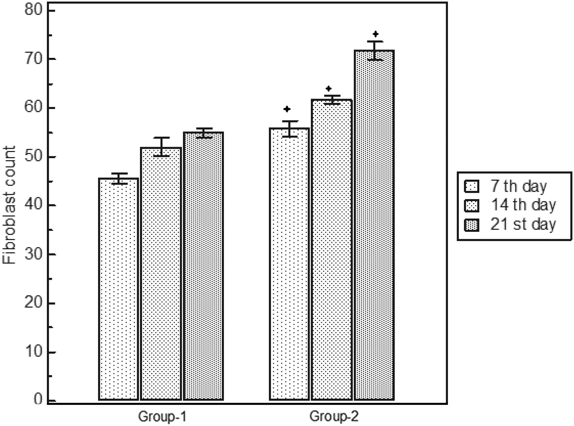
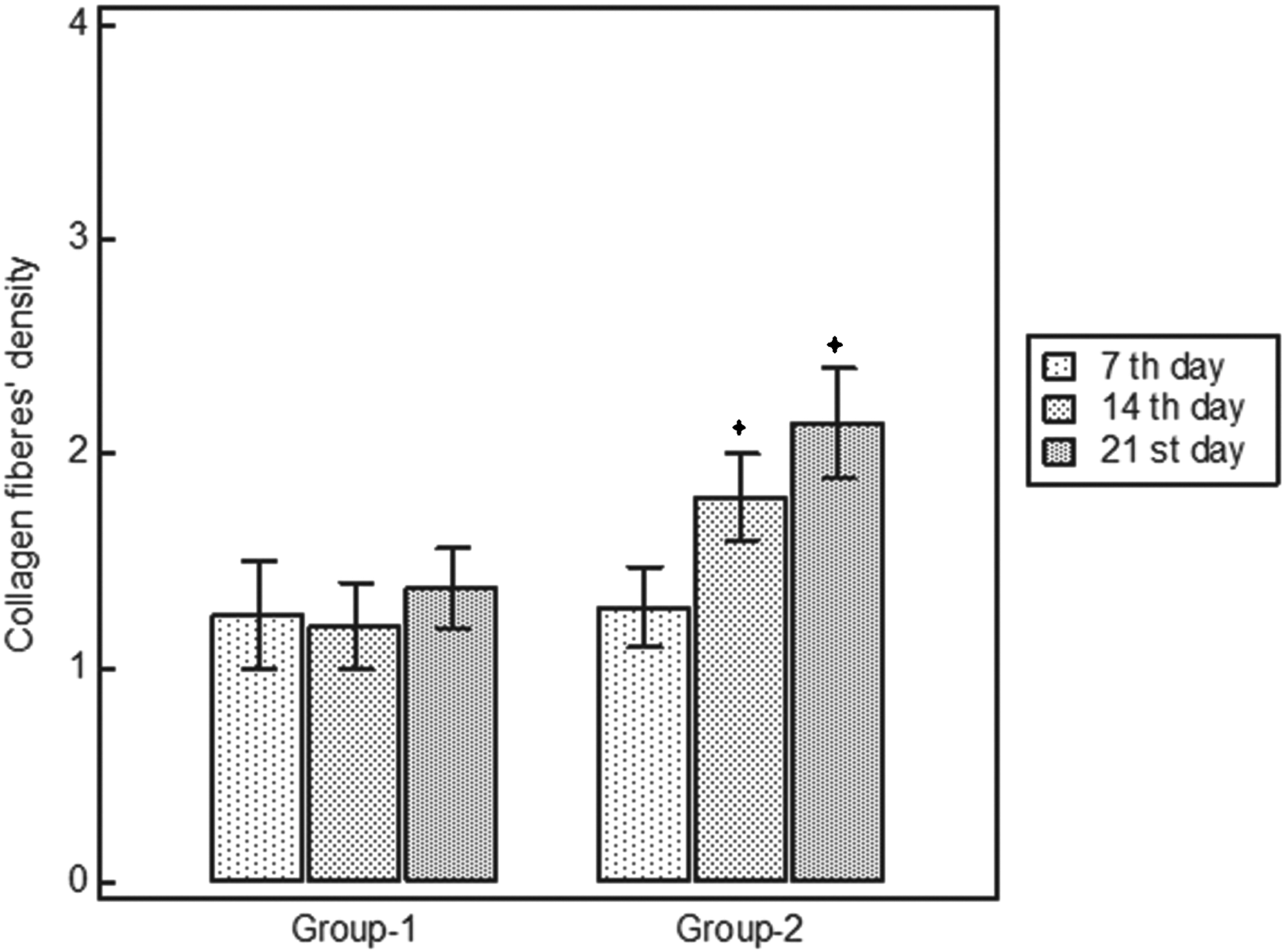
On the 7th day after wounding, the stratum corneum was already formed. but was still immature in both of the groups. Increased mitotic activity in the basal layer, the euchromatic nucleated cells in the spinous layer, and the keratohyaline granules within the granular layer were widely observed compared to the non-irradiated group. Polymorphonuclear neutrophil (PMN) cells were seen in the connective tissue of the groups (Fig. 1 [7a and b]). The fibroblastic activity was increased (Fig. 2 [7b]), the collagen fibers were still in the process of organization, and the same number of blood vessels were exhibited in the irradiated group (Fig.2 [7a and b]).
On the 14th after wounding, irradiated corneum layers exhibited mature structures (Fig. 1 [14b]). Cells within the basal layer showed increased mitotic activity as on the 7th day after wounding, and in the granulosum layer notable accumulation of keratohyaline granules were seen. A reduction was observed in the number of PMNs in group 2 (Fig. 1 [14b]). The connective tissue was characterized by blood vessels somwhat more than the non-irradiated group. In this group, the fibroblastic activity and collagen fibrils also exhibited an increase compared with the control group (Fig. 2 [14a and b]).
On the 21st day after wounding, the corneum layer assumed a mature pattern in both of the group
The semiquantitative histopathological evaluation score at 7, 14, and 21 days after wounding. Asterisk means that this group is statistically different from the control group (p<0.05).
The semiquantitative histopathological evaluation score at 7, 14, and 21 days after wounding. Asterisk means that this group is statistically different from the control group (p<0.05).
Biochemical results
With regard to oxidant and antioxidant status, significant differences were not found between the groups over the experimental period. When we compared the antioxidant status within the groups, we detected a significant decrease on the 21st day after wounding in both of the groups (Group 1: p<0.018; Group 2: p<0.017 [Fig. 5]). The total oxidant status was significantly increased on the 21st day compared with the 7th day after wounding in the Group 1 animals (p<0.028). In Group 2, we determined that the total oxidant status was significantly decreased on the 21st day compared with the 7th day after wounding (p<0.043) (Fig. 6). The biochemical Total Antioxidative Score and Total Oxidant Score are shown in Table 2.
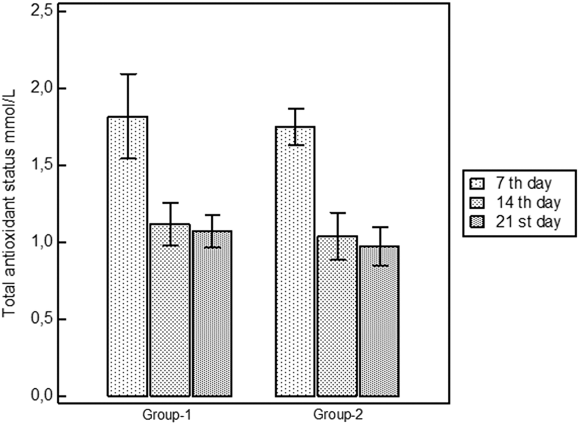
The effect of irradiation on total antioxidant status (TAS) values.
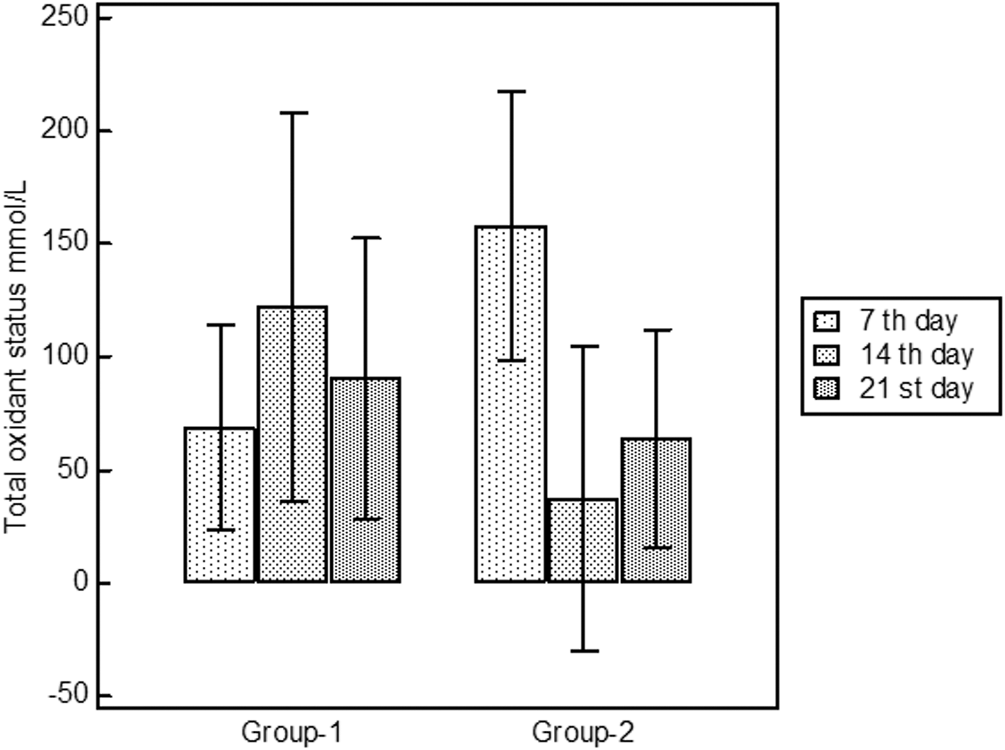
The effect of irradiation on total oxidant status (TOS) values.
TAS, total antioxidant status; TOS, total oxidant status.
Discussion
The biostimulation effect of LLLT on delayed wound healing has generally been described in models using the dorsal skin of diabetic rats. 10,23 –27 However, limited studies have investigated hard palate wound healing. 18,20,28 In contrast, extensive studies have demonstrated the benefits that laser irradiation confer on a wide range of pathological conditions, including wound healing in diabetic patients, when appropriate parameters are used. The significantly improved wound healing that LLLT promotes in both normal 1 and diabetic rats has been demonstrated in numerous studies. 29 –31 For this reason, we aimed to investigate the effects of LLLT (at a 940 nm wavelength and a dose of 10 J/cm2) on palatal mucoperiosteal wound healing in experimental diabetic rats. The laser irradiation parameters used in this study followed some instructions of manufacturer and were based on the literature. 27,29
Al-Watban et al. 29 found that a wavelength of 633 nm at a dose of 10 J/cm2 elicited the most beneficial effects on wound healing in diabetic mice. Rabelo et al. 30 determined a dose of 10 J/cm2 to be beneficial. Güngörmüş et al. 27 also concluded that LLLT (808 nm wavelength at a dose of 10 J/cm2) can have beneficial effects on diabetic wound healing when used at 2-day rather than 5-day intervals. In this study, we found that LLLT (using a 940 nm GaAlAs at 0.1 W and a dose of 10 J/cm2) had a positive effect on diabetic wound healing in rats.
The inflammatory phase of wound healing is characterized by the activation and accumulation of immune cells, particularly by polymorphonuclear leukocytes, within the wounded lesions. 4 Komesu et al. demonstrated a delayed migration of inflammatory cells to the wounded lesions in the experimental group animals compared with the control group animals on the 3rd and 7th day after treatment, by histological examination. 9 Another histological study reported that the number of inflammatory cells appeared to be less in the lesions of the experimental group than in the lesions of the control group in an experimental diabetic wound healing model. 31
In this study, we observed a more significant decrease in inflammatory cells on the 14th day after wounding in the irradiated group than in the non-irradiated group. Therefore, we conclude that the LLLT decreased the inflammatory response during wound healing. 32 Similarly, studies by Fahimipour et al. 20 and de Morais et al. 33 suggested that LLLT inhibits severe inflammatory reactions and promotes increased collagen formation.
Fibroblasts are known to play important roles in epithelialization and the synthesis of collagen tissue during the healing of skin subjected to trauma, including surgical wounds. 34 The use of LLLT has been proposed to promote the biostimulation of fibroblasts and to accelerate the healing process in vitro. 12
In our study, we detected an increase in the mitotic activity of fibroblasts in the non-irradiated group compared with the irradiated group on the 7th, 14th, and 21st days after wounding. Based on these findings, we conclude that LLLT elicits positive effects on diabetic wound healing, consistent with other studies in the literature. 35 –38
Migration of keratinocytes is critical to wound epithelialization. Grossman et al. observed the stimulation of keratinocyte proliferation following exposure to a 780 nm continuous wave (6.5 mW) diode laser at a dose of 0–3.6 J/cm2,39 . We observed the accumulation of dense keratohyaline granules in the irradiated group, at a 940 nm wavelength and a dose of 10 J/cm2.
One of the most important events in wound healing is the synthesis of collagen fibers. Previous studies showed that the synthesis of collagen was impaired or reduced in several diabetic organismal models. 40 Deficient collagen fiber production in diabetic organisms, accompanied by a number of factors, such as the increased apoptosis of fibroblasts, 5 is attributed to the inadequate collagen synthesis capacity of the resident fibroblasts. 41 LLLT has been reported to increase collagen synthesis in diabetic rats in several studies. 26
At the vascular level, treatment using a low-power laser stimulates the proliferation of endothelial cells, resulting in the formation of numerous blood vessels and the increased production of granulation tissues. 23 Importantly, the delayed healing of diabetic wounds is also characterized by impaired angiogenic and vasculogenic responses. 42 In our study, the wounded lesions of the irradiated group exhibited increased collagen synthesis and vasculogenesis compared with the lesions of the control group. Although no animal studies have been conducted using a diabetic rat model to especially examine the effect of the 940 nm GaAlAs diode laser, the findings from the current study are similar to those obtained by using different low-level lasers. 25 –27,29,31
Free radicals play a crucial role in the wound healing process, particularly during the inflammatory phase. 11 Free radicals affect all major compounds, including cellular lipids, proteins, DNA, and carbohydrates, and can cause the deterioration of cellular structure. 43
The results of previous studies have shown that during the normal healing process, although ROS production increases, LLLT can enhance antioxidant enzyme activity, minimizing the oxidative damage incurred by the wound healing process. 11,12
The balance between ROS production and the effect of the antioxidant system is believed to be directly related to healing time and the quality of the wounded tissues. Furthermore, depending upon the dose, the duration of irradiation applied to the wound, and the energy density used, LLLT has been suggested to alter the production of ROS and the antioxidant defense mechanisms. 44 On the other hand, LLLT has been reported to reduce the levels of mitochondria-derived free radicals in epithelial cells and to inhibit the wound-induced respiratory burst in rat neutrophils. 45 In addition, alterations in cellular redox state have been suggested to lead to photobiostimulatory processes. 46 Silveira et al. suggested the possibility that after the LLLT, the biochemical modulations are attributed to a decrease in the extent of the inflammatory responses and the consequent decrease in the production of ROS. 12
The pathological imbalance between the formation of free radicals and the antioxidant system has been reported in diabetic wounds. 10 It is known that low levels of antioxidants accompanied by raised levels of markers of free radical damage play a significant role in delaying wound healing.
Our study revealed a significant increase in the total oxidant status (TOS) values in the non-irradiated group, whereas a decrease in the TOS values was observed in the irradiated group, consistent with Silveira and et al. There was a significant decrease in the total antioxidant status (TAS) value in both of the groups. LLLT seemed successful in reducing the imbalance of TAS/TOS values in diabetic rats. As elevated antioxidant enzyme activity is one part of the host cellular defense system, we can conclude that the decreased TAS level in the irradiated group may be the result of the decrease in oxidative stress. Although LLLT did not enhance the cellular antioxidant status in the experimental diabetic rats, it did promote a reduction in TOS values in our study.
Conclusions
In conclusion, we suggest that LLLT can elicit beneficial effects on the diabetic wound healing process and can modulate the total oxidant status, when using a GaAlAs laser at a wavelength of 940 nm and a dose of 10 J/cm2, likely by decreasing the inflammatory response, inducing fibroblasts and collagen synthesis. The limitations of our study the result of the lack of early time points, and the lack of local tissue assessment.
Footnotes
Author Disclosure Statement
No competing financial interests exist.