
Abstract
Introduction
Although many substances are available to treat dentin hypersensitivity, 6 –9 they have turned out to be ineffective over the long term, and/or studies 10,11 have revealed contradictory results. An ideal desensitizing agent should allow occlusion of dentinal tubules without endangering the pulp, should be relatively painless, easily applied, rapid, and permanently effective, and should not discolor the teeth. 6,7
The results from research regarding the effect of lasers on the treatment of dentin hypersensitivity vary, and so do the irradiation parameters, wavelengths, and application techniques. 12 In some studies, the dentin is irradiated at low energy densities (∼4 J/cm) 13,14 with the aim of stimulating the production of tertiary dentin by the odontoblasts. On the contrary, several studies use higher energy densities in order to provoke a dentinal melting and occlude dentinal tubules, but this practice can induce significant thermal effects, if laser parameters are inadequately controlled. Studies reported that Nd:YAG, Er:YAG, CO2 and diode lasers produce an efficient desensitizing effect; 7,15 –18 however, subsequent further research seems necessary 19 to define the optimal irradiation conditions for harmlessness to pulp and tubule occlusion.
The aim of our study was to evaluate the alterations in dentin irradiated with diode laser beams (810 and 980 nm) at different parameters. We sought to establish the best laser parameter settings to achieve a reduction in the diameter of dentin tubules with the aim of finding a future clinical application for diode lasers in the treatment of dentin hypersensitivity.
Materials and Methods
The approval of the local research ethics committee is not required in our University for this kind of protocol.
Twenty-four caries free adult (18–25 years of age) human impacted wisdom teeth extracted by surgery were kept in balanced salt solution 20 at 4°C during 1 week. The external surfaces were cleaned using a scaler, and, immediately, teeth were sectioned transversely at the mid-level of the crowns at a low speed (300 rpm) using a precision sectioning 20 LC diamond blade (Isomet® Low Speed Saw, Buehler® Ltd., Lake Bluff, IL) in order to totally expose the dentin. The exposed dentinal surfaces of these discs were polished with Soft-Lex discs 3M Espe (coarse-grit disc and medium-grit) using a handpiece speed of 12,000 rpm for 20 sec. Then they were rinsed with cool water and dried with a 5 sec air blast. 21
Each exposed dentinal surface was divided into four quadrants with a 10 mm long, standard grit diamond bur (C4, 10 mm long, standard grit, Crosstech Diamond Instruments Ltd., Thailand) under cooling water.
The smear layer was removed by a 1 min application of 18% ethylenediaminetetraacetic acid (EDTA) (Ultradent Products, Inc, USA). Teeth were rinsed with distilled water and immediately irradiated at different energy densities for each kind of laser.
The first group was irradiated with the 810 nm diode (Elexxion Claros Nano, Germany), whereas we used the 980 nm diode laser (Biolitec, Germany) for the second group. The parameters used for both groups were the following: continuous, tangential, noncontact mode (the distance between the optical fiber and the irradiated surface was 1 mm), delivered energy densities per second 2547, 3184, 5092, and 6366 J/cm2 for the following output power settings: 0.8, 1, 1.6, and 2 W. The optical fiber diameter was 200 μm. Irradiation speed was 1 mm/sec. Specimens were placed on a flat surface; the optical fiber was moved by the operator tangentially (45 degree angle) at ∼1 mm/sec speed. The tangency of irradiation and the speed were controlled and appreciated by the operator with possible human error.
Half of the specimens of each group were stained with a graphite paste obtained by mixing distilled water and fine grain (particle size 25–50 μm) graphite powder (Pressol, Nuremberg, Germany) as an enhancer. Subsequently, these samples were carefully rinsed with distilled water in order to eliminate the residual graphite that could be easily removed because its particle size is larger than average diameter of dentinal tubules.
Scanning electron microscopy (SEM) analysis
The specimens were dehydrated in blue silicon (with a humidity indicator) at room temperature. At that point, they were attached to aluminum stubs and metallized with a layer of gold (25 nm thick), using vacuum evaporation in a metallizer (model SCD 005, Bautec, Berlin, Germany). 22 The samples were analyzed by SEM (Jeol JSM 840-A, Japan) and were observed under 1500x magnification.
Pulp temperature increase measurements
To assess temperature variations, 20 additional teeth were irradiated. Half of them were lased with an 810 nm diode laser and the other half were irradiated by a 980 nm diode laser. Five measurements – with and without graphite paste – were performed per tooth in each group. The irradiation parameters were: 0.8, 1, 1.6, and 2 W. Fiber diameter was 200 μm. Delivered energy densities per second were 2547, 3184, 5092, and 6366 J/cm2. Scanning speed was 1 mm/sec. Continuous and noncontact modes were used for 10 sec.
We followed the protocol used in previous studies for the measurements of pulp temperature increase during laser irradiation. 23 –29 The thickness of the dentin between the exposed dentinal surfaces and the pulp roof was 1 mm, and this measurement was further confirmed by radiography complemented by a millimeter grid.
The pulp chamber was filled with a thermoconductor paste (Prosilican thermal compound: warme Leitpaste WPN 10; Austerlitz electronic, Nuremberg 1, Germany). It was injected by a Lentulo compactor into the cameral pulp cavity to ensure optimal contact and maximal thermal conduction between the sensor tip of the thermocouple probe and the roof of the cameral pulp. The thermal conductivity of the paste amounted to 0.4 cal s−1m−1K−1. This is comparable to the thermal conductivity of soft tissues (0.2 – 0.5 cal s−1m−1K−1 depending upon hydration). 30
The temperature at the roof of the cameral pulp and that of the room were compared in order to record the variations of temperature (Δ T°C).
A type K thermocouple was used (Model TM – 946, 4 channels, Lutron, Taiwan), with an accuracy of 0.01°C. One thermocouple probe was placed in close contact with the roof of the cameral pulp. A second probe was placed at room temperature to compare temperature changes at the roof of the cameral pulp with changes in room temperature.
Once the base pulp temperature became stable after 30 sec, we started measuring the temperature variations. Pulp temperature was recorded every second for 180 sec after the end of the irradiation. Five records were repeated for each irradiation parameter.
The considered temperatures (Δt) were calculated as the difference between recorded temperatures at the roof of the cameral pulp (Tcp) and recorded room temperatures (TRT): Δt=Tcp - TRT.
The mean of recorded temperatures (Δt) and the standard deviation for each irradiation condition were calculated. Normality tests were performed using the Kolmogorov and Smirnov (KS) test.
Results
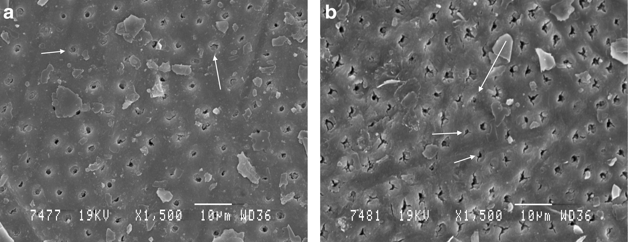
The nonirradiated control group presented open tubules, absence of the smear layer, and a regular aspect, which is a standard pattern of dentin treated with EDTA 29,30 (Fig. 1).
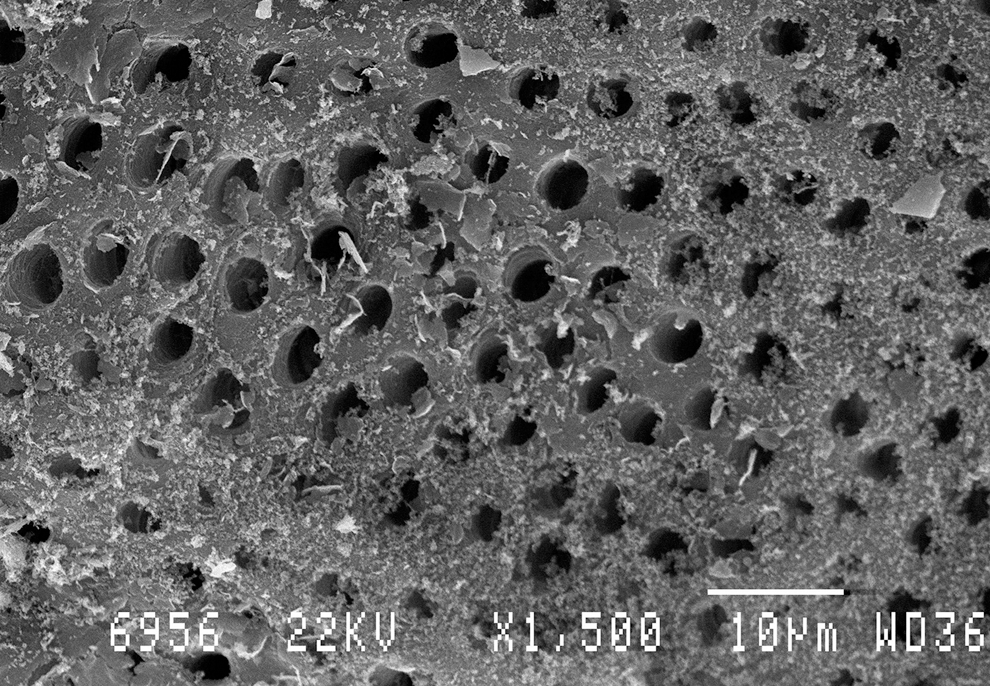
Scanning electron microscopic (SEM) view of unlased dentin (control) treated only with ethylenediaminetetraacetic acid (EDTA) (18%). The dentin is not covered by the smear layer. The tubules are open. Magnification: 1500×.
SEM analysis of the irradiated dentin surface showed surface structural changes caused by laser irradiation.
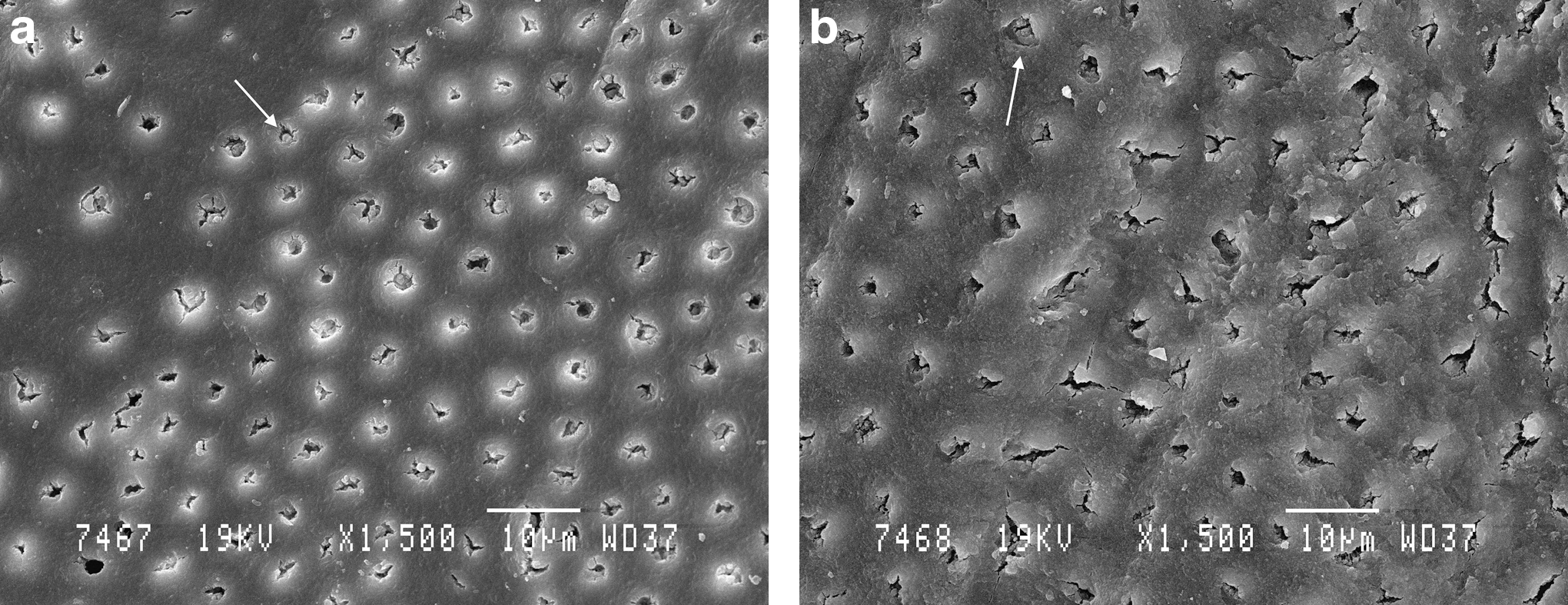
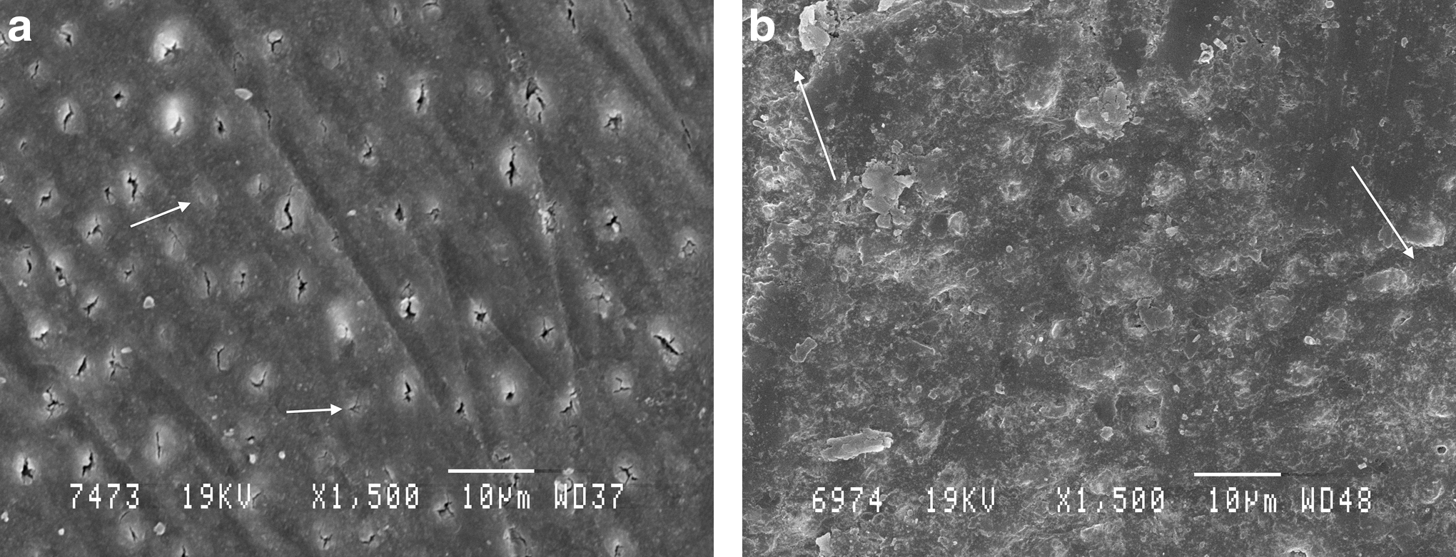
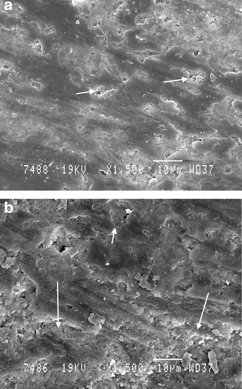
A narrowing of the dentinal tubules was observed at delivered output powers of 0.8–1.6 W for 810 nm diode laser (Figs. 2 and 3) and 0.8–1 W for 980 nm diode laser (Fig. 4).
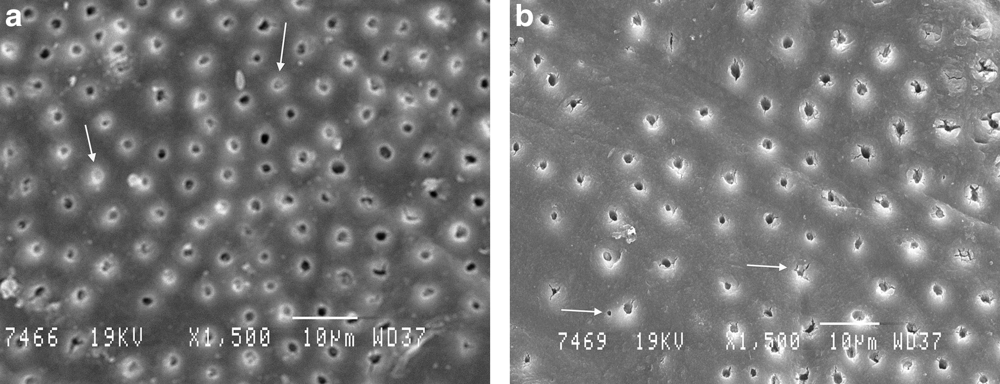
Scanning electron microscopic (SEM) views of treated dentin by diode laser (810 nm) at
Scanning electron microscopic (SEM) views of treated dentin by diode laser (810 nm) at
Scanning electron microscopic (SEM) views of treated dentin by diode laser (980 nm) at
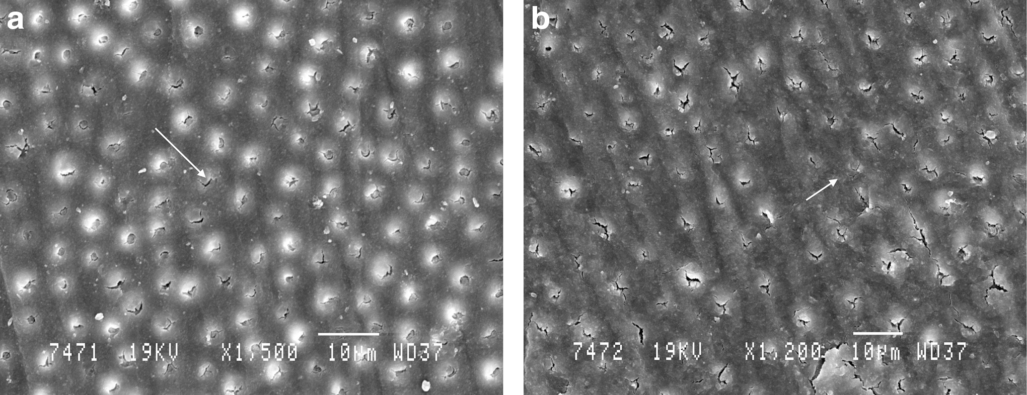
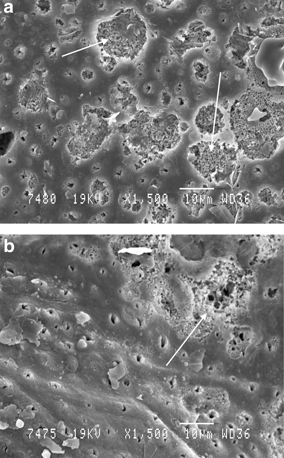
The dentin showed melting areas and a total occlusion of tubules at 2 W for 810 nm diode laser (Fig. 3) and at 1.6–2 W for 980 nm diode laser (Fig. 5). At 2 W, 980 nm diode laser irradiation provoked some areas of dentinal ablation and destruction (Fig. 5).
Scanning electron microscopic (SEM) views of treated dentin by diode laser (980 nm) at
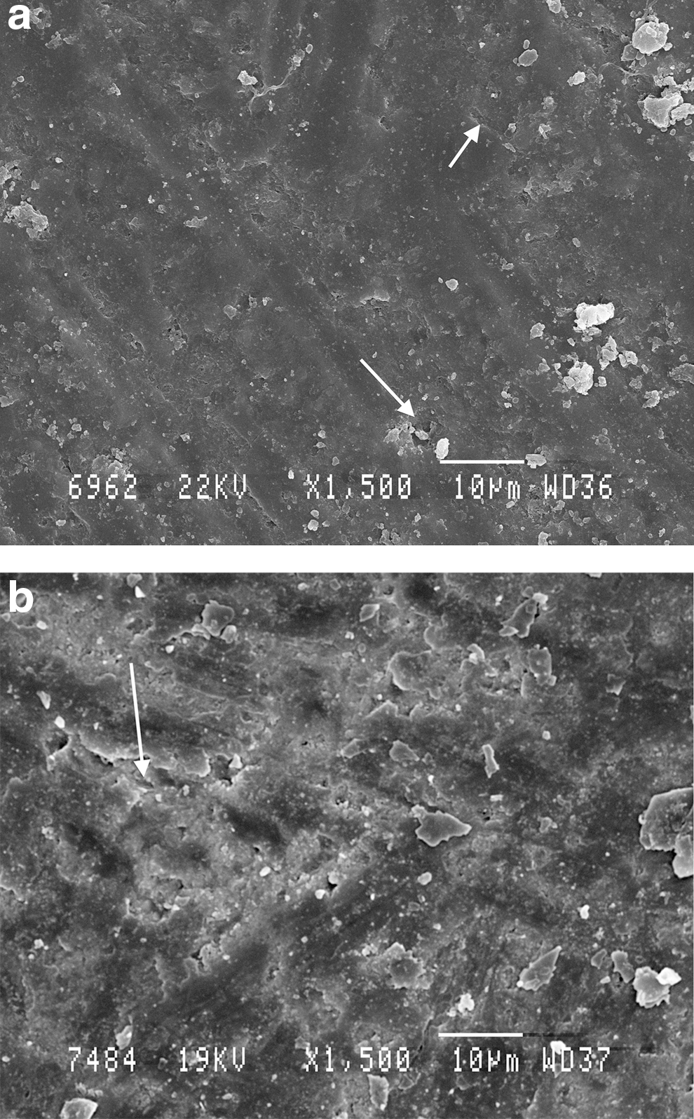
Samples stained with graphite paste
The graphite absorbed the laser beam intensely. This absorption generated an important source of heat and increased the effect of the diode beam. Therefore, the dentin surfac2e presented areas of fusion, melting, and cracks.
At 0.8–1 W, 810 nm diode laser irradiation reduced the diameter of the dentinal tubules (Fig. 6). Higher output powers (1.6 and 2 W) produced dentinal melting, craters, and loss of substance (Fig. 7).
Scanning electron microscopic (SEM) views of treated and graphite-stained dentin by diode laser (810 nm). At
Scanning electron microscopic (SEM) views of treated and graphite-stained dentin by diode laser (810 nm) at
At 0.8–1 W, 980 nm diode laser irradiation, the dentinal surface appeared irregular and scaly with a total occlusion of dentinal tubules (Fig. 8). At higher output powers (1.6 and 2 W), melted dentinal areas with loss of substances were noticed (Fig. 9).
Scanning electron microscopic (SEM) views of treated and graphite-stained dentin by diode laser (980 nm) at
Scanning electron microscopic (SEM) views of treated and graphite-stained dentin by diode laser (980 nm) at
Measurements of temperature
After 10 sec of irradiation (1 mm/sec; power range: 1–2 W), the means and standard deviations of temperature increases at the roof of the cameral pulp were between 0.8–2.30°C and 0.4–1.3°C, respectively, for the irradiation using a diode laser (980 nm) with and without graphite. The cameral pulp temperature rise ranged from 1.7°C to 3.5°C and from 0.9°C to 2°C, respectively, for the diode laser (810 nm) irradiations with and without graphite.
Pulp temperature recordings showed that the samples irradiated by 810 nm diode laser at 2 W (with graphite) presented the highest values, 3.26±0.251°C (higher than the safety level of 3 °C for pulp injury).
Tables 1 and 2 show the mean, minimal, and maximal values of pulp temperature increase for each group.
G, dentinal surface smeared with graphite.
G*, dentinal surface smeared with graphite.
All samples in each group passed the normality test (KS) with a p value>0.05.
Discussion
Diode lasers provide an abundance of available wavelengths in the visible and infrared spectrum. Near infrared (NIR) lasers are characterized by a high absorption in chromophore found in soft tissue. For this study, we selected the 810 and 980 nm wavelengths, the most commonly used wavelengths in dentistry, especially in endodontics and periodontics. 12,31 They can be modulated in continuous wave (CW) or pulsed mode. 12,31
The wavelength of a laser determines its level of absorption and interaction with the tissue. The absorption coefficient is a measure of the level of absorption that occurs in a specific tissue by a specific wavelength. A high absorption coefficient means that less energy is needed to get the same local heating effect. 12 The coefficient μa (cm−1) characterizes the absorption. The absorption coefficients of diode lasers in dental tissues are low: ∼0.1 cm−1 in dentin and 10 cm−1 in the pulp. 32
A laser wavelength of between 800 and 980 nm is poorly absorbed in water and hydroxyapatite. 12,32 This low absorption in dental tissues allows propagation, scattering, or diffused transmission of the laser radiation through the dentin, and important thermal effects. 32 –34 The energy absorbed by the dentin surface provokes a sufficient increase in temperature to obtain a melting effect and reduce or close the dentinal tubules.
In our research, the chosen parameters were selected after prior tests. According to literature, diode lasers are able to seal dentinal tubules in a far lesser degree than other lasers (Er; Cr: YSGG, and CO2) with negligible effects on desensitization. 35 A previous study showed that the irradiation of 980 nm diode laser in dentin at different output powers and delivery modes produced changes that ranged from smear layer removal to dentine fusion. 36,37
Continuous wave mode was employed because it is easier for the operator to scan the whole dentin surface in this way. Nevertheless, pulsed mode can also be useful because it enables the target tissue to cool between successive pulses, but this should be the objective of future studies. We selected the noncontact mode to protect the optical fiber from the graphite paste. The tangentially mode (45 degree angle) was preferred in the aim to avoid a direct pulp exposure by the part of the beam not absorbed by dentin.
The action mechanism of the diode laser (980 nm) in dentin substrate is approximately similar to the Nd:YAG laser (1064 nm). As both systems are in the NIR portion of the electromagnetic spectrum, 38 part of the energy is absorbed by the mineral structures of dentin such as phosphate and carbonate, disarranging the crystalline arrangement because of thermochemical ablation and provoking melting of the dentin tissue. 39,40 These transformations are more intense when higher parameters are used. 41 SEM analysis was used to verify ultrastructural changes of the irradiated dentine.
Oral tissues contain several chromophores: hemoglobin, melanin, and other pigmented proteins and (carbonated) hydroxyapatite. The absorption coefficients for the listed chromophores with regard to the wavelengths used in dental lasers is variable. Generally, pigmented tissues will better absorb visible or NIR wavelengths, whereas unpigmented tissues absorb longer wavelengths. Diode lasers are more absorbed in melanin and other pigments than in dentin.
In the present study, it was found that the diode laser beam absorption could be highly increased in a pigmented surface. We stained half of the samples with a graphite paste (graphite powder and water). The application of graphite paste enhances the effects of the diodes on the dentin surface. It provokes an important increase of temperature to reduce or close the dentinal tubules by a melting effect, but it can also provoke cracks and destruction at highest energy densities.
The wavelength of 980 nm was absorbed the most by water but the 810 nm had a greater absorption in melanin. The higher absorption by dentinal water of the 980 nm diode laser may explain its lower pulp temperature increase compared with the 810 nm diode laser. 42 –44
A 2.5°C temperature threshold for the survival of the pulp tissue was established in classical study of Zach and Cohen. 45 Nowadays, an increase in temperature of 3°C is deemed to be the maximum ceiling to not produce irreversible pulpal damage. 46
On the one hand, the temperature measurements revealed that 810 and 980 nm diode laser irradiation up to 2 W cannot be dangerous to the pulp tissues, but on the other hand, the application of graphite produces temperature elevations, which could exceed the safety level for irradiation by the 810 nm diode laser.
This in vitro study provides an approximate assessment of the temperature increase at the level of the pulp roof. The degree of water content in dentin is certainly different from in in vivo conditions. The laser beam was stopped at the surface of the exposed dentin and the thermocouple was placed at a 1 mm distance from it. The possibility of electromagnetic interference of lasers on the thermocouple is an inherent difficulty of this type of in vitro measurement of temperature increases.
Our preliminary in vitro study aimed to verify the possibility of narrowing or occluding dentinal tubules by means of diode lasers 810 and 980 nm.
Nevertheless, as demonstrated by other authors, 19 further clinical studies need to be conducted in order to confirm these in vitro results before definitive conclusions can be drawn and before use in the treatment of dentin hypersensitivity.
Conclusions
Our results confirmed that 810 and 980 nm diode laser irradiation (0.8–1 W, continuous mode, irradiation speed: 1 mm/sec for 10 sec, laser fiber diameter: 200 μm) can lead to dentinal melting and to the narrowing of dentinal tubules. Higher energy densities (1.6–2 W) produce an important destruction of the dentinal surface and hence damage the dental pulp.
The application of a chromophore (graphite paste) enhances the thermal effects of the diodes on the dentin surface; it increases the areas of fusion and destruction at high energy densities (1.6–2 W).
Author Disclosure Statement
No competing financial interests exist.