
Abstract
Introduction
Materials and Methods
Seven patients with total urinary tract calculi (four bilateral and three unilateral) who presented at our department between May 2007 and September 2012 were treated. Three of the patients with bilateral disease had forgotten stents in both sides of the ureter, and the other patient had benign prostatic hyperplasia (BPH). The three patients with unilateral disease had forgotten stents on the right side of the ureter. B-mode ultrasonics, kidney, ureter and bladder radiographs (KUB radiographs), intravenous urography (IVU), and computed tomography (CT) were performed for all of the patients before the procedure (Table 1).
The age unit is years.
Bilateral, bilateral total urinary tract calculi; Unilateral, unilateral total urinary tract calculi.
Lx/Ry means there are x stones in left kidney/ureter and y stones in the right one.
BPH, benign prostatic hyperplasia.
Patients with bilateral total urinary tract calculi all had vesical calculus, ureteral calculus, and renal lithiasis. The stone burden was defined by multiplying the width of the stones by their length. The mean size of vesical calculus was 3.6×2.9 cm, the mean size of bilateral ureteral calculus was 0.6×0.5 cm, and the mean size of the bilateral renal lithiasis was 2.5×1.8 cm. All three of the cases had mild to moderate hydronephrosis (Fig. 1a).

The other patient with bilateral total urinary tract calculi had had BPH for 8 years. The examinations showed that the patient had second degree hyperplasia of the prostate, and the vesical calculus was 3.0×2.0 cm, the size of the bilateral ureteral calculus was between 0.7×0.5 and 1.0×0.7 cm, and the largest size of the bilateral renal lithiasis was 2.5×2.0 cm.
In patients with unilateral total urinary tract calculi, all of the forgotten stents were in the right ureter. The average indwelling time of the stent was 3.6 years. The examinations showed that many stones had formed around the stents in the bladder, ureter, and kidney. The size of vesical calculus was between 1.2×0.9 and 2.2×1.3 cm, the mean size of the ureteral calculus was 0.5×0.3 cm, and the mean size of the renal calculus was 2.3×1.4 cm (Fig. 2).
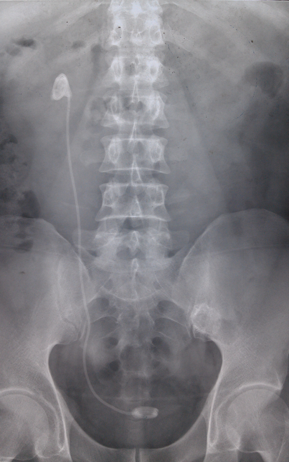
Kidney, ureter, and bladder (KUB) radiograph of a male patient, 41 years old, whose indwelling time of the unilateral stent was 3.5 years. After the operation, we found that many stones formed in the bladder and kidney around the stent, and that some small stones formed along the encrusted stent in the ureter. The largest stone in kidney is 2.4×1.5 cm, the largest stone in the ureter is 0.3×0.3 cm, and the size of stone in bladder is 1.2×0.9 cm.
Combined SWL, URL, and PCNL with holmium laser were performed to render the patients stone-free and stent-free. SWL was the first choice for management for all of the patients, and the operation was performed 1 week later. A 550 μm holmium laser fiber with an energy output of 2–2.5 J at a frequency 8–10 Hz was used in vesical calculus, 0.5–1.2 J at 5–12 Hz was used in ureteral calculus and 1.6–2.4 J at 10–15 Hz was used in renal lithiasis.
We divided the operation into two stages for the patients with bilateral total urinary tract calculi. First, the calcification at bladder and ureteral was managed with ureteroscopy (WOLF, rigid ureteroscope F8/9.8) by holmium laser lithotripsy, without stone basket. All of the calculi that we observed by ureteroscopy were crushed into ≤2 mm fragments, and the mean time of the operation was 2.5 h (Fig. 1b). For patients with BPH, we performed transurethral resection of the prostate (TURP) after the bladder and ureteral calculi were cleaned.
PCNL, standard (F18) and single-tract, was performed to resolve the calcification of the renal end in all the patients with bilateral disease as the second stage of the operation 1 week later. The mean time of the operation was 3 h, and we kept the nephrostomy tube and F7 double J tube after the operation.
For the patients with unilateral total urinary tract calculi 1 week after SWL, URL and PCNL with holmium laser were conducted for the bilateral condition, but the procedure was performed in one stage. The mean total time of the operation was ∼2.5 h.
Results
All patients were stent- and stone-free after the operation, the nephrostomy tube was removed after a mean time of 6 days, and the double J tubes were extracted smoothly using a cystoscope 2 weeks after the operation. The symptoms disappeared after the operation. The Type-B ultrasonic or KUB radiographic images were reviewed to show that the stones were almost free in the seven patients and that the hydronephrosis was visibly relieved (Fig. 1c). Biochemical tests showed that the renal function was improved or recovered.
Discussion
Patients with total urinary tract calculi are relatively rare. Various reports have documented an increased incidence of this problem, especially as a result of a forgotten double J tube in patients with a lithogenic history, and in those with chronic/recurrent stone formation or a metabolic predisposition to stone formation. 2 –8,10 Chronic lower urinary obstructive disease is a predisposing factor as well.
It was the same situation in our report; six out of seven patients had forgotten stents, and the other one had BPH.
It has been well documented that stent indwelling times are strongly and directly related to the incidence of stent encrustation and stone formation. 5,8,10 This point was confirmed in our study. Six of our patients had forgotten stents, and had a stone burden in the kidney, ureter, and bladder. The operation confirmed that the stones were stuck to the stent and formed endoluminally. The treatment for total urinary tract calculi is complex and difficult; therefore, prevention is key to curing the disease. Bultitude et al. 11 proposed that the indwelling time of a double J stent should be no more than 6 weeks (2–6 weeks). Shigemura et al. 12 found from clinical research that the discomfort symptoms could be relieved and the dose of antibiotic used to prevent and treat infections could be reduced if the indwelling time of the double J stent was <2 weeks after ureteroscopy. It is also important that the patient drink more water. 13 Some experts also advocate being stentless after ureteroscopy. In a clinical prospective study, Shao et al. found that routine stenting after ureteroscopic intracorporeal lithotripsy with a holmium laser is not necessary, as long as the procedure is uncomplicated, for distal or middle ureteral calculi <2 cm. 14
Physicians should choose the correct treatment program when stones have formed around the stent, thereby making extraction impossible. The stones should be cleared in multiple steps until the patient can eventually be rendered stent free. Violent extubation is forbidden because of serious complications, such as ureteral mucosal injury, avulsion, or ureteral rupture. 15 Six of the patients in our study presented with stone formation and a trapped stent. The body mass index (BMI) of all of these patients was <25, and some patients had mild to moderate hydronephrosis. SWL is a valuable and successful first-line treatment. Wadhera reported that patients with upper ureteral calculi and mild hydronephrosis can be effectively treated with solo SWL therapy. In patients with moderate hydronephrosis, clearance takes longer or requires secondary interventions, but the cure rate is >80%. 16 This finding is consistent with our choice of SWL for the first treatment. The shocks should be focused on the maximal area of encrustation and stones, resulting in fragmentation of the stones so that they can be separated from the double J tubes. No stones were successfully removed by SWL; therefore, we performed the surgery as the second-line treatment. Ureteroscopy and holmium laser lithotripsy can then be used as the first-step surgery for vesical and ureteral calculus. Safety guidewires should be used during ureteroscopy. None of the nine stents in our patients were successfully removed using this approach. PCNL with holmium laser is used as second-step surgery to remove the remaining stone load, particularly at the kidney and upper ureter. All of the impacted stents were removed successfully by nephrostomy. We kept new F7 double J stents in the ureters and finally inserted the nephrostomy tube. Considering the patients' surgical tolerance and serious complications that may result in long surgery times and a long duration of water pump lavage, we divided the two steps of the surgery into two procedures conducted 1 week apart in the three patients with bilateral total urinary tract calculi. The procedures in the three patients with unilateral total urinary tract calculi were performed in one session. Current clinical research had confirmed that successful management of a retained encrusted stent and the associated stone burden requires combined endourological approaches. PCNL and URL are often necessary and effective for treating a severely encrusted stent and the associated stone burden, 6,17 especially using holmium laser. Holmium laser, as a powerful and effective tool in treatment of stones, has been successfully used in urinary tract stones disease for many years, 18 –22 also including some complicated urinary tract stone disease. For multiple stones, either unilaterally or bilaterally, a single session of ureteroscopy and holmium laser lithotripsy is a favorable treatment option with a high stone-free rate. 23 The high-power holmium laser could reduce lithotripsy time significantly and cure patients with large renal stones effectively and safely, even staghorn calculi. 24 These assertion was corroborated by the successful outcome of our treatment approach.
Urinary tract stones are often caused by chronic lower urinary obstructive disease, and bladder stones associated with BPH are the most common ones. 25 However, it is rare for a patient to present with bilateral total urinary tract calculi with BPH, such as the patient in our study. For this type of patient, we treated the stone first, handling the causes and triggers of stone formation at the same time. SWL was the first-line treatment to fragment the stones and cause them to be partially released into the bladder. Surgery was the second-line treatment, and the procedure was divided into two steps performed 1 week apart. Ureteroscopy and holmium laser lithotripsy were used to address the vesical and ureteral calculus, followed by TURP. We left indwelling double J stents in both sides of the ureters after the surgery. One week later, PCNL was performed for the upper ureteral and renal stones. Many clinical studies have confirmed that the combination of endoscopic lithotripsy and TURP is the ideal treatment for bladder stones with BPH. 26,27 Our patient had bilateral ureteral and renal calculi as well as bladder stones and BPH. Therefore, we added URL and PCNL to the treatment program and obtained good results.
Conclusions
Total urinary tract calculi is a rare and complex condition. It often leads to life-threatening complications and poses a challenging task for the treating surgeon. As minimally invasive treatment modalities such as SWL, URL, and PCNL with holmium laser mature and become more effective, they are among the first choices for the treatment of severe urinary tract calculi. Our findings suggest that ureteroscopic lithotripsy and percutaneous nephrolithotomy with holmium laser could be safely and effectively performed on patients with such a complex stone burden. These approaches have broad potential for application.
We collected seven cases of total urinary tract calculi, which was rare, and treated them successfully and safety with minimally invasive process such as SWL, URL, and PCNL holmium laser step by step. We found that integrated use of SWL, URL, and PCNL could treat such complicated urinary tracts stones effectually, and that holmium laser was a good natural treatment medium.
Footnotes
Author Disclosure Statement
No competing financial interests exist.