
Abstract
Introduction
P
LEDs were developed as an alternative to lasers because they can produce effects in biological tissues similar to those of lasers. 3 Studies have shown the positive effect of LEDs on wound healing. LED therapy has been deemed to pose an insignificant risk by the Food and Drug Administration (FDA), and it has been approved for use in humans. 4
It has been shown that LED application to grafted-extraction sockets could accelerate bone formation, because of increased blood supply. 5 It was thought that the oxygen and nutrient concentrations increase in the healing sockets, thereby supporting the optimal functioning of cells such as fibroblasts and osteoblasts, and new osteoid and bone formation. 5 According to a review of the literature, LED application could have additional benefits on bone healing process following implant therapy. Therefore, in the present study, it was hypothesized that LED stimulation could accelerate the osseointegration process and reduce the waiting time before prosthetic rehabilitation.
When an implant is inserted into the bone, blood cells come into contact with the implant surface and blood monocytes secrete cytokines, growth factor, and prostaglandins. 6 These mediators, especially interleukin-1beta (IL-1β), transforming growth factor-β (TGF-β), and prostaglandin E2 (PGE2) can be detected in the peri-implant crevicular fluid (PICF), and, therefore, this fluid might be useful for evaluating the phases of healing. 7
Nitric oxide (NO) is a regulatory mediator of bone formation and resorption, and it plays an important role in the osseointegration process. NO is essential for the bone response to mechanical stimuli and in the peri-inflammatory process. 8 –10 Additionally, mechanical load on the dental implants has an impact on the NO metabolism and bone remodeling around the dental implants. 11
The aim of this study was to determine the effect of LED PBM on osseointegration after delayed implant placement by measuring the implant stability quotient (ISQ) values and evaluating the IL-1β, TGF-β, PGE2, and NO levels in the PICF during a 3-month period.
Materıals and Methods
Fifteen partially edentulous patients who presented at Gazi University Faculty of Dentistry Department of Periodontology between December 2010 and July 2011 participated in this study. The inclusion criteria were: (1) type 2 or type 3 bone quality according to the classification of Lecholm and Zarb, 12 (2) absence of metabolic bone and systemic diseases, (3) sufficient bone volume and no need for augmentation, (4) tooth extraction > 6 months before surgery, (5) adequate oral hygiene, and (6) the absence of parafunctional habits. Smokers were not excluded from the study (there were two smokers in each group). The patients were randomly assigned into two groups: the LED group (8 patients, 10 implants) received LED treatment after implant placement, and the control group (7 patients, 12 implants) did not receive LED treatment.
The study was initiated after receiving approval from the ethical committee (Ankara University Local Ethical Committee, 2011–18/3). Oral hygiene instructions were provided, and scaling of all of the teeth was performed in each patient. The full-mouth plaque index (PI), the gingival index (GI), bleeding on probing (BOP), and the pocket depth (PD) were recorded before implant surgery. The study schedules are shown in Fig. 1. Before surgery, panoramic radiographs were taken.

Study schedules. LED, light-emitting diode; RFA, resonance frequency analysis; PICF, peri-implant crevicular fluid,
Implants with an appropriate length and diameter were inserted into the bone. The same implant system was used (Xive, Dentsply-Friadent, Mannheim, Germany) in all of the patients, to avoid possible differences in healing and stability values. Before the mucoperiosteal flap was sutured, the primary stability was measured with resonance frequency analysis (RFA) presented by Meredith et al. 13 using the Osstell ISQ device (Integrations Diagnostics AB, Göteborg, Sweden). A Smartpeg™ was inserted manually into the internal connection of the implant using a plastic mount. The plastic probe of the Osstell ISQ device was brought within ∼ 2–3 mm to the top of the peg. The RFA value was measured from two different directions (buccolingual/palatinal and mesiodistal), and one low value and one high value were obtained (Fig. 2). Then, the mean ISQ values for each implant were calculated. These measurements were repeated at 2, 4, 8, and 12 weeks after implant placement.
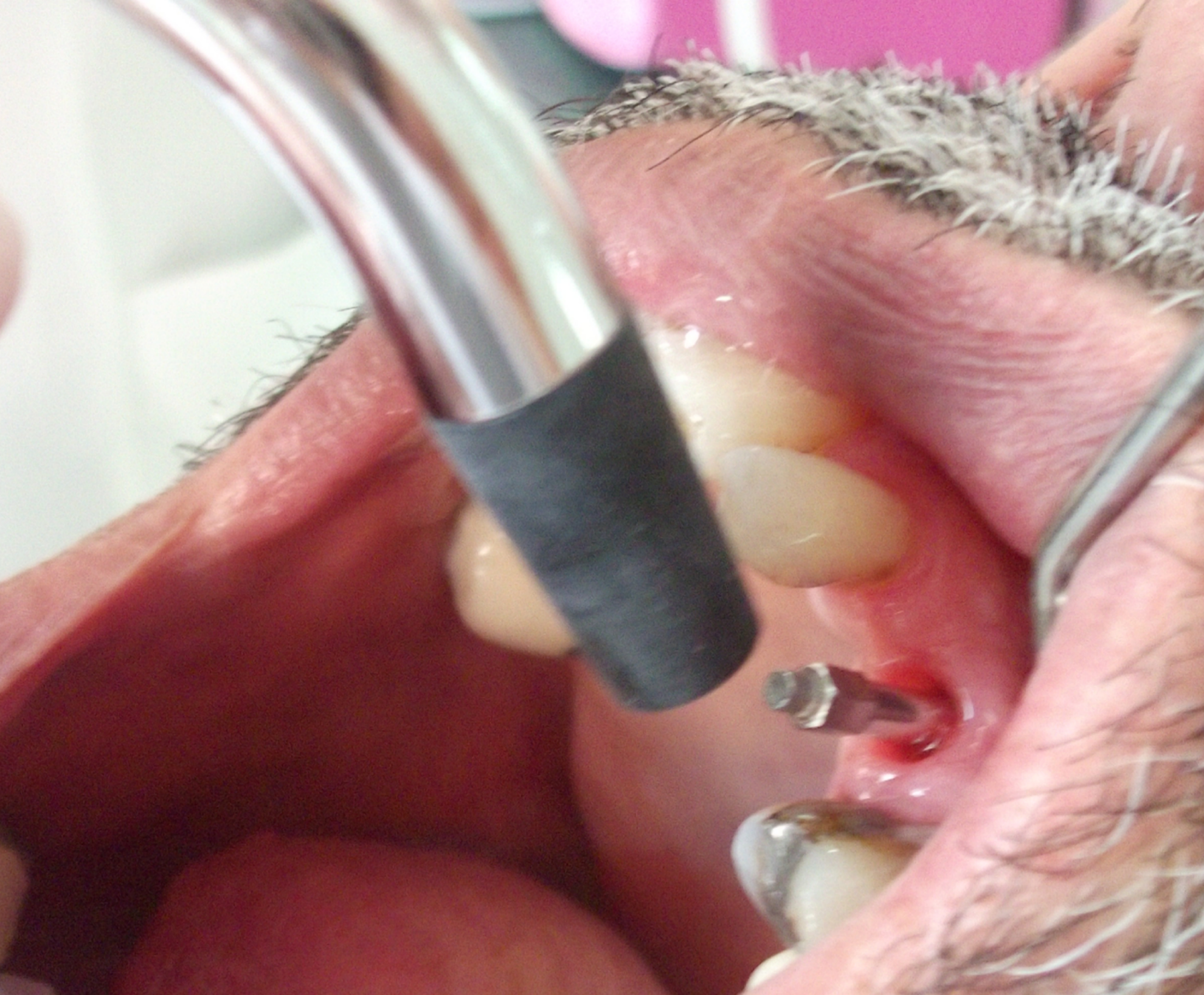
Measuring of implant stability quotient (ISQ) values with Osstell ISQ device.
During surgery, implant diameter, implant length, and bone quality were also recorded. Bone quality was assessed by the operator during drilling of the implant site based on the hand-felt perception of the drilling resistance and according to the Leckholm and Zarb 12 classification.
In the LED group, the LED device (Osseopulse™ AR 300, Biolux Research, Vancouver, British Columbia, Canada) at a wavelength of 626 nm in the NIR region was applied for 20 min over the surgical area after the mucoperiosteal flap was sutured. The treatment parameters were as follows: treatment array area, 4.80 cm2; average intensity, 38.5 mW/cm2; total power, 185 mW; total energy, 222 J; average density, 46.2 J/cm2. The LED device had eight emitters with a power of 23.125 mW each.
The LED device consisted of a headset that was adjusted on the patient's face (Fig. 3). A three point contact on the ears and nose was provided for device stability. The LED application was performed three times per week for 3 weeks during the postoperative period.

Positioning of light-emitting diode (LED) device on the face.
Clinical parameters
At weeks 4 and 12, the PI, 14 , GI, 15 PD, and BOP were recorded at the mesial, distal, buccal, and lingual/palatal aspects of the implants.
Collection of the PICF samples
The PICF samples were collected from each implant before any probing measurements were performed. Strips with any visible signs of saliva and blood contamination were discarded. Filter strips were inserted into the peri-implant crevice for 30 sec (Fig. 4). Each strip was placed in a Periotron 8000 device to determine the PICF volume. The strips were placed into Eppendorf tubes and stored at −20°C until analysis. The volume of the crevicular fluids was measured in “periotron units” and converted into microliters using the MLCONVRT. EXE program.
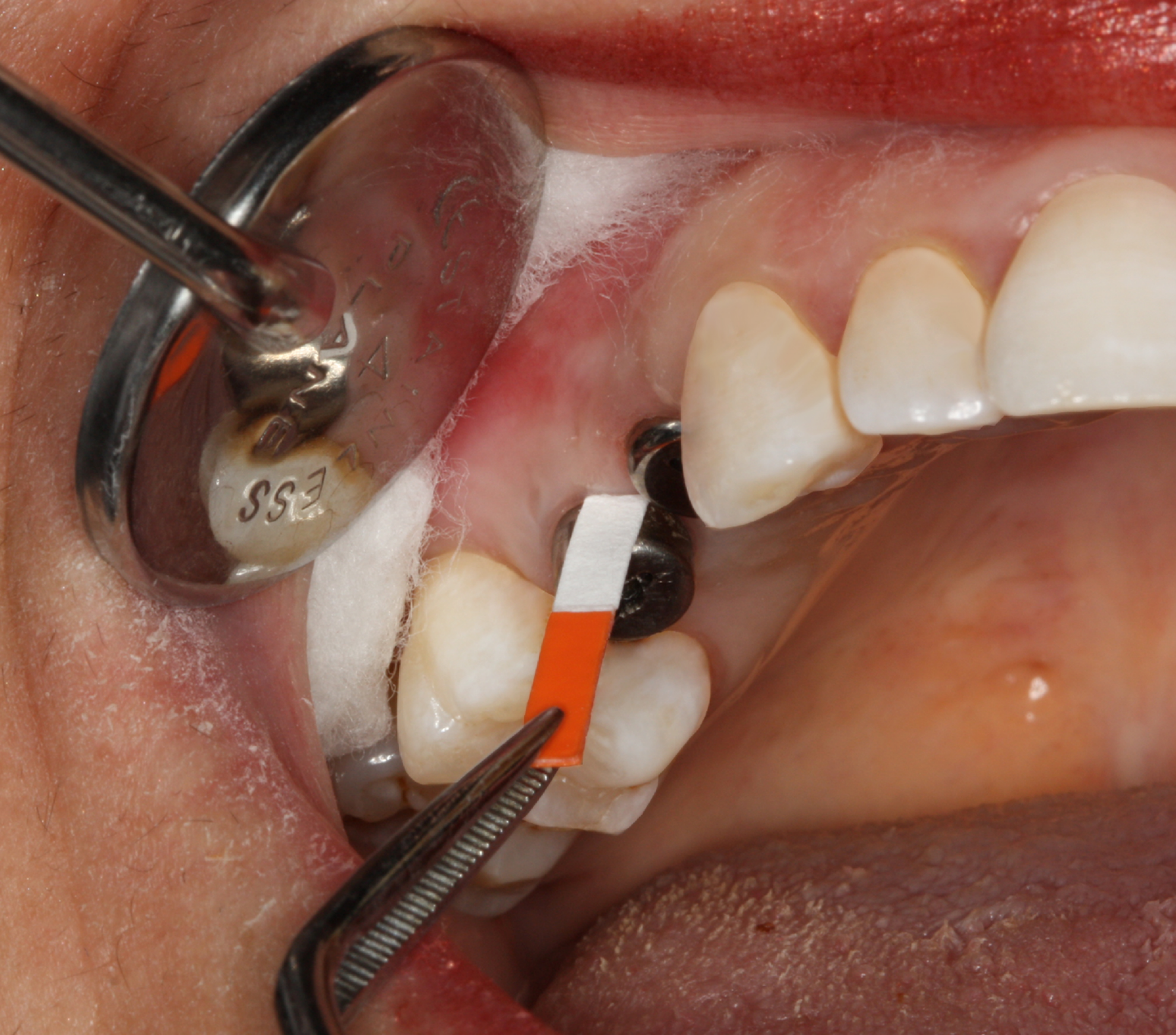
Strip inserted into the peri-implant crevice.
The PICF samples were eluted from the strips by placing them in 1200 μL phosphate- buffered solution and centrifuging at 8600 rpm, +4°C for 5 min. The supernatants were used to determine the levels of IL-1β, TGF-β, NO, and PGE2.
The IL-1β enzyme-linked immunosorbent assay (ELISA) kit (Bendermed Systems, Diagnostics, Vienna, Austria), TGF-β1 Instant ELISA kit (Bendermed Systems, Diagnostics, Vienna, Austria), Prostaglandin E2 EIA Kit Monoclonal (Cayman Chemical Company, Ann Arbor, MI), and Colorimetric Nitric Oxide Assay Kit (Oxford Biomedical Research Inc. Oxford, MI) were used according to the manufacturers' instructions. The total amount (pg) and concentration (pg/mL) of each reagent were calculated.
Statistical analysis
Data analysis was performed using the SPSS for Windows 11.5 package program. The Shapiro–Wilk test was used to determine if the data were close to a normal distribution of the continuous variables. Descriptive statistics for continuous variables are shown as the mean±standard deviation or median (25th–75th percentile), and those for nominal variables are shown as the number of observations and as percentages.
Student's t test was used to investigate the significant difference between the groups in terms of averages, and the Mann–Whitney U test was used to investigate the significant differences between the groups in terms of median values. Nominal variables were evaluated by Fisher's exact χ2 test. Spearman's correlation test was used to determine if there was a significant relationship between the continuous variables.
A dependent t test and Wilcoxon signed rank test were used to evaluate whether there was a significant difference between the measurements at weeks 4 and 12 in both groups. Changes in the ISQ levels were assessed by repeated measures analysis of variance. To determine the follow-up period, in which a significant difference was noted, a Bonferroni correction multiple comparison test was performed.
A p value of < 0.05 was considered to be statistically significant. However, in all of the possible multiple comparisons, a Bonferroni correction was made to control the type I error.
Results
No significant differences were observed in terms of age, sex, smoking habits, and bone type between the control and LED groups (Table 1). Additionally, no differences were demonstrated in the implant length or diameter and the PI, GI, PD, and BOP scores between groups (Table 1).
No significant differences were observed in the changes in the PI, GI, PD, and BOP scores near the implants from week 4 to week 12 between the two groups. The changes in these parameters were also not statistically significant in either groups.
Stability measurement
The stability values in both groups are shown in Table 2. When we compare the ISQ values in the control group between different examination periods, a statistically significant decrease was observed at each interval compared with the baseline (Table 2, p=0.005). The primary ISQ values were maintained in the LED group during the study period (Table 2, p=0.156). However, no significant differences were demonstrated in the ISQ values between the control and LED groups during any of the examination periods.
Difference between baseline and week 2 is statistically significant (p=0.018).
Difference between baseline and week 4 is statistically significant (p<0.001).
Difference between baseline and week 8 is statistically significant (p<0.001).
Difference between baseline and week 12 is statistically significant (p=0.005).
Difference between week 8 and week 12 is statistically significant (p=0.013).
No significant relationship was demonstrated between the changes in the ISQ values and bone quality in either group (p=0.814, p=0.925; control group, LED group, respectively).
Biochemical parameters
There were no significant changes in the IL-1β, TGF-β, PGE2, and NO levels in the control group from week 4 to week 12 (Table 3). In the LED group, the changes in the amounts of PGE2 and TGF-β were not found to be statistically significant (Table 3).
A slight decrease in the amount of IL-1β and an increase in the NO levels were observed in the LED group; however, these changes were not statistically significant (Table 3).
The PICF volume did not show any significant alteration in either group over time (control, LED; p=0.594, p=0.11, respectively, Table 3).
The changes in the level of IL-1β (p=0.139), TGF-β (p=0.978), PGE2 (p=0.582), NO (p=0.251) and PICF volume (p=0.251) were found to be similar between the two groups (Table 3).
There were no significant changes in the IL-1β (control, p=0.859; test, p=0.515), TGF-β (control, p=1.0; test, p=0.169), PGE2 (control, p=0.859; test, p=0.333) and NO (control, p=0.424; test, p=0.333) concentrations in either groups. In addition, none of these parameters showed significant differences in their concentrations between the two groups (Table 4).
Correlations between the parameters
There were no significant correlations between the length (r=−0.587, p=0.045; r=−0.082, p=0.821; control, LED, respectively) and diameter (r=0.583, p=0.047; r=−0.284, p=0.426; control, LED, respectively) of the implants and the baseline ISQ values in either of the groups. According to Bonferroni correction, p<0.025 was considered to be statistically significant.
When correlations between the biochemical and clinical parameters and the ISQ values were evaluated in both groups, the PI score was found to correlate with the ISQ values in the control group (r=0.787, p=0.021, Table 5).
In the LED group, the level of PGE2 correlated negatively with the ISQ values (r=0.709, p=0.022, Table 5). No other significant correlations were found between the biochemical and clinical parameters and the ISQ values in the control group.
Dıscussıon
In the present study, we evaluated whether postoperative LED application has any positive effect on implant stability and healing around implants. It has been demonstrated that LED application could prevent the loss of implant stability until implant loading.
Bone tissues have a high regeneration capacity and an ability to regain their mechanical properties and architecture. 16 However, impaired vascularization around the implant because of osteotomy leads to an increased osteoclastic activity and loss of stability. Brawn and Kwong-Hing 5 reported that the LED therapy accelerated bone formation and graft resorption in grafted extraction sockets when compared with untreated control sockets. Khadra et al. 17 have shown in their in vitro study that the attachment and proliferation of osteoblast-like cells was increased by LED application. In light of these reports, we thought that LED could improve osseointegration and reduce the healing time following implant placement, by evaluating the RFA and the PICF IL-1β, TGF-β, PGE2, and NO levels.
Different methods are currently being used to evaluate osseointegration. RFA presented by Meredith et al. 13 is a noninvasive technique to measure implant stability and osseointegration. Higher values of ISQ indicate higher implant stability. During the healing period, a good correlation was demonstrated between clinical stability assessed by the ISQ values and the biological events leading to osseointegration. 18 Several reports have shown that implant stability decreases from baseline to weeks 2–4. 18 –21 Our findings in the control group are in agreement with the results of these studies, and the lowest ISQ values were recorded at week 4. The primary stability in the control group achieved at the time of surgery could not be maintained during the study period, and a significant reduction was observed. However, a slight nonsignificant decrease in primary stability was observed in the LED group. Primary ISQ values were maintained from baseline up to the 3rd month. Similar results were reported in an animal study indicating that the stability of orthodontic miniscrews was higher when the LED device was applied, whereas lower ISQ values were obtained at the untreated control sites compared with the baseline values. 22 Our results have shown that LED therapy could prevent the loss of stability, and that adequate stability values required for prosthetic rehabilitation could be achieved.
In a previous study, implant stability did not change with time when factors such as smoking habits, implant length, implant diameter, and bone quality were considered. 23 In the present study, the ISQ values were not affected by the implant diameter and length. However, several studies have shown that the ISQ values are influenced by the jaw and the bone quality. 24 –26 Our results did not confirm such an effect; however, we are in agreement with other studies that did not demonstrate any correlation between the ISQ values and bone quality. 18,23,27 Although smoking did not seem to affect our results, smoker subjects might be considered as the limitation of the present study. The effect of laser and LED PBM on the levels of IL-1β, TGF-β, PGE2, and NO has been shown before. 28 –30 To our knowledge, there are no clinical studies on the effect of LED PBM on these biochemical markers in the PICF. Therefore, the evaluation of biochemical markers in the PICF could help us to determine the mechanism of bone healing after implant therapy, and this might support the ISQ values in our results.
An increase in the IL-1β level in the control group was demonstrated in our study. Similar results were reported by Schult-Masgau et al. 31 and Petković et al., 32 who showed that when the healthy situation around the implants was impaired, there was an increase in the IL-1β levels. In the present study, the PI scores around the implants were increased from week 4 to week 12. This increase in the PI score also might have caused an increase in the IL-1β levels. Some previous studies support our findings. 33,34 In contrast to the results obtained in the control group, the IL-1β levels were decreased in the LED group. Choi et al. 35 reported an anti-inflammatory effect of the LED application at 635 nm wavelength that caused the inhibition of IL-6 and IL-8 secretion. Desmet et al. 1 showed a decrease in the IL-1β levels following LED therapy. These findings were in accordance the decreased level of IL-1β in the LED group. This decrease can also be attributed to the proinflammatory effect of LED therapy. In both groups, the TGF- β levels were increased, similarly to the previous studies. 31,36,37 TGF-β levels are increased in soft tissues surrounding the implant following implant therapy. 31 Whelan et al. 29 evaluated wound healing in diabetic mice after LED therapy, and showed that LED therapy induced an increase in theTGF-β levels in the wound area.
In the present study, a negative correlation was found between the PGE2 level and changes in the ISQ levels from baseline to week 12 in the test group, whereas no similar correlation was observed in the control group. A decrease in the PGE2 levels in the LED group might have caused an increase in the ISQ values. The finding of inhibition of PGE2 release by LED therapy in a study by Lim et al. 30 supported the anti-inflammatory effect of LED. This result was in accordance with our findings.
NO in the PICF is involved in bone turnover and the remodeling phase, and also in the peri-inflammatory process. Tözüm et al. 38 evaluated the nitrite levels to study the NO metabolism at 3, 6, 9, and 12 months following implant therapy, and these levels were found to be decreased from month 1 to month 3. In the present study, the NO levels were found to be decreased in the control group in accordance with previous studies. 39 On the contrary, there was a slight nonsignificant increase in the NO levels in the LED group. Barolet reported 4 that when respiratory chain activity was impaired, LED therapy could activate respiration by pumping NO from the cells to the extracellular matrix. This might explain the increase in the PICF NO levels, and support the effect of LED therapy on osseointegration.
In both groups investigated in the present study, a decreased PICF volume suggested that implant health was maintained during the study period. Because there were no inflammatory conditions around the implant, all of the changes in the biochemical markers seemed to reflect the effect of LED therapy only.
Conclusions
LED therapy provided maintenance of implant stability during the 3-month period. The changes in the biochemical parameters suggested that LED therapy has an effect on the tissues surrounding the implant, and that it could affect bone healing positively. LED therapy could prevent loss of stability and hence, it may allow for loading implants during the earlier period.
Footnotes
Author Disclosure Statement
No competing financial interests exist.