
Abstract
Introduction
Laser is now established as a suitable device for the selective and precise removal of caries. Enamel and dentin can easily be removed by using lasers with different wavelengths. 3 When lasers are compared with the high speed drill, they are shown to reduce the loss of healthy enamel and dentin, and present a painless and less fear-producing dental treatment for patients. 4 –8
Erbium-based lasers have been used in dental treatments, and the energy levels of these lasers have made some changes in the structure of the teeth. The changes in the structure of the teeth could help in the mechanical bond, in removing the smear layer, and in opening the dentin tubules. The Erbium-based lasers are able to cut enamel and dentin with the same efficiency and speed as the high speed drills. Another advantage of these lasers is that they stimulate the formation of a secondary dentin, have an antibacterial effect, and allow pain-free cavity preparation and caries removal without the need for anesthetics. 4,5,9
The Erbium, chromium:yttrium–scandium–gallium–garnet (Er,Cr:YSGG) laser system can be used for cutting both enamel and dentin, soft tissue procedures, hemostasis, and coagulation. It has been used for endodontic treatments, removal of the carious lesions, cavity preparation, increasing acid resistance of the enamel, removal of smear layer and debris, and root canal sealing. Some studies have suggested that the Er,Cr:YSGG laser allows a sufficient cutting and ablation with minimal or no thermal damage to adjacent tissue, and does not cause pulpal inflammations. 10 –18 Compared with the conventional high speed drill, a similar Ca/P ratio and Knoop hardness number of the cavity floor were reported when lasers were used in cavity preparations. 10 –18
To date, there have been many investigations aiming to examine the morphology of the resin–dentin interface of various adhesive systems on flat dentin surfaces or cavities prepared with high speed drills in permanent teeth. However, a small number of studies have been made to determine the quality of Er,Cr:YSGG laser-prepared dentin surfaces for composite resin bonding in primary teeth. Therefore, the purpose of this study was to analyze and compare the resin–dentin interface in cavities prepared with Er,Cr:YSGG laser or bur in primary teeth. The tested null hypothesis was that there was no difference between Er,Cr:YSGG and bur preparation in terms of interfacial micromorphology, with or without acid-etching procedure.
Materials and Methods
Extracted human primary molars (n=20) were used in this study. The teeth were stored in distillated water and used within 1 month.
Cavity preparations
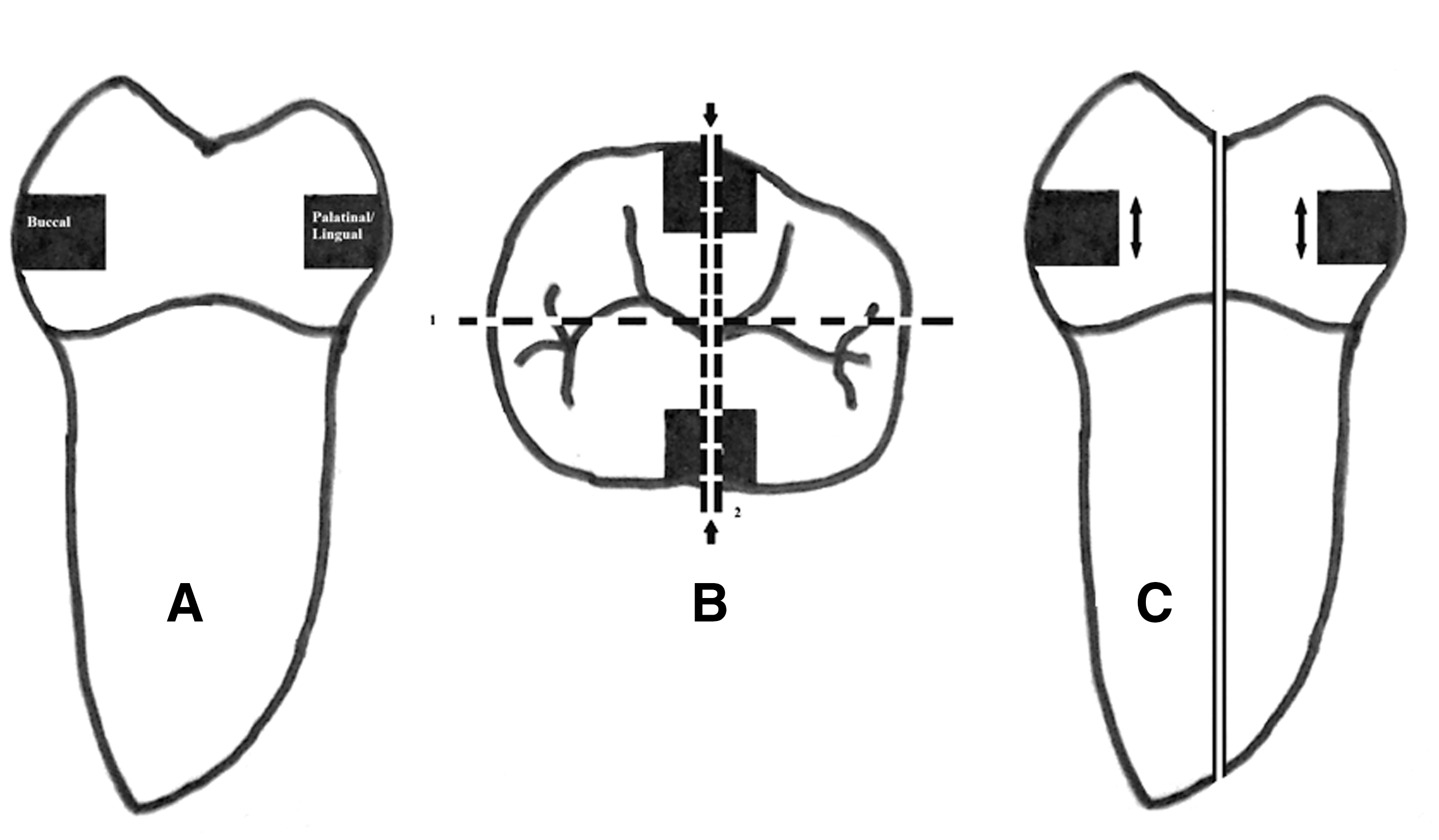
Ten teeth were selected randomly and class V cavities (3×2×1.5 mm) were prepared with Er,Cr:YSGG laser (Waterlase MD, Biolase Technology Inc., San Clemente, CA) on buccal and palatinal/lingual surfaces (Fig. 1A). The power output was set at 6.0 W (85–90% air and 80–85% water) for enamel and 3.5 W (70% air and 65% water) for dentine preparation. Therefore, 10 primary molar teeth were used and 20 cavities were obtained. Laser was used in a defocused mode with a working distance of 1.5–2 mm.
The remaining 10 teeth were used for the bur-preparation group; class V cavities were prepared with diamond bur (Shofu Inc., Kyoto, Japan) as in the laser group. Then, all palatinal/lingual cavities were acid etched, rinsed, and gently dried. Acid etching procedure was not performed on buccal cavities. Therefore, four groups were made.
Group 1 (G1): Er,Cr:YSGG laser
Group 2 (G2): Er,Cr:YSGG laser and acid etching
Group 3 (G3): Bur
Group 4 (G4): Bur and acid etching
Adhesive system and bonding procedures
Single Bond (Adper™ 3M ESPE, St. Paul, MN) was applied to the cavities and light-cured with Hilux Ultra Dental Curing Light (600 mW cm2, 450–520 nm, Benlioglu, Turkey), then a composite resin (Filtek™ Z250, 3M ESPE, St. Paul, MN) were placed to the cavities according to the manufacturer's instructions. The composites were light cured for 20 sec and stored in distilled water for 24 h. The teeth were polished with flexible polishing disks, embedded into acrylic resin. All teeth was sectioned in the x and y direction with a slow speed saw (Mecatome T201 Presi, France) under water cooling to obtain 1×1 mm sticks from the center of the cavities (Fig. 1A–C). The sticks were immersed in ammoniacal silver nitrate solution for 24 h in a dark chamber. At the end of 24 h, the sticks were washed under water for 5 min and placed in photodeveloping solution for 8 h. The sticks were washed with running water for 5 min and stored in distilled water for 1 month at room temperature.
Scanning electron microscopy/ energy dispersive X-ray spectroscopy preparation (SEM-EDX)
After being embedded into acrylic resin, the sticks were polished with wet silicon carbide papers and finished with a diamond paste. After being conditioned with 5% phosphoric acid for 5 sec, the sticks were immersed in ethanol solution for 10 sec, coated with a thin layer of gold, and analyzed in SEM (Jeol 6060, Japan).
Results
In G1 and G2, the surfaces were wavy (Fig. 2), whereas the surfaces were smooth in G3 and G4 (Fig. 3). Microcracks were observed in some of the lased cavities (Fig. 2). In G1 and 2, the dentin tubules were exposed, and the lack of smear layer and gaps was noticeable in the resin–dentin interface (Fig. 4). In G3, there were gaps and a smear layer (Fig. 5), but in G4, there were no smear layer or gaps (Fig. 6).
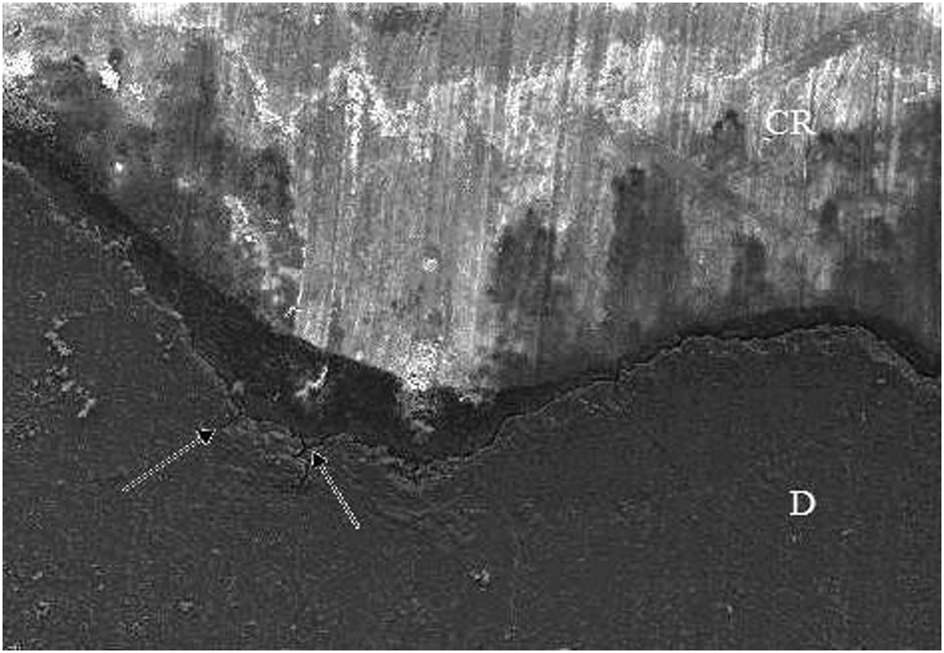
Rugged surface and microcracks were observed in laser cavities (×250). CR, composite resin; D, dentin. Black arrow shows a microcrack.

A smooth surface was observed in a bur cavity (×250). CR, composite resin; D, dentin.
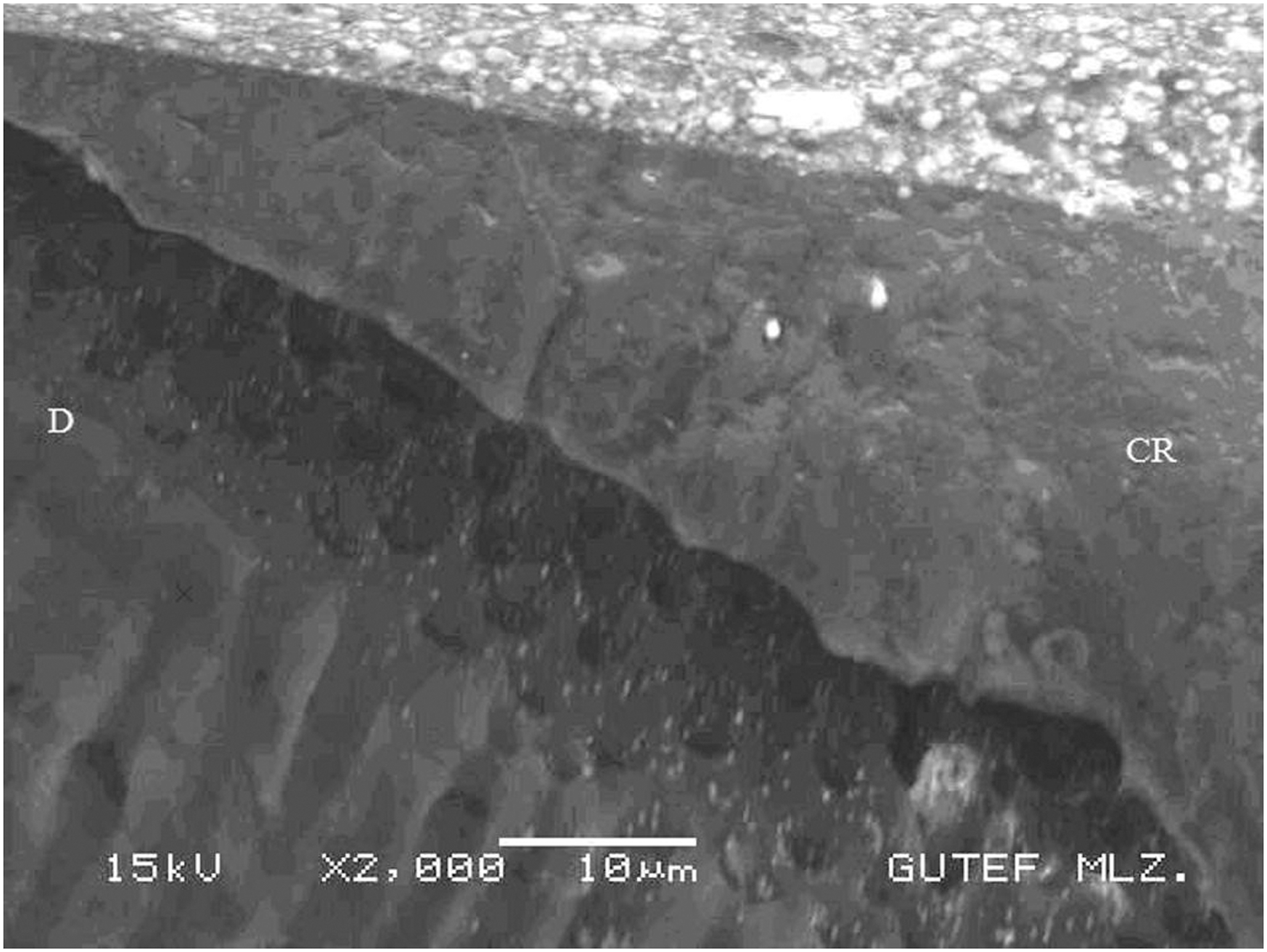
Note the exposed tubules. No smear or gap were observed (×2000). D, dentin; CR, composite resin.

Gaps and smear layer were observed in G3 (×2000). D, dentin; CR, composite resin.
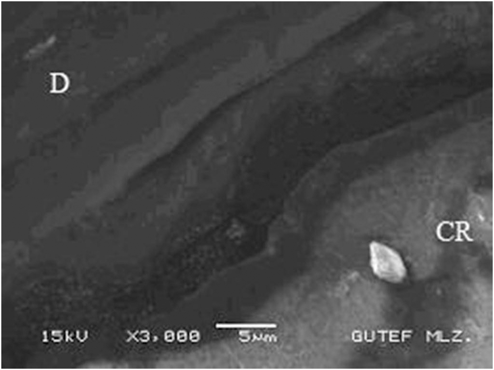
No gaps or smear layer were seen in G4 (×3000). D, dentin; CR, composite resin.
In G2, there were increased resin tags (Fig. 7). In G1, there was a thinner hybrid layer, and in some parts, the absence of the hybrid layer was observed. The hybrid layer was thicker in G2 than in G4. Some resin tags were broken in cavities that were prepared with laser (with or without acid etching) (Fig. 8). In the acid-etched groups (2 and 4), the silver ions were seen not only in the hybrid layer but also in dentin tubules (Fig. 9).

Increased resin tags were noticeable in laser and acid etch group (×3000).
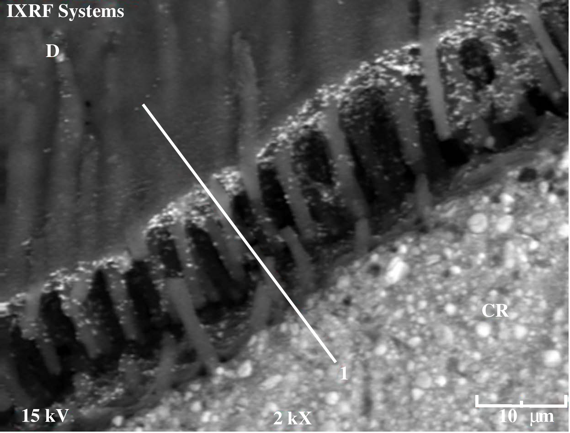
Some of resin tags were broken in G1 and 2. D, dentin; CR, composite resin.
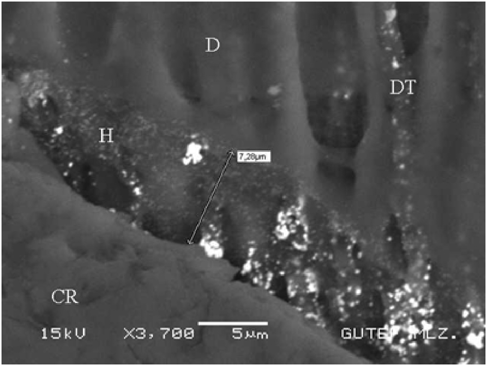
Ag ions were in dentin tubules. D, dentin; H, hybrid layer; DT, dentin tubule; CR, composite resin.
Discussion
The null hypothesis was rejected. There were differences among the groups. Er,Cr:YSGG and bur preparation created differences in interfacial micromorphology regardless of acid etching procedure.
The elapsed time for cavity preparations in the use of the laser was longer than for the high speed drill, which corresponded to other studies. 17,19
The mechanism of Er,Cr:YSGG laser's ablation effect was attributed to the violent microexpansion of water droplets after efficiently absorbing the laser energy, subsequently forming hydrokinetic forces that could quickly ablate the dental hard tissues. 20 The reasons for the energy parameters that were used in this study were based on case studies. When dentin surfaces were irradiated >3.5 W (i.e., 4 or 4.5 W) microcracks were seen under SEM and a frequency of 20 Hz was set prefabricated and could not be changed, which were the reasons for selecting 3.5 W and 20 Hz energy parameters. Second, the roughest surface and the highest ablation efficiency were seen at the 80% air pressure level under SEM, and flattened surfaces were seen in other air pressure levels; therefore, 80% air pressure level was selected. Third, maximal water pressure level was selected to avoid charred or carbonated dentin surfaces. A scaly, irregular and rugged appearance of dentin was observed in G1 and G2 (Fig. 2). Absence of smear layer and exposed dentinal tubules were seen, and melting or carbonation was not seen in the selected energy parameters under SEM. The peritubular dentin protruding from the surrounding intertubular dentin was possibly the result of the higher mineral content and the lower water content of peritubular dentin. 18,21,22 Widened dentin tubule orifices were seen when acid was applied after Er,Cr:YSGG laser irradiation, because of removing the mineral content of the dentin. Flat surfaces were seen after acid etching. 18,23
Previous research has shown that Er,Cr:YSGG laser is an effective device for cutting dental hard tissues, 3,4,7,14,15,17,20 and that the dentin surfaces prepared by Erbium-based lasers are similar in micromorphology. 13 These surfaces were rough, irregular, and there is a lack of smear layer. In agreement with the authors, there was no smear layer, and opened dentin tubules were observed in the lased cavities; the resin–dentin interface was rugged in the laser groups because of the microexplosions produced by the laser. 18,24,25 The irradiation effect of the laser was affected by various factors, such as variations in the energy level, frequency, and time of application, all of which might have caused microcracks in the dentin surfaces. 26
In G3, gaps and a smear layer were seen. However, no gaps or smear layer was observed in G4, possibly related to acid etching.
When single bond was applied to lased cavities, wider, shorter, funnel-shaped resin tags were seen, and they were greater in number when compared with the bur groups.
In the present study, gap formation was observed in the laser groups, and the reason for these gaps could be a result of collagen alteration. Benazzato and Stefani 27 reported that when air–water spray was used after laser ablation, it denatured dentinal collagen fibers in deep regions of dentin, and modified dentinal collagen structurally in the intertubular area. However, in the first portion of the dentinal tubules, collagen seemed to indicate a possible hybrid layer formation. On the other hand, De Munck et al. 28 observed that the adhesives bonded significantly less effectively to lased than to bur-cut enamel/dentin. The authors stated that the laser irradiation could inhibit hybridization, and had negative effects on the dentin/adhesive systems interface. Acid etching hampers the hybridization of dentin because applying acid after laser irradiation inhibits the provision of sufficient exposure of collagen fibers. 24,29 –33
Conclusions
In the present study, laser and bur cavities were performed in primary teeth. Based on the findings of this research, it may be concluded that acid etching was recommended after laser preparations, to have better adhesion.
Footnotes
Author Disclosure Statement
No competing financial interests exist.