
Abstract
Introduction
Laser radiation is absorbed by photoreceptors located in mitochondria, producing a series of changes in cellular processes, such as excitation of the cellular respiratory chain. 9 The stimulation of the respiratory chain alters the redox potential of the cytoplasm, accelerating electron exchange and increasing adenosine triphosphate (ATP) production. Therefore, an increase in cellular metabolism is observed, leading to an increase in DNA synthesis, RNA, and proteins. 1 Additionally, laser irradiation stimulates the fusion of adjacent mitochondria, leading to the formation of giant mitochondria, resulting in the ability to provide higher levels of respiration and energy (ATP) to cells. 10,11
Researche has demonstrated that LLLT applied before or after exercise can significantly attenuate the increase in creatine kinase (CK), enhance muscle antioxidative capacity, reduce the inflammatory reaction after exercise, decrease cell apoptosis, and biomodulate muscle recovery between exercise sessions. 7,12 –18 In this context, many authors have investigated the effects of LLLT on fatigue levels and on physical performance in different populations. 4,8,12 Ferraresi et al. 4 studied the effects of 808 nm laser, with a total energy of 50.4 J, on muscle performance in physical strength training in young male volunteers. The authors observed that laser therapy produced a significant increase in the maximal load supported by the quadriceps in exercised volunteers, and improved muscle performance compared with strength training only. Moreover, Toma et al. 18 demonstrated that LLLT (808 nm wavelength, 100 mW output, energy 7 J) applied on the quadriceps of elderly women was able to increase the number of repetitions of knee flexion-extensions after a protocol of fatigue. Similar results were found by de Almeida et al., 19 using the 808 nm laser, and Leal et al., 20 using the 904 nm laser, who demonstrated decreased post-exercise skeletal muscle damage and inflammation, and significantly delayed muscle fatigue, enhancing skeletal muscle performance in rats.
As mentioned, much evidence showed that LLLT could constitute a promising resource to improve muscle performance associated with physical exercise, 4,18,20 which could be relevant to rehabilitation and sports medicine. 8 In this context, such responses would be important in the improvement of muscle performance, providing fatigue delay, faster lactate removal, increased tissue metabolism, and higher levels of cellular energy. In athletes and the elderly, these benefits would lead to better performance and less occurrence of lesions and muscle pain, in a strength program.
However, there is no consensus in the literature related to the best laser parameters to be applied in association with physical exercise, and there are few data investigating the association between LLLT and resistance exercise training. Therefore, it was hypothesized that LLLT may contribute to increased muscle performance, improving efficiency during a resistance training program in rats. For these reasons, the present study aimed to evaluate the effects of 808 nm laser on biochemical markers and the morphology of skeletal muscle in rats, when applied after a resistance training protocol.
Methods
Experimental design
Thirty male Wistar rats (8 weeks of age and weighing 300–350 g) were used in this study. They were maintained under controlled temperature (22±2°C), with light–dark periods of 12 h, and with free access to water and commercial diet. All animal handling and procedures were strictly conducted according to the Guiding Principles for the Care and Use of Laboratory Animals. The animal experimental plan was reviewed and approved by the Experimental Animal Ethical Committee of the Federal University of São Paulo (0314/10) and the national guidelines for animal care were observed.
Rats were randomly distributed into three groups (n=10 each group): control group (CG), trained group (TG), and trained and laser irradiated group (TLG).
Resistance training protocol and determination of the maximal load
The resistance training protocol was consisted of a climbing exercise using a training support ladder apparatus (1.1×0.18 m, 2 cm grid, 80 degree inclination) with a housing chamber (20×20×20 cm) at the top of the ladder. The load apparatus was fixed on the proximal portion of the tail with a self-adhesive foam strip. A Velcro strap was wrapped around the foam strip and fastened. If necessary, a stimulus with tweezers was applied to the animal's tail to initiate movement. The training started with two familiarization sessions with 24 h between them, without any weight attached to the tails of the animals. These sections had the aim of teaching the rat to climb the ladder, and consisted of three consecutive climbings to the top, with an interval of 60 sec of resting.
One day after this familiarization, the test to determine the maximal load supported by each animal was performed. This consisted of four to eight ladder climbs while carrying progressively heavier loads. For the initial climb, the load carried was 75% of the animal's body mass. After this, an additional 30 g of weight was added, until a load was reached with which the rat could not climb the entire length of the ladder. Failure was determined when the animal could not progress up the ladder after three successive stimuli to the tail. The highest load successfully carried the entire length of the ladder was considered the rat's maximal carrying capacity for that training session (initial maximal load [IML]). The next training session consisted of four ladder climbs with 50%, 75%, 90%, and 100% of the rat's previous maximal carrying capacity, determined in the previous session. During subsequent ladder climbs, an additional 30 g load was added, until a new maximal carrying capacity was determined. 21
This training protocol was repeated three times per week for 5 weeks, totaling 15 sessions of training. After the last treatment session, the test was performed again to determine the final the maximum load (FML) and lactate levels. Forty-eight hours after the last test, the euthanasia of the animals was performed through carbon dioxide asphyxia. The right and left tibialis anterior (TA), blood, and liver were removed for analysis.
Low-level laser irradiation
Laser treatment was performed three times per week, for 15 sessions. A Ga-Al-As laser (Thera laser, DMC Equipamentos, São Carlos, SP, Brazil), 808 nm, 100 mW, continuous wave, 0.028 cm2, 3.57 W/cm2, 33 sec, 120 J/cm2, 3.3. J per point was used.The irradiation was performed after the resistance exercise protocol, at one point in the middle region of the TA belly of both legs, through the punctual contact technique.
Analysis
Evaluation of body mass and maximal load
Rats were weighted at the first and last session of training to measure the body mass. The initial and final maximal load was obtained in the test to determine the maximal load supported by each animal at the beginning and at the end of the protocol.
Lactate blood evaluation
Lactate levels in blood were measured using a lactimeter Accutrend Plus® (Roche-Diagnostics GmbH, Mannheim, Germany). Blood samples were collected from the tail vein of each animal at rest, and at the beginning and end of the experimental period (initial baseline lactate [IBL] and final baseline lactate [FBL]). Similarly, blood samples were taken immediately after the protocol to determine the maximal load, also at the beginning and the end of the experiment (initial peak lactate level [IPL] and final peak lactate level [FPL]). After the collection, blood samples were immediately placed on the lactimeter tape, which was inserted in the lactimeter device for analysis.
Glycogen analysis
Glycogen was extracted from 100 mg of liver and left TA samples collected as described previously. Aliquots were treated for glycogen breakdown, pelleted with ethanol and saturated Na2SO4, and resuspended for final measurement of glycogen determination by colorimetry with phenol and sulfuric acid. The absorbance was acquire by spectrometry at 490 nm (Spectramax®, São Paulo, Brazil).
Histological quantitative analysis
At the end of the experiment, rats were euthanized individually by carbon dioxide asphyxia, and the right TA was removed for analysis. Muscles were immediately washed after removal with saline and then fixed in 10% buffered neutral formalin solution. After fixation, the muscle tissue was processed by embedding it in paraffin. Posteriorly, tissue was sectioned (5 μm) and stained with toluidine blue. For muscle fiber morphometry, the cross-sectional area (CSA) of 100 randomly selected fibers was measured in the middle belly of each TA muscle, using a light microscope (Axioplan 2, Carl Zeiss, Jena, Germany) equipped with a digital camera (AxioCam HRc, Carl Zeiss, Germany) and the AxioVision 4.7 software (Carl Zeiss). 22 A blind procedure was used for measurements.
Statistics
Data are expressed as the mean±standard error of the mean (SEM). Shapiro–Wilk's and Levene's tests were applied to evaluate the normality and homogeneity of the results, respectively. Comparisons among experimental groups were performed by analysis of variance (one way ANOVA), and the Tukey's post-test used to compare individual groups. A p value<0.05 was considered significant. All analyses were performed using a statistical software (v. 6.0).
Results
Body mass evaluation and maximal load
Table 1 shows the data of the body mass evaluation at the beginning and the end of the experiment. There were no significant differences in mass among the experimental groups. Moreover, similar findings were observed in the initial and final maximal load between TG and TLG.
CG, control group; TG, training group; TLG, training and laser group; IBM, initial body mass; FBM, final body mass; IML, inicial maximal load; FML, final maximal load. p≤0.05.
Lactate evaluation
Figure 1 shows the baseline resting lactate levels. Similar findings were observed for all experimental groups before the resistance exercise program. At the end of the experiment, the baseline lactate of CG was significantly higher than for TG and TGL. Furthermore, TGL showed a significantly lower value of lactate levels at rest, at the end of the experiment, than did TG.

Resting lactate evaluation. Initial baseline lactate (IBL); final baseline lactate (FBL). *p≤0.05 vs. trained group (TG); **p≤0.001 vs. control group (CG).
No significant diference was observed in the peak lactate levels between the trained groups, both at the beginning and at the end of the experiment (Fig. 2).
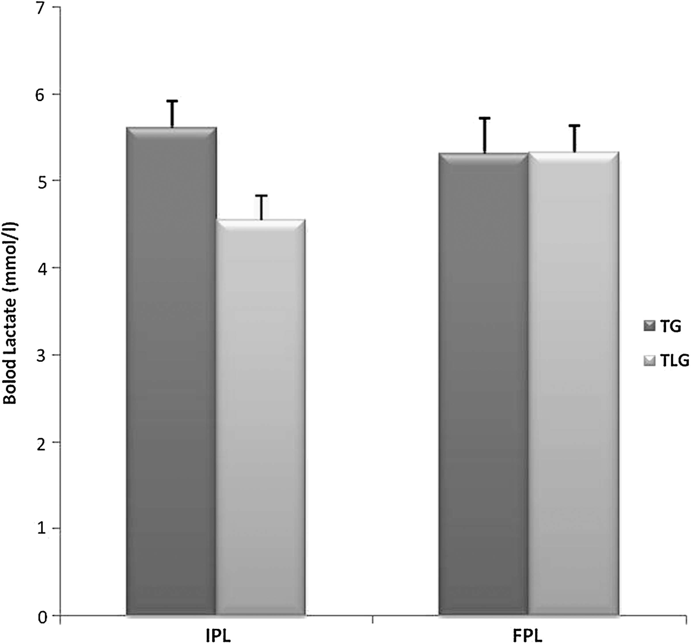
Peak lactate evaluation. Initial peak lactate level (IPL); final peak lactate level (FPL).
Glycogen analysis
A significantly decrease in the muscular glycogen concentration was observed in TG and TGL compared with CG (Fig. 3). Muscular glycogen concentration was higher in TGL than in TG. Furthermore, no significant difference was found in the hepatic glycogen concentration among groups.

Muscle and hepatic glycogen concentrations. *p≤0.05 vs. control group (CG) and † p≤0.05 vs. trained group (TG).
Morphometry of CSA
Morphometric analysis revealed that the resistance training protocol associated with laser irradiation produced a significant increase in the CSA compared with the other groups (Fig. 4).

Morphometry of muscle fiber cross-section area. CG, control group; TG, trained group; TLG, trained and laser group. Histological analysis:
Discussion
This study aimed to evaluate the effects of 808 nm laser on biochemical markers and the morphology of skeletal muscle in an experimental rat model, applied after a resistance training protocol. It was hypothesized that laser irradiation could improve muscle performance in exercised rats. The main findings of this study showed reduction in the resting lactate levels, a decrease in muscle glycogen depletion, and an increase of the CSA of TA muscle fibers in TGL.
Several therapeutic approachs have been investigated to optimize human muscle performance in physical exercise. 16,19 Among them, LLLT may be an effective alternative to increase tissue bioenergetics and the availability of cellular energy, increasing the performance during aerobic exercise. 8 LLLT associated with several physical exercise protocols has shown positive results in delaying skeletal muscle fatigue, decreasing muscle damage, and improving muscle strength. 16,17,19
Blood lactate concentration is one of the most often measured parameters during performance testing in athletes, and is also used as a variable to determine muscle recovery after exercise. 23 It has been suggested that increased levels of serum lactate are associated with an intracellular acidification of skeletal muscle that contributes to muscle fatigue. 21 In the current study, a lower lactate concentration was found in TGL at the end of the experiment, indicating that LLLT was able to improve physical condition, and promoted lactate removal. These findings corroborate those of Leal et al., 17 who found that specific doses of laser irradiation were able to reduce markers of fatigue and muscle damage, improving skeletal muscle recovery in athletes after an exercise protocol. In clinical settings, LLLT also presented positive results, decreasing muscle damage and blood lactate change post-exercise. 11 In addition, De Marchi et al. 5 demonstrated that laser irradiation applied before a progressive-intensity running exercise program decreased CK and lactate dehydrogenase (LDH) enzyme concentration, protecting skeletal muscle against exercise-induced damage and improving muscle performance. The beneficial effects of laser therapy on lactate removal may be related to its positive effects on microcirculation, improvement of mitochondrial respiratory chain, and mitochondrial function, enhancing ATP synthesis. 12
Furthermore, glycogen depletion was measured in this study. Glycogen is the predominant source of glycolysis for ATP production during exercise, and its metabolism has been widely investigated. 23 It is well established that prolonged muscle contraction results in decreased muscle glycogen concentration, 24 which also contributes to muscle fatigue and subsequent decrease in physic performance. 25 Similar results for hepatic glycogen concentration were found for all groups in this current study. Interestingly, the muscular glycogen concentration was higher in TGL than in TG. These results associated with a lower lactate concentration demonstrated that LLLT was able to modulate some of the physiological process related to exercise, and that it might be promising to delay the development of skeletal muscle fatigue and to improve post-exercise recovery.
Furthermore, the CSA was increased in TGL compared with the other groups. Previous studies have already demonstrated that LLLT increased the number of cells per muscle fiber and enlarged muscle fiber diameter in different experimental models. 22,26,27 Ferraresi et al. 4 showed that the association of strength training and LLLT increased muscle volume of the thigh of the dominant limb compared with strength training only. It seems that laser therapy acts on the activation of muscle satellite cells (myogenic regulatory precursor cells) and modulates myogenic transcription factor expression, such as myoD E myogenin, markers of muscle growth and hypertrophy. 28 This led us to infer that the regulation of these events by LLLT may contribute to new muscle fiber formation and promote the increase in muscle hypertrophy in trained and irradiated rats.
As this study was limited to the biochemical marker analysis and muscle morphology, the investigation of cell and molecular pathways involved in the positive action of LLLT in exercised rats remains to be undertaken.
Conclusions
In summary, the results of the present study indicate that LLLT decreased resting lactate concentration and improved muscle fiber morphology, which may have contributed to improvement of muscular performance in the exercised rats compared with the unirradiated animals. Further investigations are required to investigate possible response mechanisms that may explain the positive outcomes obtained when examining laser irradiation after a resistance protocol. Such future studies will undoubtedly contribute to a better understanding of the safety and efficacy of LLLT in sports medicine.
Footnotes
Author Disclosure Statement
No competing financial interests exist.