
Abstract
Introduction
After the water molecules and OH- groups present in the organic components of dental hard tissues absorb the radiated energy scattered by the Er:YAG laser, a sudden increase in temperature occurs. 4 This heating effect vaporizes water molecules, which causes an immediate increase in pressure within dental hard tissues such as enamel and dentin. 4 The resultant volumetric expansion induces microexplosions, with ejection of microparticles of dental hard tissues. 1 –4,26 This process, called ablation, leads to morphological changes in dental hard tissues. 1,4,5,7,20 –24,27
Because of this mechanism of action, for effective results with an Er:YAG laser, water molecules must be present in the field of application. It is well known that an external water supply is as important as the water content of the substrates for efficient ablation with the Er:YAG laser on enamel or dentin surfaces. 28 –30 Without an external water spray, vaporization and dissipation of water molecules will rapidly occur because of the low water content of these tissues. 31 Drying out of the surface of enamel or dentin causes a significant reduction in the ablation efficiency of the Er:YAG laser. 5,26,32 –34 This situation raises the risk of undesirable structural changes such as cracks, 35 carbonization, or melting, 26 in the surrounding tissues. Several researchers have shown that using Er:YAG laser ablation with an external water supply prevents an excessive temperature rise (>5.5°C for 1 min) and the resultant irreversible thermal damage. 36,37 Moreover, controlled temperature increases, which prevent the inadequate bonding associated with fused and recrystallization regions, 32,38 –41 are not harmful for the dental pulp and other tissues adjacent to the irradiated area. 1,42,43
Although the necessity of an external water supply for effective and safe Er:YAG laser ablation of enamel and dentin surfaces is known, 28 –30 the effect of water flow rate on ablation efficiency has not been clearly demonstrated. In the literature, there are conflicting reports concerning the effects of water flow rate on Er:YAG laser ablation efficiency. 5,29,30,32,36,38,44 Whereas Hibst and Keller 36 suggested a 1–2 mL/min water flow rate for efficient and safe ablation with the Er:YAG laser at the power energy setting of 150–250 mJ (0.3–1 W), Armengol et al. 38 reported that a water flow rate of 1.4 mL/min at the power settings of 4 Hz and 140 mJ provided adequate ablation without any thermal damage. According to Kim et al. 32 a water flow rate of 6.75 mL/min yielded better ablation efficiency of the enamel surface than that obtained with other tested water flow rates. Coluci et al. 30 stated that the water flow rate used with the Er:YAG laser had no effect on the shear bond strength (SBS) of composite resin. Therefore, the aim of the present study was to evaluate the effect of water flow rate on enamel surface roughness and SBS of orthodontic brackets after Er:YAG laser ablation. The null hypothesis was that there was no difference between different water flow rates with respect to enamel surface roughness and SBS of orthodontic brackets.
Materials and Methods
Eighty sound human upper premolar teeth, extracted for orthodontic reasons, were used in this study. Teeth with structural defects on the buccal surface, such as caries, fissuring, macroscopic fractures, or cracks, attritions, any restorations, abrasions, or staining, were excluded. All teeth were stored at room temperature in distilled water containing 0.2% thymol to inhibit microbial growth for ∼2 months. After the teeth were cleaned with rubber cups, they were polished with fluoride-free pumice. Before embedding the teeth in a self-curing acrylic resin block, they were washed and dried with an oil-free air spray.
The buccal surfaces of teeth in group 1 (control) were etched with 37% phosphoric acid gel (3M Unitek, Monrovia, CA) for 20 sec, rinsed with a water spray for 15 sec, and dried with a moisture- and oil-free air spray until a dull, frosty appearance was obtained on the enamel surface.
The laser-irradiated groups (2, 3, 4) were classified according to the water flow rate used during Er:YAG laser irradiation. The buccal surfaces of the teeth, where the bracket base would be placed, in each laser-treatment group were uniformly irradiated with the Er:YAG laser (Light Walker, Fotona, Slovenia) at power settings of 1.2 W, 10 Hz, 120 mJ, and 100 μs. The beam spot size was 0.63 mm2, energy density was 19 J/cm2, and power density was 190.4 W/cm2. The laser was applied with a noncontact handpiece parallel to the occlusal surface, and positioned 8 mm from the buccal surface of the teeth. The water flow rates in the three laser-irradiated groups (2, 3, 4) were 25, 36, and 48 mL/min, respectively (Table 1). After laser irradiation, all enamel surfaces were dried with a moisture- and oil-free air spray.
One specimen from each different surface treatment modality was used for visual observation of the differences in enamel surfaces by scanning electron microscopy (SEM) (Jeol JSM 6390 LV, Tokyo; Japan) at × 1000 magnification. Ten observers performed the evaluations of SEM images in a blinded manner.
For all groups, a thin layer of Transbond XT (3M Unitek) light-cured adhesive primer was applied to the enamel surface. Seventy-six upper right first premolar metal brackets (Master series; American Orthodontics, Sheboygan, WI) were used in the present study. The surface area of the bracket base was 10.27 mm2. The bracket, with Transbond XT paste on its base, was placed to the center of the middle third of the buccal surface. A sharp scaler was used to remove resin remnants around brackets. A light-emitting diode (LED) (Demi, Kerr, Orange, CA) was used for 20 sec to polymerize the composite beneath the brackets.
All teeth were stored in distilled water at 37°C for 24 h and thermocycled for 500 cycles (5–55°C), with a dwell time of 30 sec. A Universal Testing Machine (AGS-X; Shimadzu, Japan) at a constant crosshead speed of 1 mm/min was used to record the SBS of brackets bonded onto the different-conditioned enamel surfaces, until failure occurred. The buccal surfaces of all teeth embedded in self-curing acrylic were oriented with the testing machine such that tensile force was applied to the bracket in parallel to the long axis of the teeth. The SBS values of all brackets were recorded in megapascals (MPa).
Composite resin remnants on the enamel surface after shear testing were examined with a light stereomicroscope (M165C; Leica Microsystems, Wetzler, Germany) at a magnification of × 10 to determine the fracture pattern in each group. The patterns were then classified into four types according to the amount of composite remaining (0=no composite left on enamel surface, 1=≤ 50% of the composite left on the enamel surface, 2=> 50% of the composite left on the enamel surface, 3=all composite left on the enamel surface) (Table 2).
Statistical analysis
The SPSS version 14.0 for Windows (SPSS Inc., Chicago, IL) was used for all statistical analyses. Tests of homogeneity of variances were performed with Levene's test. Because the results of Levene's test determined that the data had a nonhomogeneous distribution, the Kruskal–Wallis test was used to compare the SBS values of each group, and p<0.01 was considered statistically significant.
Results
The results of the Kruskal–Wallis test indicated that there were no statistically significant differences among groups (p=0.630). The SBS values of all groups were very close. The mean SBS value of group 4 was the highest (13.94±4.1 MPa) among the tested groups (Table 3). The SBS values of groups 1, 2, and 3 were 13.77±6.5, 12.14±5.4, and 13.28±2.2 MPa, respectively (Table 3).
Group 1 served as control.
p>0.01 based on Kruskall–Wallis test.
Fracture patterns of all groups are shown in Table 2. The type 2 fracture pattern was found in 60% of the specimens in group 1; the same type was found in 40%, 50%, and 65% of the samples in groups 2, 3, and 4, respectively.
The SEM photomicrographs of enamel surfaces for each group are presented in Fig. 1A–E. Figure 1A shows the enamel surface of an untreated tooth. Figure 1B shows the characteristic appearance of an enamel surface classified as a type 1 etching pattern per Silverstone et al., 45 with the holes in the center of the enamel prism after 37% phosphoric acid etching. The etching pattern in group 2 was similar to that in type 3 (Fig. 1C), with damage patterns unrelated to enamel prism structure, 45 microcracks, and a more irregular surface pattern on the enamel surface than in the other laser-irradiated groups, with a water flow rate of 25 mL/min. In groups 3 and 4, the etching patterns after laser irradiation were similar to those in type 1, with the exception of more irregular and nonhomogeneous holes in the center of enamel prisms.
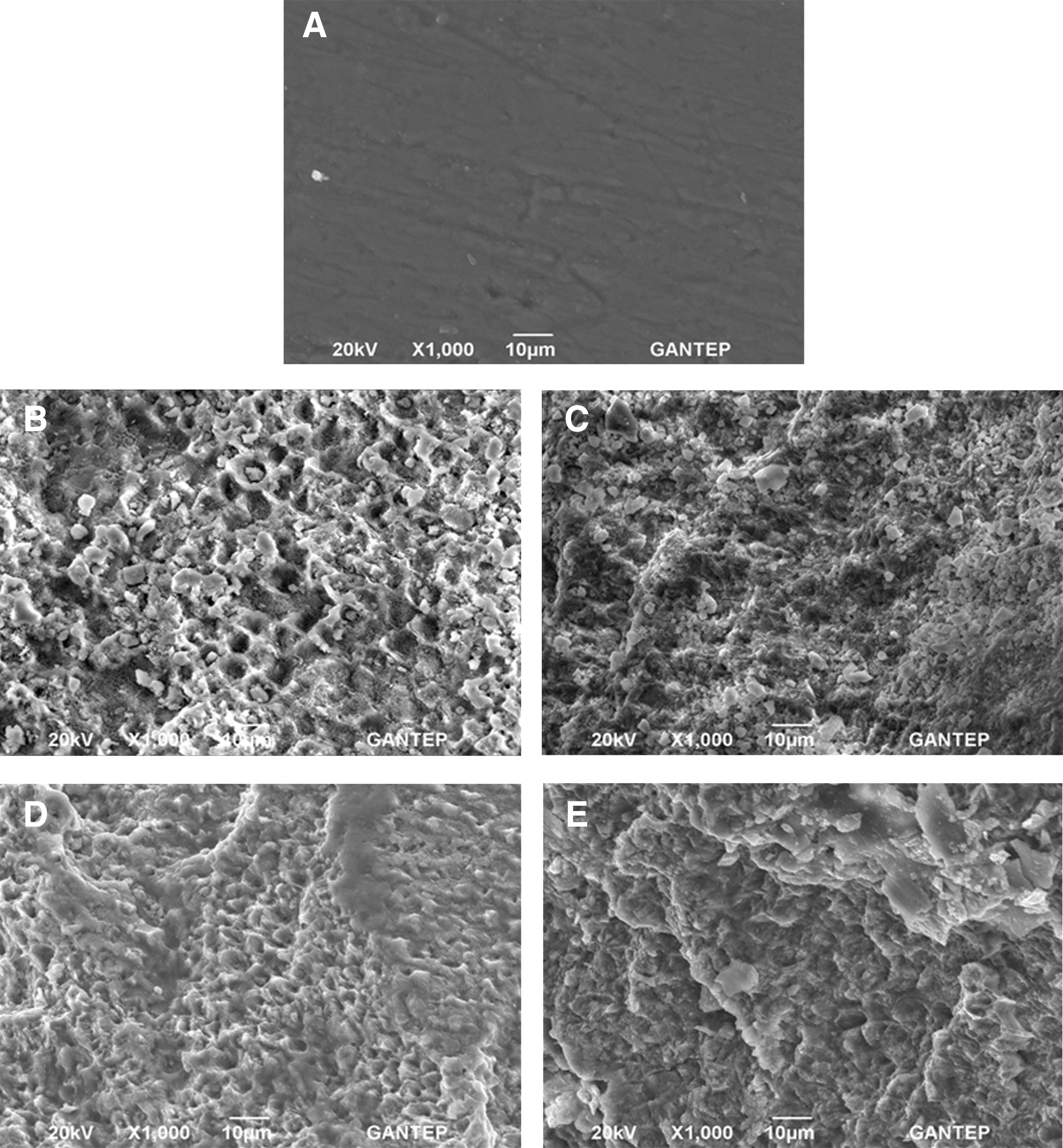
SEM images of enamel surfaces (original magnification ×1000).
Discusson
The SBS values obtained among laser-irradiated groups, in which different water flow rates were used (25, 36, and 48 mL/min), were similar to those obtained in the acid-etched group (group 1). The highest SBS value was obtained from group 4 (13.94±4.1 MPa) followed by group 1 (13.77±6.5 MPa), group 3 (13.28±2.2 MPa), and group 2 (12.14±5.4 MPa); however, no significant differences were found among groups (p=0.630).
Some reports have suggested that Er:YAG laser application does not ablate the enamel surface efficiently, 46,47 causing lower bond strength than that obtained for enamel etched with 37% phosphoric acid. 46,48,49 However, others have reported that the Er:YAG laser could be used to etch the enamel surface. 25,26,50 In the present study, the SBS values of all groups (12.14–13.94 MPa) were within the clinically acceptable range (10.7–14 MPa) 6,51 –53 suggested to sustain forces that typically emerge during orthodontic treatment.
There is no widely accepted setting for water flow rate with Er:YAG laser ablation applied to the enamel surface. Several authors have suggested different water flow rates to yield better ablated enamel surfaces. 20,32,54 Visuri et al. 54 stated that there were no statistically significant differences between Er:YAG laser ablation rates obtained with a water flow of 6.13 mL/min and those obtained with 11.09 mL/min at a fluence of 60 J/cm2, with the exception of results obtained with a water flow rate of 5.45 mL/min. Although they increased the water flow rate twice at fluences>90 J/cm2, the ablation rates in enamel did not change. 54 Colucci et al. 29 reported that Er:YAG laser ablation (80.22 J/cm2, 300 mJ/4 Hz) with a water flow rate of 2.0 mL/min, caused a higher amount of mass loss from the enamel surface, as compared with groups with water flow rates of 1.0 and 1.5 mL/min. They also concluded that the more the water flow rate was increased, the fewer structural thermal changes occurred. 30 Additionally, Kim et al. 32 suggested that Er:YAG laser (at 250 mJ pulse energy) with a water flow rate of 1.69 mL/min provided better ablated surfaces for both enamel and dentin. They also found that a water flow rate of 6.75 mL/min (at a 400 mJ pulse energy) resulted in the most optimally ablated enamel surface among the tested groups. 33 In the present study, the SEM photomicrographs of all groups except group 2, in which the water flow rate was 25 mL/min, presented patterns similar to type 1. The SBS values of the three experimental groups were in a very close range (13.28–13.94 MPa). Although there were no statistically significant intergroup differences in SBS values, the lowest SBS value (12.14 MPa) was obtained in group 2. This finding may be related to the relatively lower water flow rate of 25 mL/min used for the Er:YAG laser in group 2. Because adequate external water supply is necessary for an efficiently ablated surface without any thermal damage, a flow rate that is too low would have a negative effect on adhesion. 6,28 –30,55 –57 Therefore, it is reasonable to assume that prevalent microcrack formation, which might occur because of the relatively inadequate water flow rate of 25 mL/min used in group 2, decreased SBS values, because of the lack of optimally ablated enamel surface for the composite resin to interlock. 47 Therefore, the null hypothesis was rejected.
The fracture patterns of the groups (Table 2) supported the results of the SBS test. In group 4, 65% of the specimens showed a type 2 fracture pattern. This indicated that>50% of the composite was left on the enamel surface after the SBS test, implying that the bond strength between composite and enamel was higher than that between composite and bracket. This frequency of occurrence of the type 2 pattern in group 4 was followed by those in groups 1, 3, and 2. This was an expected result, as the SBS values of all groups were in the same order, indicating that the higher the SBS value, the higher the amount of remaining composite on the enamel surface.
Limitations
The main limitation of this in vitro study is the inadequacy of the thermocycling process in reproducing of variations of the oral environment, such as temperature and pH changes, humidity, and dental plaque. Although tensile force was applied to the bracket in parallel to the long axis of the teeth by the universal testing machine in the laboratory, brackets were exposed to different combinations of tensile, shear, and rotational force vectors in the oral cavity. Therefore, the results of this in vitro study give some useful information to clinicians about new surface roughening techniques; however, it is difficult to make direct correlation between results of this in vitro study and a clinical situation. Further in vitro and in vivo studies are required to indicate the applicability of such a technique in daily clinical practice.
Conclusions
Within the limitations of the present study, it can be stated that Er:YAG laser application with an appropriate water flow rate can be an alternative method to conventional acid etching with 37% phosphoric acid, to provide an efficiently ablated enamel surface. Furthermore, according to the results of the present study, Er:YAG laser ablation with a water flow rate of 48 mL/min resulted in better enamel surface alterations without any thermal damage to yield higher SBS values.
Footnotes
Author Disclosure Statement
No competing financial interests exist.