
Abstract
Introduction
K
Meanwhile, GNPs were suggested to be used in vivo in cancer plasmonic photothermal therapy (PPTT), in which laser is used to heat GNPs. 7,8 It is mandatory to consider the effect of laser on GNPs and the effect of laser GNPs interaction on biological tissues. GNPs, when irradiated with a second harmonic pulsed Nd:YAG laser (532 nm), showed a diameter reduction, and their shape became almost a perfect sphere. This phenomenon was limited to pulsed laser and was interpreted by heating of GNPs within the time of irradiation, the large absorption cross-section of GNPs, and the thermal insulation of irradiated GNPs in water. 9,10 The shape change was suggested to be secondary to melting, and the size reduction was suggested to be secondary to vaporization. 11 The maximum diameter of GNPs after laser irradiation depended upon the absorbed laser energy. 10 The average diameter of 8 nm GNPs, irradiated with 532 nm laser, decreased as the laser shot increased, and the smallest possible diameter decreased with the laser fluence. 12 Toxicity caused by nanometer dimensions is a major concern, as GNPs are expected to be widely used in biomedical applications. 13
We demonstrated in a previous work that by spectroscopy and transmission electron microscopy (TEM), the irradiation of 15 nm GNPs with 532 nm pulsed laser for 3 and 5 min resulted in the decrease of average GNPs diameters from 15 nm to 4 and 3 nm, respectively. 14 When primary cultures of rat kidney cells (RKCs) were incubated with GNPs irradiated with pulsed laser, a significant decrease in RKC viability occurred. The decrease in cell viability was quantified colorimetrically by trypan blue and MTT assays. Here, we conducted a preliminary analysis of the subcellular toxic events leading to this decrease in RKC viability. We studied cell membrane integrity by detecting lactate dehydrogenase (LDH) release, the induction of apoptosis by the alkaline phosphatase (ALP) assay, and the occurrence of oxidative stress by the glutathione (GSH) assay. This study is a step in a continuum, aiming to investigate the possible side effects of on exposure to laser pulses. Therapeutic applications will follow if it becomes evident that these materials are safe enough to be applied to living organisms.
Materials and Methods
Preparation and characterization of GNPs
We prepared GNPs by citrate reduction of Gold (III) chloride trihydrate (HAuCl4.3H2O) according to the Turkevich method 15 modified by Frens. 16 We characterized the prepared GNPs by UV-visible spectrophotometer (T80+, PG instruments, UK), TEM (JEOL, JEM 1230 EM, Japan), and particle size analyzer (Malvern Zetasizer Nano ZSPXRD, Malvern Instruments Ltd., UK).
Irradiation of GNPs with pulsed laser
We prepared a stock solution (10 nM concentration) of 13 nm GNPs in deionized water. We exposed 2 mL of the stock solution, while agitating in a quartz cell, to laser pulses from a Q switched Nd:YAG laser (Brio, Quantel, France) operating with its second harmonic generator (SHG) to provide laser pulses of 532 nm wavelength, 50 mJ/pulse energy, 5 ns duration, and 10 Hz repetition rate (frequency). The wavelength was selected as it falls around the absorption peak of the prepared GNPs. The laser beam cross section was determined by the burned pattern printed on a thermal recording paper and was typically 0.2 cm2. The laser pulse energy measured by a power meter (model AC5001, SCIENTECH joulmeter, USA) was 50 mJ. The energy was measured in front of the quartz cell. We irradiated samples for 1, 3, and 5 min. After laser irradiation, we used the absorption spectroscopy, TEM, and Zetasizer again to detect modifications in GNP size. The contrast power of TEM was set at its default (50%) and kept unchanged during the whole study, so as not to modify the results in respect of GNPs sizes after laser irradiation. We diluted laser-irradiated GNPs to 1, 2, and 4 nM in fresh culture media. Equal volumes (2 mL) of the stock solution of 13 nm GNPs diluted to 1, 2, and 4 nM in the same culture media were taken as the unirradiated samples. Prior to incubation with cells, media containing both laser-irradiated and unirradiated GNPs were filtered through a 0.2 μm pore-size sterile filter to avoid any contamination that might hamper the growth of primary culture of RKCs.
Rat primary kidney cell culture
Our research protocol was approved by the Ethical Committee at the Faculty of Veterinary Medicine, Cairo University. The animal care protocol was in compliance with the guidelines of The Canadian Council of Animal Care. 17 We dissected kidneys from three adult male rats, 100–120 g in weight, obtained from the animal house at The Ophthalmology Research Institute, Guiza, Egypt. After the animals were euthanized, the kidneys were meticulously dissected and placed in alcohol. Kidneys were transferred to culture media [Dulbecco's Modified Eagle's Medium (DMEM) containing 10% fetal bovine serum (FBS) and 1% penicillin streptomycin; Lonza Bioproducts, Belgium], their capsules removed and cut into small fragments. Using a glass homogenizer containing culture media, we converted kidney fragments into cell suspensions. We diluted 10 μL of cell suspensions in 190 μL phosphate-buffered saline (PBS) (pH=7.4), and counted cells using a hemocytometer (Clay Adams, New York, USA). We placed appropriate volumes of cell suspensions into tissue culture plates and incubated them in a humidified atmosphere (95% air and 5% CO2) at 37°C in the incubator (Thermo Forma, II 3110, USA).
Subcellular toxicity studies
We plated RKCs in flat-bottomed microtiter plates (Greiner Bio-One, Germany) and grew them to 70–80% confluence. Media were replaced with fresh filtered ones containing either laser-irradiated or unirradiated GNPs. We re-incubated the plates for different time intervals (24, 48, and 72 h). Three different tests/metabolic assays were performed: LDH release assessment, ALP assay and intracellular GSH assay. For each test, we gradually raised the concentration of irradiated or unirradiated GNPs from 1 to 2 and 4 nM. Cells incubated for equal time intervals in culture media without GNPs were used as a negative control, and rat serum or appropriate materials provided by standardized tissue culture protocols were used as positive controls. All assays were repeated three times.
LDH assay
After incubation for 24, 48, and 72 h, respectively, the culture media were aspirated and centrifuged at 3000g for 5 min in a Beckman benchtop GS15R centrifuge (Beckman Coulter, USA) in order to obtain a cell-free supernatant. The LDH in media was determined using a diagnostic kit (Bio System, Barcelona, Spain). As described, 18 aliquots of media and warm reagents were mixed, and the absorbance was recorded using a spectrophotometer. LDH activities from cell cultures incubated with laser- irradiated and unirradiated GNPs were compared with those from the negative control.
ALP activity assay
ALP activity was determined following the modified method of King and King (1954). Cells were prepared with deionized H2O followed by sonication using a sonicator (Virtis, virsonic 475, SP Scientific, Gardiner, NY, USA) for 2 min, and then centrifuged at 2400g for 10 min as described by Nakayama et al. 19 The ALP activity was measured, using the colorimetric determination of alkaline phosphatase assay kit, at λ=510 nm according to the manufacturer's recommendations (Biomerieux, France).
GSH assay
Cells were seeded in ∼0.51 mL of culture media as 1×10⁵ cells per well in 24 well flat-bottomed microtiter plates, and incubated in a CO2 incubator at 37°C until forming a confluence sheet. Media from each well were discarded and replaced with 1 mL of fresh media containing GNPs. The microtiter plate was re-incubated for the test period. Cells were centrifuged at 2000g for 10 min at 4°C, and the supernatant was discarded. Cell pellets were homogenized in cold buffer and centrifuged at 10000g for 15 min at 4°C. The supernatant was then transferred to a new tube and used for the GSH assay. The GSH level was calculated according to the kit manufacturer's recommendations (Bio System, Barcelona, Spain). GSH concentrations in cultures incubated with media containing irradiated or unirradiated GNPs were calculated relative to the negative control.
Statistical analysis
Values of cell viability were expressed as the mean±SD. Significance was calculated by Student's t test using SPSS program (version 15, SPSS Inc. Chicago, IL), and p values<0.05 were considered significant. Two way ANOVA test was used for analysis of ALP activity data.
Results
Effect of irradiation of GNPs with pulsed laser
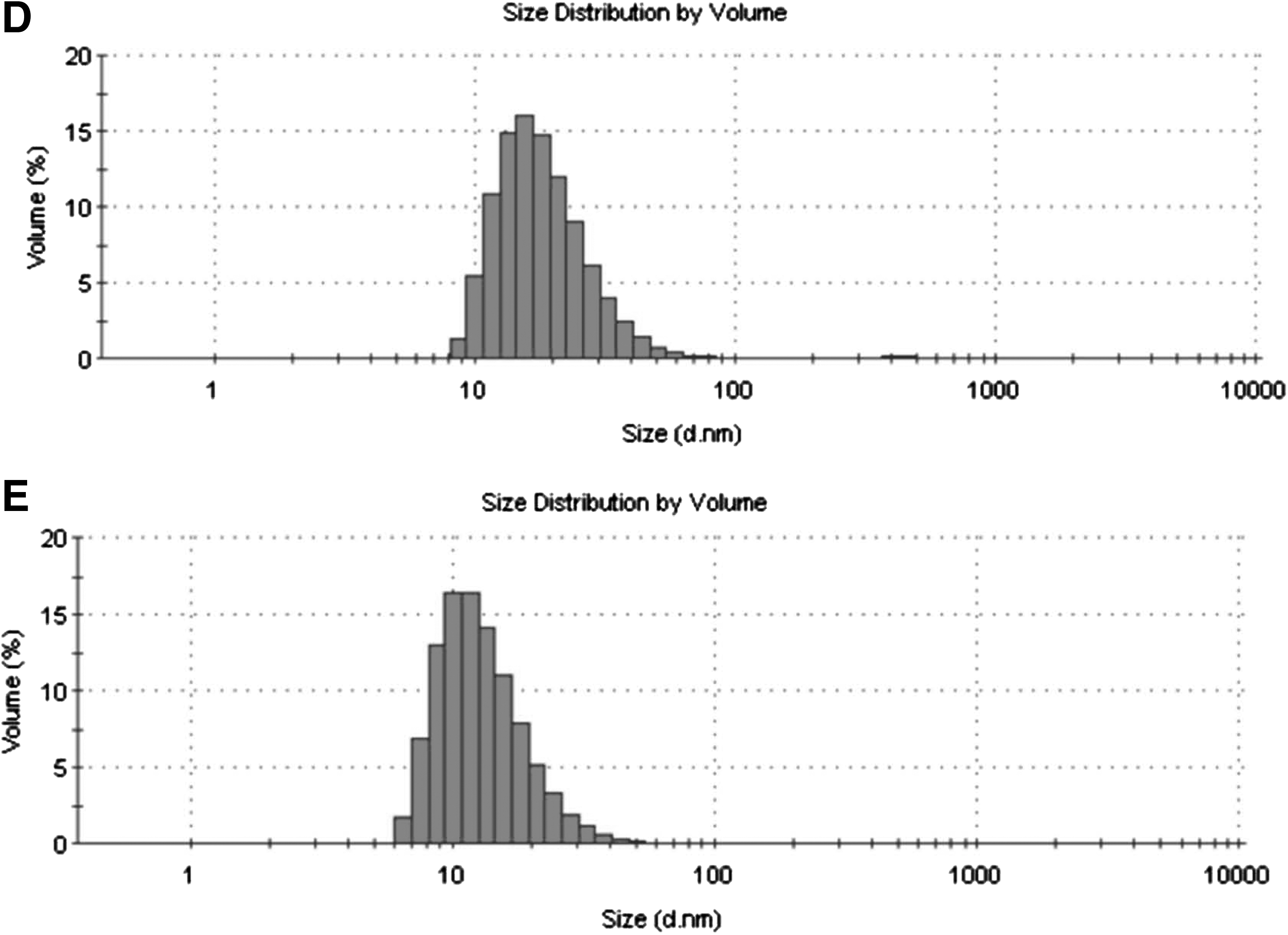
Figure 1 demonstrates the TEM images (A and B), absorption spectra (C) and Zetasizer curves (D and E) of 13 nm GNPs before and after irradiation with pulsed laser. Laser-irradiated GNPs showed a reduction in both optical density and mean particle diameter. The effect varied according to the time of irradiation. The decrease in optical density was ∼22% after 5 min of irradiation. Absorption spectra showed blue shifts from 520 nm to 518, 514, and 513 nm after pulsed laser irradiation for 1, 3, and 5 min, respectively (Fig. 1C). Analysis of TEM micrographs (Fig. 1A, B) and Zetasizer curves (Fig. 1D, E) demonstrated that 95% of GNPs underwent a reduction in size from 13 to 5 nm after 5 min of pulsed laser irradiation.
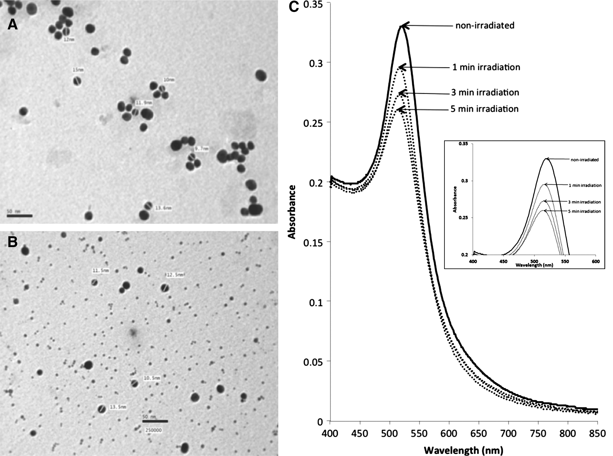
Transmission electron micrographs (TEM) of 13 nm gold nanoparticles (GNPs) in aqueous medium
Subcellular toxicity studies
LDH assay
When RKCs were incubated with unirradiated 13 nm GNPs and with GNPs irradiated for 1 min, we did not observe any significant increase in LDH (concentration of GNPs and incubation time increased from 1 to 4 nM and from 24 to 72 h, respectively) (Fig. 2A, B). When RKCs cells were incubated with GNPs irradiated for 3 min with 532 nm pulsed laser, we observed a significant increase in LDH after 72 h of incubation with 2 and 4 nM of GNPs (126% and 140%, respectively) (Fig. 2C). When RKCs cells were incubated with GNPs irradiated for 5 min, we observed a significant increase in LDH activity after 72 h of incubation (126%, 130%, and 141% for incubation with 1, 2, and 4 nM of irradiated GNPs, respectively) (Fig. 2D).
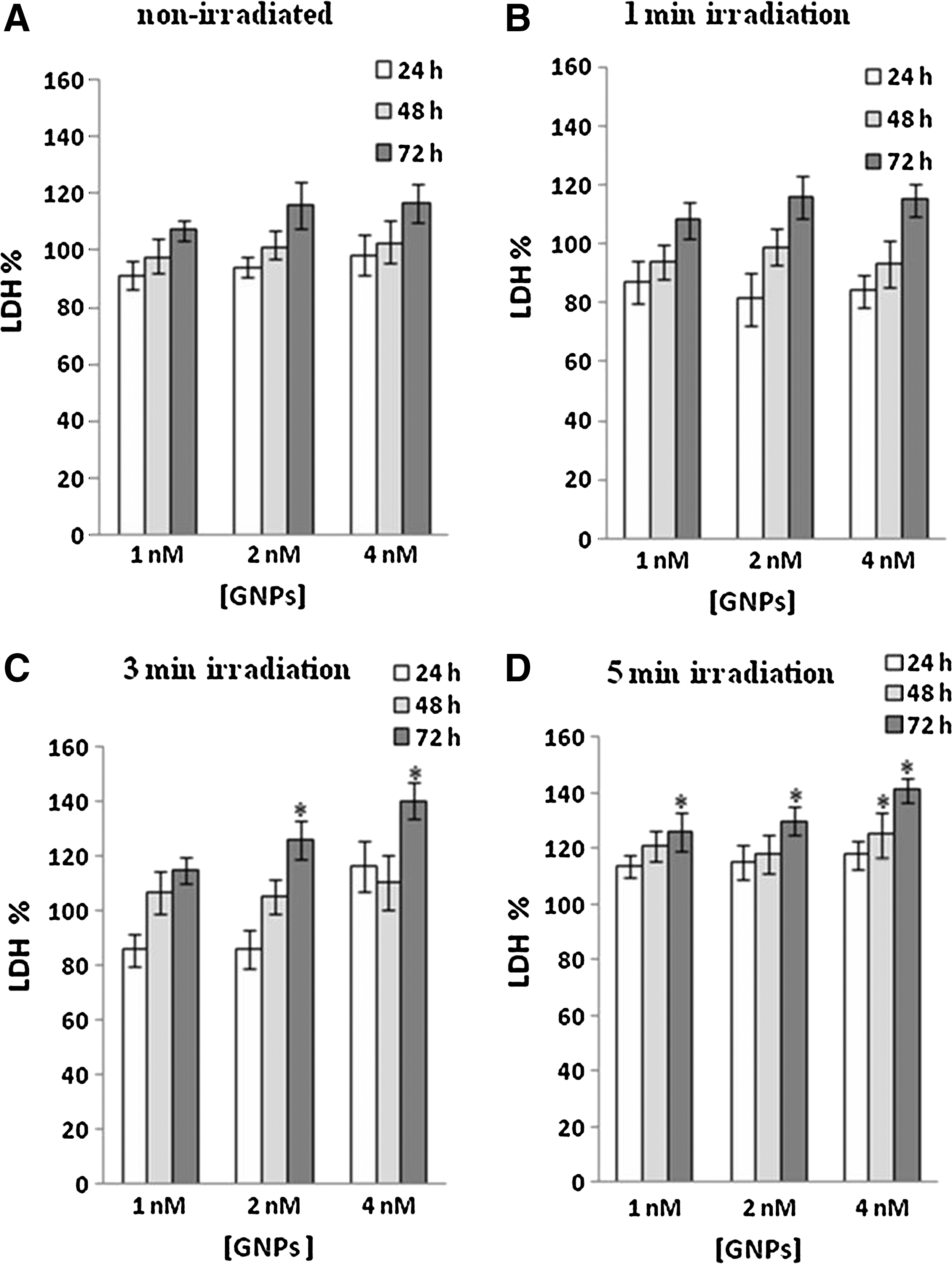
Percentage of lactate dehydrogenase (LDH) release from rat kidney cells (RKCs) (mean value±SD) measured after incubation with 1, 2, and 4 nM gold nanoparticles (GNPs) for different incubation times (24, 48, and 72 h) for unirradiated GNPs
ALP activity assay
When RKCs were incubated with unirradiated 13 nm GNPs, we did not observe any significant increase in ALP activity (Fig. 3A). When RKCs were incubated with laser-irradiated GNPs (irradiation time=1 min), we observed a significant increase in ALP activity after 48 h of incubation with 2 and 4 nM of irradiated GNPs (130% and 135%, respectively) (Fig. 3B). When RKCs were incubated with irradiated GNPs (irradiation time=3 min), we observed a significant increase in ALP activity after 48 h of incubation with 2 and 4 nM of irradiated GNPs (166% for both concentrations) (Fig. 3C). When RKCs were incubated with irradiated GNPs (irradiation time=5 min), we observed a significant increase in ALP activity after 48 h of incubation with 2 and 4 nM of irradiated GNPs (162% and 160%, respectively). After 72 h, ALP activity increased significantly only after incubation with 4 nM of irradiated GNPs (154%). This change was lower than that after 48 h of incubation with the same concentration of irradiated GNPs, reflecting a possible tolerance effect (Fig. 3D).
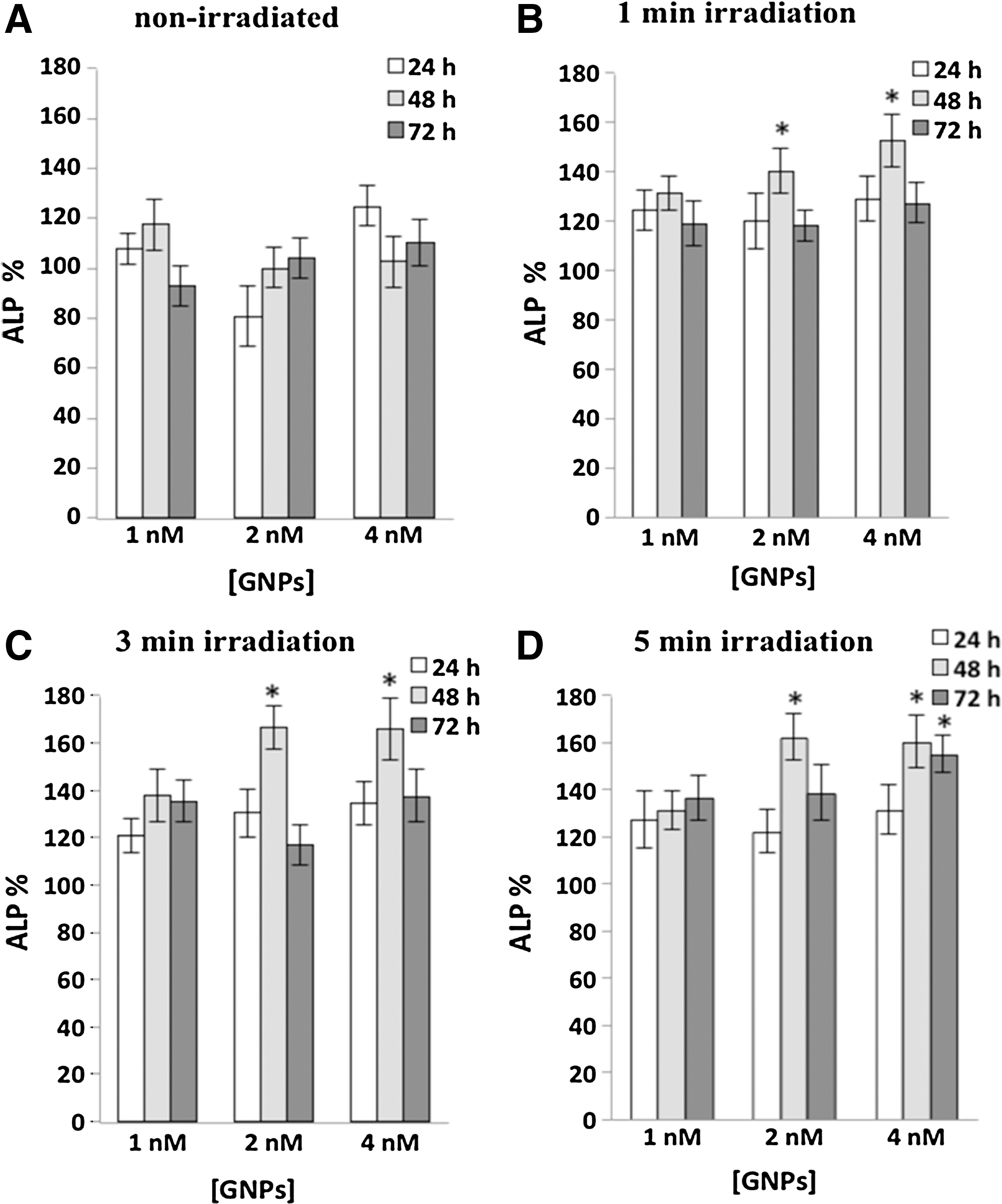
Percentage of alkaline phosphatase (ALP) of rat kidney cells (RKCs) (mean value±SD) measured after incubation with 1, 2, and 4 nM gold nanoparticles (GNPs) for different incubation times (24, 48, and 72 h) for unirradiated GNPs
GSH assay
When RKCs were incubated with unirradiated 13 nm GNPs, intracellular GSH did not increase significantly (Fig. 4A). When RKCs were incubated with laser-irradiated GNPs (irradiation time=1 min), we observed a significant increase in intracellular GSH after 24 h of incubation with 2 and 4 nM of irradiated GNPs (139% and 144%, respectively) (Fig. 4B). When RKCs were incubated with laser-irradiated GNPs (irradiation time=3 min), we observed a significant increase in intracellular GSH after 24 h of incubation with 1, 2, and 4 nM of irradiated GNPs (139%, 141%, and 139%, respectively) (Fig. 4C). When RKCs were incubated with irradiated GNPs (irradiation time=5 min), we observed a 141% increase in intracellular GSH when the concentrations of irradiated GNPs were increased to 2 and 4 nM only after 24 h of incubation (Fig. 4D).
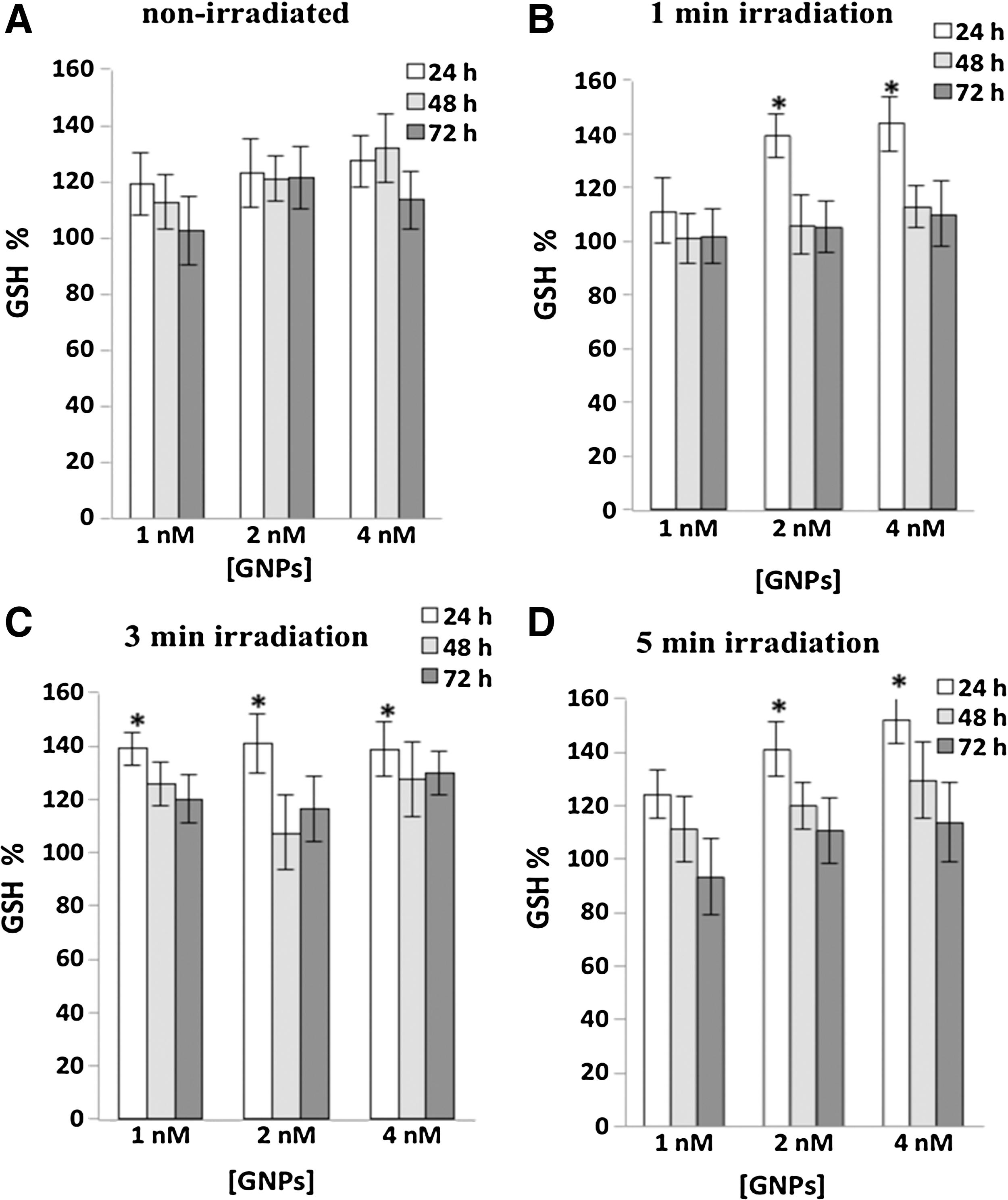
Percentage of glutathione (GSH) of rat kidney cells (RKCs) (mean value±SD) measured after incubation with 1, 2, and 4 nM gold nanoparticles (GNPs) for different incubation times (24, 48, and 72 h) for unirradiated GNPs
Discussion
Although elemental gold proved to be safe on living organisms, gold at the nanoscale level – being chemically and physically different – needs to be investigated for biocompatibility before in vivo applications. 20 As little is known about the potential deleterious effects of the end- and byproducts of exposure of nanomaterials to laser, 21 and to maximize the potential benefits of nanomaterials in the realm of PPTT, the interactions of nanomaterials exposed to laser irradiation with biologic systems should be exploited.
In agreement with those of Dammer et al. 22 our results show that GNPs, exposed to 532 nm pulsed laser, underwent size reduction. Under the applied laser parameters, where a power meter was used to assess the laser beam, the obtained GNPs sizes were reproducible. We detected 2, 6, and 7 nm blue shifts in the absorption band of GNPs after 1, 3, and 5 min irradiation with 532 nm pulsed laser respectively indicating a shift in GNP average diameter from 13 nm to 10, 6, and 5 nm, respectively.
In a previous work, we have illustrated the decrease in viability of RKCs incubated with GNPs irradiated with 532 nm pulsed laser. 14 The smaller sized GNPs were probably implicated in the observed decrease in viability. As trypan blue and MTT assays gave general overviews on cell damage, we used ALP, LDH, and GSH assays to give more insight into the subcellular toxic events in RKCs incubated with laser-irradiated GNPs.
Our results showed an overall increase in LDH release from RKCs upon increasing the concentration of laser-irradiated GNPs and increasing the incubation time of irradiated GNPs with RKCs. This may be attributed to a number effect. As GNP sizes decrease, the number of GNPs in the culture media and GNPs taken up by RKCs should increase with concentration and time of incubation. Coradeghini et al. 23 showed by inductively coupled plasma mass spectrometry (ICP-MS) that the number of GNPs taken up by BALB/3T3 cells increases with higher concentrations and with exposure time, and that the number of 5 nm GNPs taken up by cells was always higher than the number of 15 nm GNPs per cell. Uboldi et al. 24 studied the release of LDH from human alveolar cells upon incubation with 9.5–25 nm GNPs. They found that after 24–48 h, GNPs induced a mild LDH release. In addition, after 72 h of exposure to GNPs, they observed a dose-dependent release of LDH in the supernatant. The amount released was significantly higher than that after shorter exposure times (24–48 h) 24 . Although our results agree with the aforementioned ones, they do not agree with those of Arnida Malugin and Ghandehari, 25 who studied the effect of GNPs (rods and spheres, 30–90 nm in diameter) on human prostate cancer cells by LDH assay. They observed no LDH leakage upon increasing the concentration of GNPs to 34 nM. The discrepancy can be attributed to differences in cell lines and/or concentrations, shapes, and sizes of GNPs.
A pronounced increase in ALP activity is an indicator for apoptosis. 26 We observed that GNPs irradiated for 1, 3, and 5 min led to an increase in ALP activity after 48 h, denoting an induction of apoptosis. However, this increase in ALP activity disappeared after 72 h, except after incubation RKCs with 4 nM GNPs exposed to laser pulses for 5 min. Disappearance of ALP activity increase may indicate the emergence of a tolerance effect. Moreover, the time frame of ALP activity increase did not coincide with the decrease in cell viability observed by trypan blue and MTT assays, 14 which indicates that apoptosis is not the single or major factor implied in reduction of RKCs viability.
Fan et al. 3 found that reactive oxygen species (ROS) level in human bone marrow mesenchymal stem cells and human hepatoma carcinoma cells increased 1.5-fold by 15 nm GNPs. Their results suggested that GNPs may induce cell death from excessive ROS formation. Dichlorofluorescin (DCFH) was used by these authors to evaluate intracellular ROS formation where nonfluorescent fluorescein derivatives emit fluorescence when oxidized by ROS, the fluorescence being directly proportional to the concentration of ROS. 27 Under normal conditions, ROS are easily neutralized by antioxidant defenses such as GSH while under conditions of excessive production, for example, during exposure to GNPs, the natural antioxidant defenses are overwhelmed. During oxidative stress when GSH is depleted while oxidized glutathione (GSSG) accumulates, cells respond to the drop in the GSH/GSSG ratio by mounting protective responses. 28 The GSH assay is, therefore, another method to determine the oxidative stress upon exposure to nanoparticles. 29 As our study was qualitative rather than quantitative, we relied only on the GSH assay. We found an elevation of GSH in cells incubated for 24 h with GNPs irradiated for 1, 3, and 5 min by pulsed laser. This may indicate an early emergence of ROS. In general, the elevation of GSH disappeared after 48 and 72 h. Early increases followed by downregulation of GSH can be explained by cell adaptation against external stress. 30 In addition to the well-documented enzymatic plateau, GNP uptake by cells may also reach a plateau within specific time intervals. 31,32 The time intervals depend upon the cell type and the state of the cell cycle. 33 Unlike the increase in LDH activity, GSH elevation did not coincide in time with the decrease in viability shown by the trypan blue and MTT assays. 14 This may indicate that the cell membrane disruption, rather than the oxidative stress, is the major factor leading to the decrease in viability observed upon incubation of RKCs with GNPs irradiated with pulsed laser.
Nanoparticle cytotoxicity most likely follows endocytosis, but it is possible that it stems from interactions at the cell membrane, even though the particles are endocytosed. 34 The impact of Zeta potential on endocytosis of GNPs is also obvious. 35 However, it is mostly affected by the capping material and/or opsonization. Our team is conducting an undercurrent study focusing on these issues. Figure 5 tries to explain the suggested origin of subcellular toxicity upon exposure of 13 nm GNPs to 532 nm pulsed laser.
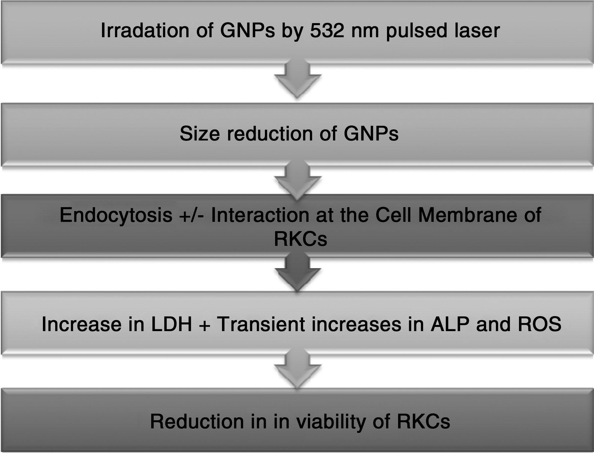
Suggested origin of subcellular toxicity upon exposure of 13 nm gold nanoparticles (GNPs) to 532 nm pulsed laser. GNPs, gold nanoparticles; RKCs, rat kidney cells; LDH, lactate dehydrogenase; ALP, alkaline phosphatase; ROS, reactive oxygen species.
Although the cytotoxicity of gold nanoparticles depends primarily on their size, 23, 34 , the “critical toxic size” for each nanoparticle is not yet determined, as all the size ranges were not tested on different cell types. Our work will be continued with confocal microscopic studies, using stains specific to different cell organelles to focus on the exact location of GNPs of various sizes in kidney cells and in other cell types.
Conclusions
RKCs incubated with non–laser-irradiated 13 nm GNPs did not show any significant subcellular toxicity as detected by ALP, LDH, or GSH assays. However, the steady increase in LDH release and the tolerance effect shown in ALP and GSH assays upon exposure of the same cells to GNPs irradiated by pulsed laser, indicate that the decrease in viability is mainly attributed to cell membrane disruption, rather than to apoptotis or an emergence of ROS. This effect is probably the result of smaller-sized GNPs generated after exposure of 13 nm GNPs to laser pulses. Exposure of GNPs to laser pulses to elicit photothermal effects may lead to subcellular harmful events on normal kidney cells.
Footnotes
Acknowledgement
We would like to thank Dr. Taher Ahmed Salah El Din, Head of the Nanotechnology Laboratory, Agriculture Research Center, Guiza, Egypt for his kind help and support.
Author Disclosure Statement
No competing financial interests exist.