
Abstract
Introduction
A
In previous studies, many factors have been involved in the pathogenesis of CNV, including Bruch's membrane damage, inflammation, and imbalance between the stimulators and inhibitors. 5 –8 Hence, CNV animal model was established by laser photocoagulation, transgenic animal expressing hPk1 or CC chemokine receptor-3, subretinal injection of adeno-associated virus-mediated VEGF, and mechanical removal of retinal pigmented epithelial (RPE) cells. The most widely used animal model is built by laser photocoagulation based on the study of Ryan et al., in which argon laser was used in 44 rhesus monkeys with the incidence of CNV being 90% and that of fluorescein angiographic leakage being<10%. 9 The structure of the retina and macula with fovea of the monkey is the most similar to those of humans, but it is expensive and difficult to large-scale use of monkeys. 5 Then, laser photocoagulation was extended to established CNV in rats, 10 with the incidence of CNV being 60%, and in mice, 11 with the incidence of CNV being 81%. By now, viewpoints on the pathogenesis of CNV and the angiogenic mechanism of CNV in addition to the genetic loci that control the size of the laser-induced CNV have been obtained from mouse laser-induced CNV,as the retinal blood vessels of the mouse (and other rodents) make imaging and intervention easy. 12 Kiilgaard 8 reported the incidence of CNV, to be 83% by diode laser in a pig model, which more closely mimics humans and is easier to study than rodents. Moreover, on account of different laser parameters, the incidence, occurrence, and duration of CNV are different. In order to make a reproducible and proper animal model for CNV, we need to establish CNV in BN rats with a Krypton laser.
Materials and Methods
Materials
Twenty-five male BN rats weighing between 180 and 200 g were involved in this study. Two eyes of one rat without any laser photocoagulation were randomly selected as the control group, and the other 48 eyes of 24 rats were selected as the experimental group with laser photocoagulation. All experimental procedures employing animals adhered to the Association for Research in Vision and Ophthalmology Resolution on the Use of Animals in Research, and were approved by the Animal Care Committee in Tianjin Medical University General Hospital. The ethics council of Tianjin Medical University General Hospital authorized the study, which adhered to the tenets of the Helsinki Declaration.
Methods
Laser-induced CNV
The rats were anesthetized for all procedures with intraperitoneal 846 compound anaesthetic (0.5 mg/kg). The pupils were dilated with 1% tropicamide. The fundus was visualized with a −53D cornea contact lens with the help of 1% methylcellulose solution. A Krypton laser (Coherent Novua 2000) with a 647.1 nm wavelength was used (350 mW power, 0.05 sec exposure, 50 μm spot size). Eight argon laser spots were applied to each fundus in a circle around the optic disc. Formation of an air bubble on the retina indicated the rupture of Bruch's membrane.
FFA
Eight eyes of four rats were randomly selected to receive examination by FFA 3, 7, 14, 21, 28, and 56 days after laser photocoagulation. FFA was performed by taking serial fundus photographs with a TRC-WT3 camera (Variable Angles Retinal Camera, Topcon TRC-WT3) after intraperitoneal injection of 0.5 mL/kg of 10% fluorescein sodium. Leakage was defined as the presence of a hyperfluorescent lesion that increased in size with time in the late-phase angiogram. Angiography was graded in a masked fashion by two examiners using reference angiograms. When the two scores for a lesion did not coincide, the higher score was used. Angiograms were graded as follows according to Murata standard: 0 meant no leakage, 1 meant slight leakage, 2 meant moderate leakage, and 3 meant prominent leakage. 1
Histopathology analysis of CNV lesions
At various times after angiography, rats were euthanized with an overdose of chloral hydrate. The eyes were enucleated and fixed in 10% formaldehyde solution for 1 week. They were dehydrated through a series of graded alcohols including 60%, 70%, 80%, and 90% for 24 h, and then dehydrated through anhydrous alcohol for 20 min. Eyes were then transferred into dimethylbenzene, dehydrated for 20 min, and processed for paraffin embedding three times. Once embedded, 5∼6 μm serial sections of the tissues were cut through the entire lesion, stained with hematoxylin and eosin, and examined by light microscopy.
Transmission electron microscopy
One eye was randomly selected 3, 7, 14, and 21 days after laser photocoagulation. The eyes were fixed in phosphate-buffered 2.5% glutaraldehyde solution. The eyes were dissected, each burn was identified and isolated under a dissecting microscope, and the tissue strips were post-fixed with 1% osmium tetroxide. The tissues were dehydrated through a series of graded alcohols, permutated by trimethylene oxide embedded two times, embedded in epoxy resin EPON-812, and serial semithin sections were cut by 1 μm, stained with toluidine blue, and examined by light microscopy. For some lesions, ultrathin sections were prepared with an ultramicrotome (LKB-NOVA, 700Å), counterstained with uranyl acetate and lead citrate, and examined with a transmission electron microscope.
Maximal thickness of CNV lesions
In hematoxylin-eosin–stained sections, a spindle-shaped subretinal fibrovascular scar was obvious, and could be distinguished from adjacent normal structure. The maximum vertical meridian passing through this spindle-shaped scar was measured as the thickness. Twenty-five sections of each lesion were measured, and the highest thickness was chosen for analysis and comparison. This measurement was made with an eyepiece reticule after calibration with a stage micrometer with 10 μm divisions.
Statistical analysis
χ2 test was used to compare the leakage score by FFA. For analysis of CNV thickness, Q tests and mono-agent analysis of variance were used. For all analyses, a computer software (SPSS10.0 for Windows) was used, with p<0.01 considered to be statistically significant.
Results
FFA
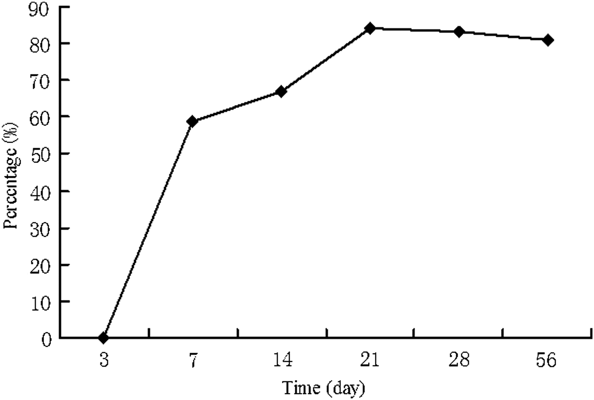
The changes of staining intensity were detected by FFA after photocoagulation. No leakage was found on day 3 (grade 0), the leakage appeared in burns on day 7 (grade 1), the intensity of fluorescein staining gradually increased on day 14 (grade 2), reached a peak on day 21 (grade 3), (p<0.01) and remained stable after day 21 (p>0.05) (Figs. 1 and 2, Table 1).
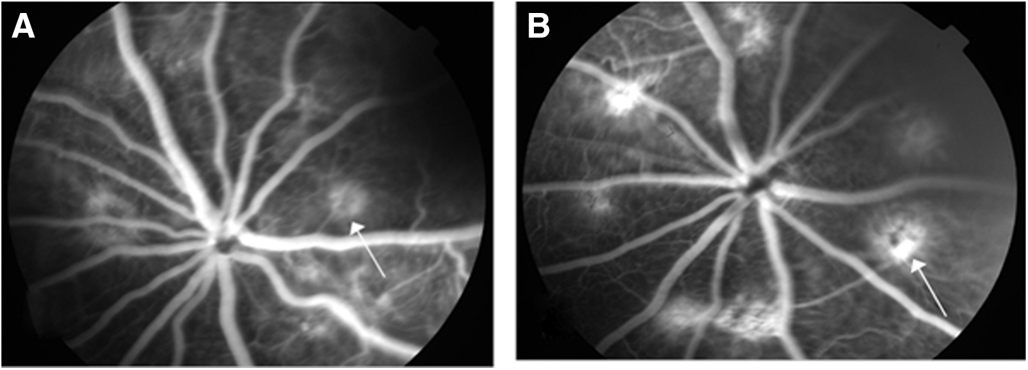
The leakage after photocoagulation at different time points.
The change of leakage after photocoagulation at different time points (p<0.01).
N, number; P, percentage.
Histopathology
Light microscopy
Three days after laser treatment, Bruch's membrane and the RPE layer were disrupted in the center of laser burns, and all layers of the choroid were destroyed within the laser burn. In association with this tissue, there were many neutrophilic granulocytes and pigment-containing macrophage-like cells, and at the borders of the burn there were many RPE cells (Fig. 3A). Seven days after laser treatment, retinal edema was found, newly formed vessels with wide lumens extended from the choroids into the subretinal space, where they became partially enveloped by RPE cells (Fig. 3B). Fourteen days after laser treatment, retinal edema decreased, fibroblasts and collagen fibers increased, RPE cells migrated and proliferated, and large collections of vascular channels were seen in the subretinal space (Fig. 3C). Twenty-one days after laser treatment, burns had shown significant fibrovascular proliferation (FVP), and a large amount of newly formed vascular channels was found. (Fig. 3D). Fifty-six days after laser treatment, a notch from the outer retina layer to the inner retina layer within the laser burns was shown, with a great deal of fibrocytes in the CNV, and FVP was kept stable.

Histopathology analysis of choroidal neovascularization (CNV) lesions at different time pionts.
Transmission electron microscopy
Three days after laser treatment, incomplete vascular channels were surrounded at the retina side of choroids (Fig. 4A). Seven days after laser treatment, newly formed vascular channels were observed, in which adhered endothelial cells and red cells, and the lumens between RPE cells were widened (Fig. 4B). Fourteen days after laser treatment, the retina side of the choroid had shown small endothelial cell axons in the capillary vessels (Fig. 4C) Twenty-one days after laser treatment, agglomeration of capillary vessels in which many sausage-like red cells were observed, was found between melanin cells in choroids (Fig. 4D)

Transmission electron microscopy of choroidal neovascularization (CNV) at different time points.
Maximal thickness of CNV lesions
The thicknesses of CNV were 25.22±3.56, 45.50±4.08, 62.52±3.25, 62.00±3.68, and 61.08±4.06 μm, 7, 14, 21, 28, and 56 days after laser photocoagulation, respectively. The maximal thickness of CNV lesions increased from day 7 to day 21 (F=468.96, p<0.01), and kept stable after day 21 (p>0.05) (Fig. 5).

The change of the maximal thickness of choroidal neovascularization (CNV) lesions after photocoagulation at different times(p<0.01).
Complications
Except subretinal hemorrhage (three eyes) in laser burns, no other indication was found, such as vitreous hemorrhage, retinal detachment, choroidal detachment, and retinal surface member formation.
Discussion
AMD is the most important cause of vision loss in elderly patients. 1 The major cause of severe vision loss in AMD is CNV. The key point of treatment is to inhibit the formation and development of CNV. In order to explore new treatment strategies, it is important to establish an animal model, which should be safe, effective, stable, and, highly similar to humans.
Angiogenesis is a multistage process. Exudative AMD involves pathological angiogenesis that originates from the choroid beneath the retina to form CNV, containing abnormal blood vessels that can leak fluid and blood. CNV is a result of pathological angiogenesis. Angiogenesis is an invasion process that requires proteolysis of the extracellular matrix and proliferation and migration of endothelial cells, with simultaneous synthesis of new matrix components. The fluid and blood released from CNV can damage the structure and function of the overlying retina, usually in the central macular area, leading to the loss of central vision. 3,13
Krypton laser photocoagulation can induce the formation of CNV through three biological effects, which include heat, mechanism injury, and photochemical effects. The retina develops a proliferative rehabilitative process combined with inflammation in burns after laser photocoagulation. In our study, three days after laser photocoagulation, neutrophilic granulocytes and pigment-containing macrophage-like cells infiltrated, and the degenerated and necrotic retina and choroid were observed under light microscope, which confirmed the existence of inflammation after laser injury. Proliferative RPE cells were seen under transmission electron microscope, which indicated that the proliferative rehabilitative process happened in the photocoagulation areas. Three aspects were combined in the formation of CNV, including protease and various growth factors that participated in CNV formation released from active macrophages, growth factors secreted by immigrated proliferative RPE cells, and the secretion of growth factors by the stimulation of microvessel emphraxis and local retina tissue ischemia and anoxia. 14,15
The changes of leakages from laser dots were detected by FFA after laser photocoagulation. No leakage was found on day 3, and the leakage appeared in burns on day 7 (59%). The intensity of fluorescein staining gradually increased on day 14 (67%), reached a peak on day 21 (84%), and remained stable after day 21.
A same result of the changes of CNV after laser photocoagulation was obtained through histopathology study. Three days after laser treatment, Bruch's membrane and RPE layer were disrupted in the center of laser burns and all layers of the choroid were destroyed within the laser burn. In association with this tissue, there were many neutrophilic granulocytes and pigment-containing macrophage-like cells, and at the borders of the burn there were many RPE cells. Seven days after laser treatment, retinal edema was found, and newly formed vessels with wide lumens extended from the choroids into the subretinal space where they became partially enveloped by RPE cells. Retina edema decreased, fibroblasts and collagen fibers increased, RPE cells migrated and proliferated, and large collections of vascular channels were seen in the subretinal space 14 days after laser treatment. Burns that had shown significant FVP and a large amount of newly formed vascular channels were found 21 days after laser treatment. A notch from the outer retina layer to the inner retina layer within the laser burns was seen 56 days after laser treatment, with many of fibrocytes in CNV.
Under the transmission electron microscope, incomplete vessels were surrounded at the retina side of the choroids 3 days after laser treatment. Seven days after laser treatment, newly formed vascular channels were observed at the retinal side of the choroids, in which endothelial cells and red cells were adhered, and the interspaces between RPE cells were widened. Fourteen days after laser treatment, the retinal side of the choroid had shown small endothelial cell axons in the capillary vessels. Agglomeration of capillary vessels in which many sausage-like red cells were observed was found between melanin cells in choroids 21 days after laser treatment.
The maximal thickness of CNV lesions is an important measurement index, which means the maximum vertical meridian passing the central part of the lesion from RPE layer to the inner limit membrane. 16,17 Coincident with the results of FFA and histopathology, the maximal thickness of CNV lesions increased from day 7 to day 21, and kept stable after day 21.
Except for occasional subretinal hemorrhage in laser burns, no other indication was found in this study, such as vitreous hemorrhage, retinal detachment, choroidal detachment, or retinal surface member formation. Therefore, this CNV model induced by Krypton laser in rats is safe.
Several models of CNV with different animals and laser parameters have been described, and each has shown a different occurrence time and incidence of CNV. The laser-induced CNV model in monkeys occurred at 1 week peaked at 4 weeks reported by Zahn et al. 18 As Kiilgaard reported, CNV in pigs induced by laser occurred on day 7 with the incidence of 83% on day 21. 10 Other researchers reported that CNV in mice occurred within 1 week and lasted for 4 weeks, and that the incidence was 63.6% 19 Although the models in mice have some benefits, such as relatively low price and easier to raise, there were many disadvantages, including that the eyes were small, difficult to operate on, and quite different in retinal structure from those of humans. In this study, we have produced and characterized a model of laser-induced CNV in BN rats, except the advantages of mimicking human retinal structure, relatively easy manipulation, and high incidence of CNV (84%), which appeared within 1 week, reached a peak on day 21, and lasted for 56 days.
In addition, we also observed that the fluorescein leakage entirely surrounded the photocoagulation spots and the CNV mostly occurred in the margin of photocoagulation spots, whose ranges were all in the lesion zones. The same results were also reported by Ryan. 20 These experimental study results will, we hope, serve as useful suggestions for estimating the formation and development of CNV by fluorescein leakage areas in the clinic.
The previous study results revealed that laser used in establishing a CNV animal model included argon laser, semiconductor laser, YAG/green laser, diode laser, and Krypton laser. The mechanism of laser-induced CNV is the rupture of Bruch's membrane by laser photocoagulation. 21 –24 Bora 25 reported that CNV appeared 5 days after argon laser injury in a mouse model, and the incidence was 87%. Takahashi 26 found that CNV was induced on day 7 after semiconductor laser in a mouse model. Inomata 27 successfully made CNV in a wild-type C57B/6 mouse model 7 days after YAG/green laser, and the incidence was 85.7%. Kiilgaard 8 reported the incidence of CNV was 83% by diode laser in a pig model. Shen's study result revealed that CNV appeared 2–3 weeks after Krypton laser injury in pigmented rats, and the incidence was 46.4%. 24 In our study, the CNV appeared 7 days after Krypton laser, and remained stable until 56 days, with an incidence of 84%. CNV animal model induced by Krypton laser in our study was stable and long lasting, and had a high success rate compared with the previous methods.
Although a laser-induced model is widely used for the study of CNV, it is controversial whether it mimics the most common form of naturally occurring CNV in AMD. In our study, the results revealed that the essential process such as the breakup of the basement membrane, the migration and proliferation of vascular endothelial cells, the translation of fibroblasts into fibrocytes, the increase of collagenous fibers, and scar formation were similar to previous studies, which confirmed the mechanism of CNV formation. 5,28,29 In the AMD patients, drusen deposits are a high risk factor for the occurrence of CNV; however, in the laser-induced model, CNV occurs after laser spot. 30 Also, BN rats do not have a fovea, where CNV occurs often in AMD patients.
Conclusions
In summary, CNV models were successfully induced by Krypton laser in rats, which are stable, long lasting, and highly successful. The experimental model study results will, we hope, serve as reliable information for further study on CNV
Footnotes
Author Disclosure Statement
The authors have stated that they do not have a significant financial interest or other relationship with any product manufacturer or provider of services discussed in this article. The authors also do not discuss the use of off-label products, which includes unlabeled, unapproved, or investigative products or devices. No competing financial interests exist.