




Abstract
Introduction
N
Laser technology has been considered a new potential strategy in caries prevention since the first studies conducted by Stern and Sognnaes 5 and Stern et al., 6 in which they demonstrated that acid resistance of enamel increased when irradiated by ruby laser. In general, the use of laser technology for caries prevention has been controversial, because there are several variables that could be modified (type of laser, wavelength, irradiation parameters, type of demineralization induced in samples) and the final result in the studies depends upon the interaction of all of them. 7
In particular, the effectiveness of Er:YAG laser (λ=2940 nm) has been questioned. 8,9 However, several studies have demonstrated that the treatment with this laser prevents enamel demineralization, 10 –14 including quite scarce information regarding the effects on deciduous enamel. 11 Castellan et al. 11 concluded that Er:YAG laser can be an alternative tool for enhancing deciduous enamel acid resistance; nonetheless, key factors to consider such as the morphological and chemical changes produced by irradiation have not been studied. In fact, some researchers have reported that enamel surface of permanent teeth suffered collateral damages (fractures, craters, exposed prisms) as a result of the thermal phenomena that occur during irradiation, even at energy densities usually considered subablative. 15 –17 Additionally, there are studies supporting the idea that Er:YAG laser irradiation improves the incorporation of fluoride from topical products to dental enamel, 12 and the combination of both treatments leads to a greater reduction in mineral loss when it is subjected to an acid challenge. 12,18
For these reasons, the aim of the present study was to evaluate in vitro morphological and chemical changes on human deciduous enamel produced by Er:YAG laser irradiation, fluoride application, and combined treatment, as well as the effects induced by acid dissolution on treated surfaces.
Materials and Methods
Tooth selection and sample preparation
The protocol of this study was reviewed and approved by the Research and Ethics Committee of the Autonomous University of the State of Mexico. Deciduous molars extracted for prolonged retention without obvious decay or evidence of fluorosis, fractures, or fillings were obtained with the patients' informed consent. Immediately after extraction, teeth were collected in a 0.2% thymol solution and transported to the laboratory. The specimens were cleaned with tridistilled water, traces of soft tissue were removed with a scalpel, and the crown was separated from the remnant root by means of a carbide disc. Crowns were gently brushed with a soft brush (Sulcus, Oral-B, Mexico) and finally rinsed with tridistilled water. They were stored at 4°C in 0.2% thymol solution before the analysis.
Afterwards, the crowns were rinsed with tridistilled water and air dried. They were observed under the stereoscopic microscope, and the ones that showed fractures were discarded. Specimens were scanned with a laser fluorescence caries detection system DIAGNOdent® pen (KaVo, Biderach, Germany) and 40 were selected for the study after showing values between 0 and 13 (healthy teeth). Each crown was fixed with a thermoplastic epoxy resin (Allied, USA) to a glass slide placed on a hot plate (Corning, USA) and a mesiodistal central cut was performed using a low speed diamond wheel saw (South Bay Technology Inc., USA), under constant tridistilled water irrigation. One block with an enamel square area of 3 x 3 mm was obtained from each buccal and lingual surface. Each enamel block was considered as the experimental unit. Subsequently, the samples were cleaned for 5 min in separate containers filled with tridistilled water in the ultrasonic bath (Quantrex Q140, L&R Ultrasonics, NJ, USA) and they were air dried.
The diagram of the experimental design is shown in Fig. 1. Scanning electron microscopy (SEM) and energy dispersive X-ray spectroscopy (EDS) were conducted during three stages: before treatment (BT), after treatment (AT), and after acid dissolution (AAD). All procedures were performed in isolated experimental units.
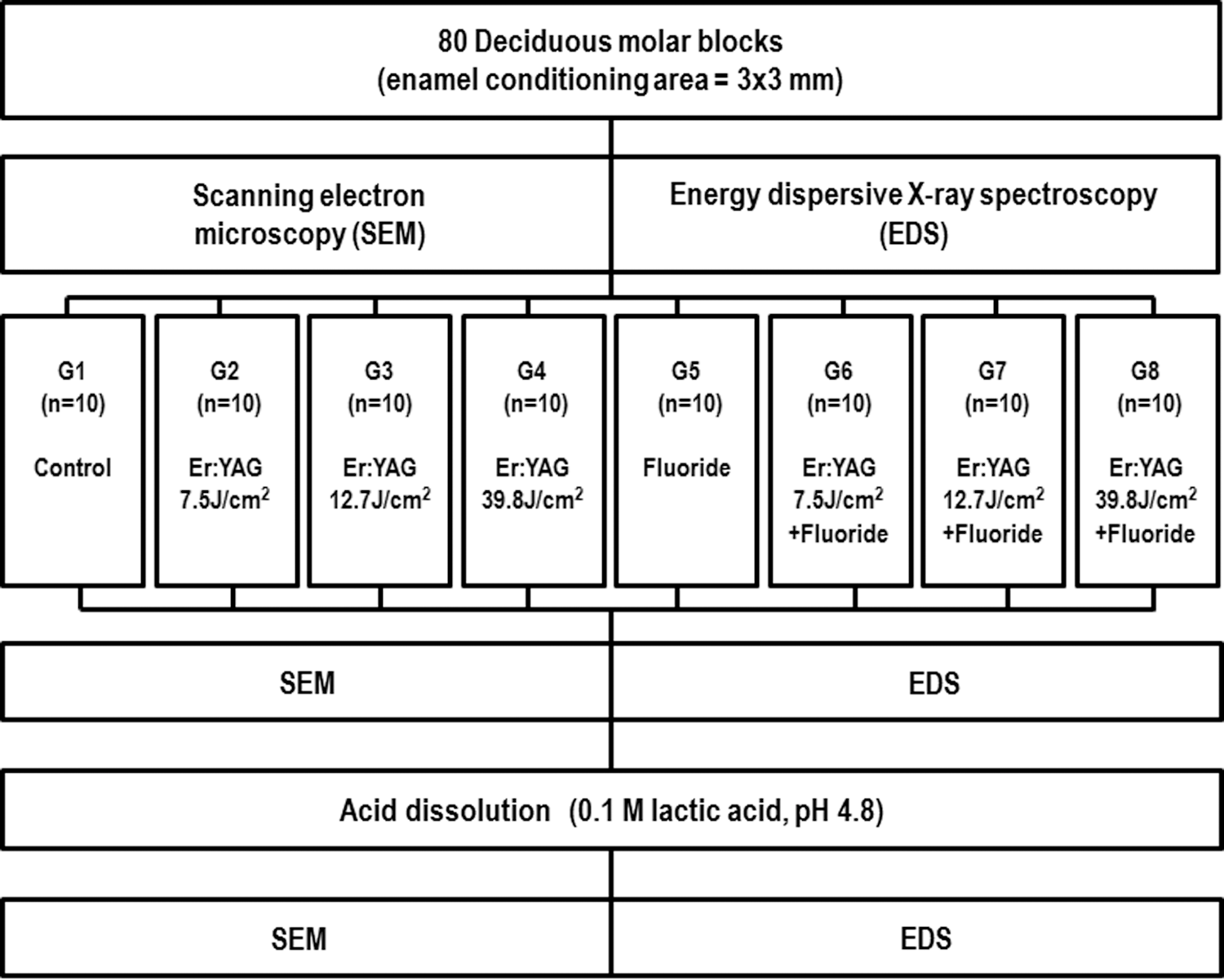
Diagram of the experimental design.
SEM
The blocks were fixed to aluminium stubs with double-sided adhesive carbon tape (SPI Supplies, USA). The analysis was performed using a scanning electron microscope (JEOL, JSM-6510LV, Japan) in the low vacuum mode at 10 Pa of chamber pressure, with an electron acceleration voltage of 25 kV and detecting backscattered electrons. The morphology of the enamel surface was observed at a magnification of 400x. The scaler tool of the image software (INCA, Oxford Instruments, Oxfordshire, United Kingdom) was used to trace a cross from the corners of the square sample, and the exact center of the cross was examined. To ensure satisfactory inter-examiner reproducibility of morphological findings, three examiners were calibrated with enamel SEM images selected from our research files prior to the start of SEM analysis (κ 0.95).
EDS
The whole area visualized under SEM at a standardized magnification of 100x was analyzed to determine the atomic percentages (at%) of carbon (C), oxygen (O), fluorine (F), trace elements (sodium+chlorine+magnesium), calcium (Ca) and phosphorus (P) using an X-ray detector system (Oxford Instruments, 7582, U.K) attached to the microscope.
Surface treatments
Eighty block samples were randomly assigned to eight groups (n=10) and enamel surface was conditioned as shown in Table 1.
Er:YAG laser irradiation
The irradiation of the specimens was performed using an Er:YAG laser system (OpusDuo AquaLite EC, Er:YAG+CO2, Lumenis, Yokneam, Israel) in the nonfocused and noncontact modes, with a wavelength fixed at 2.94 μm, at a pulse repetition of 7 Hz, and a pulse duration of 400 μsec. Energy levels were calibrated using the calipers of the equipment, and the energy delivered was measured periodically with a power meter (LaserMate-P, Coherent Co., Santa Clara, CA, USA). The surface was scanned once by hand (13 sec) with the sapphire tip of the laser perpendicular to it, at a working distance of 1 mm and with tridistilled water irrigation (5 mL/min).
Fluoride application
Enamel surface was dried with a compressed-gas duster (Falcon Safety Products, Inc., NJ, USA). Then, the teeth were treated individually during 4 min with 1.23% APF topical foam (Butler, Sunstar Americas, Inc., IL, USA). After, the samples were rinsed with tridistilled water for 1 min and then air dried. For G6, G7, and G8, fluoride was applied immediately after Er:YAG laser irradiation.
Acid dissolution
Samples were coated with an acid-resistant varnish, except on the enamel area designed for conditioning. Each specimen was immersed individually in plastic tubes containing 2 mL of a 0.1 M lactic acid solution with a pH of 4.8 and incubated at 37°C for 24 h. 19 After this time, the samples were removed and rinsed with tridistilled water.
Statistical analysis
All data were analyzed using the SPSS 18.0 statistical package (SPSS Inc., Chicago, IL, USA). The Kolmogorov–Smirnov test was performed to estimate the distribution of the data. Subsequently, the one way analysis of variance (ANOVA) was used to compare among groups; when significant differences were found, Bonferroni or Tamhane's T2 post-hoc tests were applied, depending upon Levene's test of homogeneity of variance.
A repeated measures ANOVA was performed to determine the differences among stages (before treatment, after treatment, and after acid dissolution). The level of significance was stated at p≤0.05 in all statistical analysis.
Results
SEM surface analysis
Morphological changes of deciduous enamel produced by Er:YAG laser, fluoride, and acid dissolution can be seen in Fig. 2. Before laser treatment, teeth showed some defects such as grooves, fractures, and even exposed prisms. After irradiation, the adverse effects were more accentuated at higher energy densities.
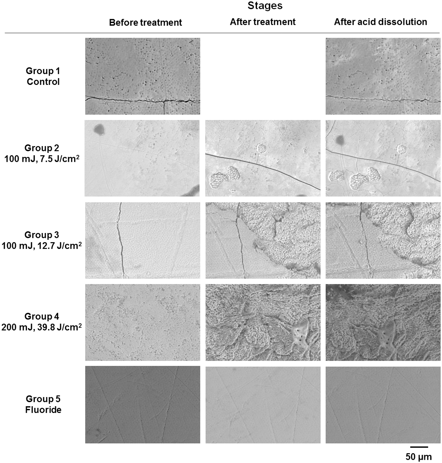
Representative scanning electron microscopic (SEM) micrographs of enamel surfaces for control, irradiated, and fluoride groups. Before treatment, the teeth showed smooth surfaces or microporosities, as well as some defects. After treatment, signs of damage were observed on irradiated enamel, including craters, completely exposed prisms, fractures, and melting. No morphological changes were observed with fluoride application and after acid dissolution. Original magnification, 400x.
However, surface morphology was not modified after fluoride application, or after the acid dissolution process.
EDS evaluation
The chemical composition of deciduous enamel surface determined before treatment, after treatment, and after acid dissolution is expressed in at% (means and standard deviations) (see Tables 2 and 3).
Capital letters in a row are for the comparison by chemical element of different groups in the same stage. Same capital letters follow means that do not differ statistically.
Bonferroni test, p<0.05; **Tamhane's T2 test, p<0.05.
Lower-case letters in a column are for the comparison by chemical element of different stages in the same group. Same lower-case letters follow means that do not differ statistically [repeated measures analysis of variance (ANOVA), p<0.05].
EDS, energy dispersive X-ray spectroscopy; BT, before treatment; AT; after treatment; AAD, after acid dissolution.
Capital letters in a row are for the comparison by chemical element or molar ratio of different groups in the same stage. Same capital letters follow means that do not differ statistically.
Bonferroni test, p<0.05; **Tamhane's T2 test, p<0.05.
Lower-case letters in a column are for the comparison by chemical element or molar ratio of different stages in the same group. Same lower-case letters follow means that do not differ statistically [repeated measures analysis of variance (ANOVA), p<0.05].
EDS, energy dispersive X-ray spectroscopy; BT, before treatment; AT, after treatment; AAD, after acid dissolution.
There were statistically significant differences in all analyzed elements, highlighting the following findings. Table 2 shows a decrease AT in C at% in G3, G4, and G8, more evident in groups irradiated at the highest energy density, also AAD. Additionally, in G5, there was an increase in C at% after fluoride application. A decrease in O at% was produced in groups treated with fluoride (G5–G8). Relative to F at%, G5–G8 showed an increase, which was higher in G7. AAD, the values of these groups dropped to 0.00. An increment in trace elements following treatment is displayed in G3, G4, G6, and G7.
Table 3 illustrates the calculated Ca/P molar ratio, as well as the content of such elements. Statistically significant differences were found in G4, G7, and G8. The Ca at% was increased AT in these groups, and values remained without modifications AAD. With regard to P at%, G4 and G8 had an increment AT and G7 showed a higher value AAD. The Ca/P molar ratio was superior AT in both groups irradiated at 39.8 J/cm2 (G4 and G8), and no changes were observed AAD. This ratio was also increased after the combined treatment of G7. Subsequent to surface treatment, G4, G7, and G8 had Ca/P values higher than the one obtained by the control group, but similar to G5 value.
Discussion
In the present work, we have studied the morphological and chemical changes of deciduous enamel surface induced by Er:YAG laser irradiation, fluoride topical application and combined treatment, as well as the effects induced by acid dissolution on treated surfaces.
In the first stage, teeth were subjected to SEM and EDS analysis, a procedure recommended by the authors as a baseline to evaluate the original morphological and chemical characteristics of enamel surface. At this stage, SEM observation showed defects such as grooves and slightly exposed prisms, which could be attributed to the natural wear processes. 20,21 The presence of scarce microfractures before treatment could be the result of exposure of the tooth to masticatory or traumatic forces in the oral cavity, dental extraction process, or section of the tooth during sample preparation.
The irradiation parameters were chosen according to the results of a pilot study conducted in our laboratory and designed after literature review. 10 –12,22,23 Water irrigation was used as a cooling agent to limit the temperature rise in dental tissues, 24 and to avoid the formation of undesired chemical phases more susceptible to acid dissolution. 13 Additionally, the topical fluoride used was APF, because it could reduce enamel decalcification, 25 and increase the formation of calcium fluoride (CaF2). 26
Morphological changes AT were more evident at higher energy densities. At 7.5 J/cm2 detachment of material in small isolated zones (shallow craters) was produced. Although Apel et al. 22 considered this energy density subablative for permanent teeth, this may not be applicable to deciduous enamel, because it has other characteristics including a thinner thickness, and a lower content of Ca and P; 27 therefore, a predominantly “prismless” outer layer, 28 whereby deciduous enamel could be less resistant to the laser thermal effect than permanent enamel.
Lased surfaces with an energy density of 12.7 J/cm2 showed more extended and numerous craters than those produced by laser irradiation at 7.5 J/cm2. Nevertheless, the formation of new fractures was not observed in either of the two densities.
At the highest energy density, lased teeth showed a totally modified surface with prisms completely exposed, fractures, and melting. Even at the macroscopic level, specimens of G4 and G8 showed a rough surface and a white color contrasting with the original tooth hue. In our study, the enamel morphology of specimens lased at 39.8 J/cm2 showed severe damage, which does not justify its use for caries prevention per se. For preventive purposes, dental enamel is not intended to be ablated or melted, only structural and chemical changes are expected. 29 Furthermore, the formation of rough surfaces could influence bacterial adhesion and plaque accumulation, 12,15,30 and also fractures could be points to initiate demineralization in deeper areas. 29
Otherwise, fluoride application and the acid dissolution process did not result in morphological modifications. Nonetheless, both procedures produced chemical changes as measured by EDS, a useful analysis tool for this purpose. According to the results of chemical composition obtained by EDS, the C at% decreased AT in G3, G4, and G8. Samples of G4 and G8 (the highest energy density) showed a significantly decrease with respect to other groups, even AAD. This results could be explained by a carbonate reduction and modification of the organic matter in Er:YAG-irradiated enamel, as reported by Liu and Hsu. 31 Additionally, the reduction of carbonates decreases the solubility of hydroxyapatite, because carbonate substitutions in the enamel surface give rise to less stability and, consequently, higher solubility. 32
On the contrary, for G5, the C content increased after fluoride application, probably because of the intake of organic compounds from the applied APF, such as glycerin, sodium saccharin, and polaxamer. This supports the findings of G7 regarding the unaccentuated reduction of the C content after combined treatment. Even though the irradiation process could have liberated carbon, when APF was applied, enamel reintegrated this element. However, the analytical technique used in our study is not capable of differentiating between carbon from the inorganic carbonates and carbon from the organic compounds inside the adamantine matrix. By contrast, the results of G8 could have been influenced by the ablation phenomenon, with possible changes in physical and chemical properties of the enamel, making it less akin to C and F uptake.
A decrease in O at% was observed in the groups with fluoride application, probably because of the release of this element resulting from the acid base reaction of hydroxyapatite 33 with the phosphoric acid of the APF product.
The F content AT was higher for G7 than for G5, G6, and G8. This could be explained because the energy density employed in G7 generated a surface with morphological and chemical characteristics that promoted the adsorption of F, as observed by SEM and EDS. Nevertheless, all the groups that acquired F after APF application lost it AAD. This suggests that the majority of the F atoms were not incorporated into the hydroxyapatite structure. The abovementioned idea may be supported by the fluoride mechanisms described by other authors. 12,34 When fluoride is topically applied, initially there is a deposition of CaF2 on the enamel surface, which serves as a reservoir of this element, and only a small amount of F gets firmly bound to enamel forming fluorapatite, an almost insoluble compound. 12 Afterwards, during an acid attack, CaF2 deposited on enamel releases F to perform its anticariogenic action. 34
Also, the higher deposition of fluoride in G7 could be associated with the P at% increase AAD, probably caused by a withholding of the phosphates in the apatite structure. Nevertheless, additional studies are required to clarify it.
Trace elements increased in the irradiated groups at higher energy densities, as well as in combined treatment groups, except for G8. However, as expected, they remained<1.0 at%.
The Ca at% increased in enamel when irradiated at 39.8 J/cm2 and after the combined treatments of G7 and G8; these values remained without change even AAD. The P at% was increased AT in G4 and G8, and AAD in G7. Despite the increment in P at% in G4 and G8 AT, the Ca/P molar ratios were increased as a result of a great increment in the Ca at%. Additionally, G7 had also a significant increase in Ca/P molar ratio AT, at the expense of Ca at%.
The Ca/P molar ratio values achieved for G4, G7, and G8 AT were higher than those obtained by control group, and close to 1.67, which is the stoichiometric ratio for pure hydroxyapatite Ca10(PO4)6(OH)2. Also, AAD, the ratio in these groups was higher than BT.
Because the Ca/P molar ratio has been considered a reliable mineralization indicator that allows establishment of behavior patterns, independent of variations of other elements in the teeth, the findings in G4, G7, and G8 suggest chemical changes that enhance the mineral content of the enamel structure. Because enamel Er:YAG laser irradiation results in both chemical and morphological changes, research in this field is focused on studying irradiation parameters appropriate for the supposed use, in this case for caries preventive purposes in combination with fluoride. The conditions employed in G4, G7, and G8 resulted in propitious chemical changes; however, morphological changes were adverse.
Conclusions
Morphological changes of Er:YAG laser irradiated enamel surface represented from mild to severe damages according to the used energy densities and applied protocol. Therefore, the conditions employed in this study are not recommended for the prevention of dental caries in deciduous teeth.
Er:YAG energy density influenced chemical changes in enamel to enhance its structural mineral content. The energy density of 12.7 J/cm2 caused a decrease in the C at%, as well as increased F uptake and an increase in the Ca/P molar ratio when combined with fluoride.
Acid dissolution removed fluoride from enamel surface, as shown by EDS analysis.
Footnotes
Acknowledgments
The authors are thankful to the personnel and patients of the Pediatric Dental Clinics of the Universidad Autónoma del Estado de México (UAEM) and of the Centro de Especialidades Odontológicas del Instituto Materno Infantil del Estado de México (IMIEM), for their support in the collection of the deciduous molars.
Author Disclosure Statement
No competing financial interests exist.