
Abstract
Introduction
A
PACT involves the combination of visible light, usually laser or light-emitting diodes (LEDs) and a photosensitizer (PS). 2 The PS is a compound that is capable of absorbing light of a specific wavelength and transforming it into useful energy. 2 This energy is in the form of free radicals of singlet oxygen, and is capable of damaging all types of biomolecules and of killing microbial cells by reacting with cellular components such as proteins, nucleic acids, and lipids. 3 Light and drug are nontoxic by themselves; hence, only cells bound by PS and receiving light are affected by “photodynamic action.” 3 PACT treatments of bacteria including Streptococcus mutans, 4,5 Streptococcus sanguinis, 6 Porphyromonas gingivalis 7 and Actinobacillus actinomycetemcomitans 6 have shown positive outcomes.
Toluidine blue has been shown to have bactericidal effects against gram-positive and gram-negative bacteria. 8,9 Characteristics such as low toxicity to host cells associated with hydrophilicity, simple molecular structure, and efficacy on singlet oxygen production make this compound the most studied dye in the PACT field. 10,11 Although this agent is very promising, its use is subject to lengthy clinical and legislative assessment. Furthermore, potential for staining teeth, resin-based restorations, and toxicity represent clinical limitations. 9
Therefore, more immediate benefits could be attained from PS that potentially have fewer side effects. 12 Several investigations have highlighted the use of curcumin as a feasible PS in PACT application. 5,13 –15 Curcumin has been used as spice in traditional Indian cuisine and medicine as well. 16,17 In addition, curcumin is the major constituent of turmeric powder, exhibiting a wide range of pharmacological effects, including anti-inflammatory, anticarcinogenic, and anti-infection activities. 18 Its effective use in oral disinfection and prosthetic decontamination have been attested to by recent studies. 14,19 Additionally, its antimicrobial property by lipid membrane peroxidation could be enhanced by light at a proper wavelength. 20,21 Curcumin absorbs light in the blue wavelength range (300–500 nm) of the visible spectrum, which has a lower penetration depth into the tissue compared with red light, because of scattering and absorption by biomolecules; therefore, the use of curcumin in PACT is restricted to topical use for superficial wounds (e.g., skin and mouth). 20 This fact could benefit the dental field, because of decontamination of the oral cavity by reducing the number of bacteria present over hard tissues, attested to by a recent investigation. 14 In addition, great effectiveness, low cost, and simple manipulation represent some advantages when using this PS. 22
Some reports have shown the use of a broad-visible light (400–700 nm) (e.g., halogen lamps, tungsten filament lamps, and white LEDs) to photoinactivate some micro-organisms involved with endodontic infections, 23 dental caries, 24 and mycoses. 25 Overall, these studies used long incubation and exposure times as well. The investigation of Gonzales et al. 25 photosensitized samples of Metarhizium anisopliae and Aspergillus nidulans after an incubation time of 30 min in methylene blue and toluidine blue, and, subsequently, exposure to a 300 W halogen lamp at 50 mW/m2 for 30 and 60 min, respectively. A 2-log10 and 4-log10 reduction was achieved after exposure of Escherichia coli and Enterococcus faecalis to irradiation of a white LED for 30 min (15 mW/cm2; 13 J/cm2) in the presence of toluidine blue (62.5 μM). 23 A tungsten filament lamp (400 W; 22.5–22.7 mW/cm2) in combination with different PS at 22 μM achieved a bacterial reduction of 48 h old S. mutans biofilms ranging from 1.5-log10 to 2.2-log10 after irradiation for 15 min. 24
Even though the use of a noncoherent broad visible light is claimed to be a limitation because of its long exposure time to photoactivate the studied PS, none of the previous reports have investigated the PACT effect of a white light in the presence of curcumin and toluidine blue aiming to photoinactivate cariogenic microorganisms. In addition, in this study we used a novel noncoherent broad-spectrum visible light source at a high set power that delivers high power intensity, resulting in an extra-short exposure time.
Therefore, the aim of this study was to verify the antimicrobial effect of PACT mediated by curcumin and toluidine blue exposed to a novel broad-visible light device at high set power and intensity on planktonic suspensions of S. mutans.
Materials and Methods
Bacterial strain
The micro-organism used in this study was S. mutans UA 159 (ATCC 700610). An aliquot was inoculated in buffered tryptone yeast extract (TSBYE) containing 1% (wt/vol) glucose and grown overnight, at 37o C, 5% CO2. Then, the bacterial suspension was centrifuged at 4000 rpm for 5 min, and the supernatant discarded. The cell pellet was resuspended in 20 mL of sterile phosphate-buffered saline (PBS). The cell numbers were adjusted at an optical density (OD) of 1.5 at 540 nm, 26 equivalent to ∼2×10⁹ colony-forming units (CFU)/ mL.
PS
In this investigation, curcumin (Sigma Aldrich, St Louis, MI) was dissolved in dimethyl sulfoxide (DMSO) to obtain a stock solution at 750 μM. After this, this solution was diluted in deionized water to obtain final concentrations at 0.075 μM; 0.75 μM, and 7.5 μM (keeping the final concentration of DMSO at 0.25%). In the same manner, a 1000 μM stock solution of toluidine blue (Sigma Aldrich, St Louis, MI) was prepared in deionized water and subsequently dissolved at concentrations of 0.25 μM, 2.5 μM, and 25 μM. Both stock solutions were kept in the dark, and were diluted in deionized water immediately before the experiments.
Light source
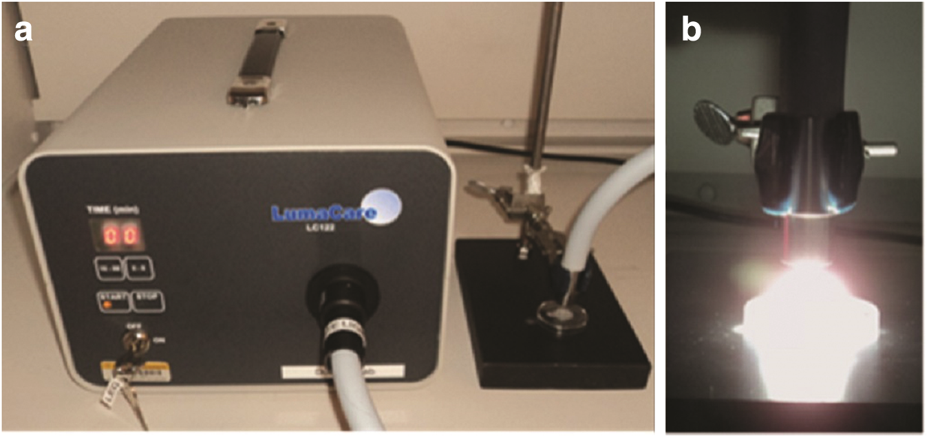
A single source of a noncoherent light that produces the full spectrum of visible light was used (LumaCare LC-122 A, LumaCare Medical Group, Newport Beach, CA). This light offers interchangeable probes at specific frequencies that are connected with a simple interlocking connection. An exploratory range test had been performed previously (data not shown) using different concentrations of curcumin and toluidine blue exposure to different dosimetries of lights at specific wavelengths (blue and red LED, respectively) established by published works, 5,27 aiming to define the protocol to be applied by white light. This light probe (beam diameter=12 mm) presented a wide spectrum from 400 to 700 nm at 550 nm (±150 nm) of central wavelength with a power intensity of 3410 mW/cm2 and set power of 3930 mW. The dosimetry tested was 42 J/cm2 (12.2 sec) and the work distance to activate both PS was fixed at 5 mm (distance between the light source and cell line surface) avoiding thermal effect (LumaCare™ manufacturer instruction), which had been previously tested (data not shown).
Photodynamic therapy application study design
S. mutans suspensions were incubated with concentrations of the tested PS. Curcumin solution was kept in the dark with S. mutans suspension for 60 sec (incubation time) 14 whereas toluidine blue was kept for 5 min. 13 After the incubation time, the suspensions were irradiated for 12.2 sec to achieve the tested dosimetry (42 J/cm2) with the samples receiving the total energy dose of 21 J. PACT groups were treated with both PS and light (C+WL+ and T+WL+, where C=curcumin, T=toluidine blue, and WL=white light). To determine whether PS alone induced any toxic effects on bacterial viability, bacterial suspensions were exposed to PS under identical conditions to those described, but no exposure to light (treated only with C or T: Group C+WL− and T+WL−). Exposing cells to irradiation determined the isolated effect of white light with no previous exposure to PS (treated only with white light: Group C−WL+ and T−WL+). S. mutans suspensions with no exposure to either PS or white light (C−WL− and T−WL−) were used as control groups. Figure 1 illustrates the application of PACT.
After the treatment application, aliquots of the treated suspensions were used to perform 10-fold serial dilutions, and the diluted samples were plated onto 5% defibrinated sheep blood agar (Sigma Chemicals, Co.) and then incubated at 370C, 5% CO2 for 48 h to investigate the number of viable microorganisms. After incubation, the total number of CFU was determined and number of CFUs per milliliters of suspension (CFU/mL) was obtained and transformed into logarithm (log10).
Statistical analysis
In order to verify the differences among the studied groups, the variable reduction in viable bacterial colony counts was analyzed by two way ANOVA and Tukey's test. The p value was<0.05 for statistical significance. BioEstat 5.0 software for Windows (Sociedade Civil Mamiraua, Manuas, AM, Brazil) was used for data analysis.
Results
Curcumin at the concentration of 0.75 μM exposed to 42 J/cm2 (12.2 sec) of white light resulted in a bacterial log reduction of 5.37 log 10 (±0.22) when compared with the control group (p<0.05).
The exposure of 42 J/cm2 of the white light in the presence of toluidine blue at 25 μM concentration (T+WL+) also achieved a substantial photokilling bacterial rate (p<0.05) with a reduction in log of 5.41 log10 (±0.26). Figures 2 and 3 demonstrate the effect of white light-PACT mediated by curcumin and toluidine blue, respectively. The application of light (C−WL+ and T−WL+) and both PS (C+WL− and T+WL−) by themselves did not reduce the log10 CFU when compared with control groups (p>0.05).
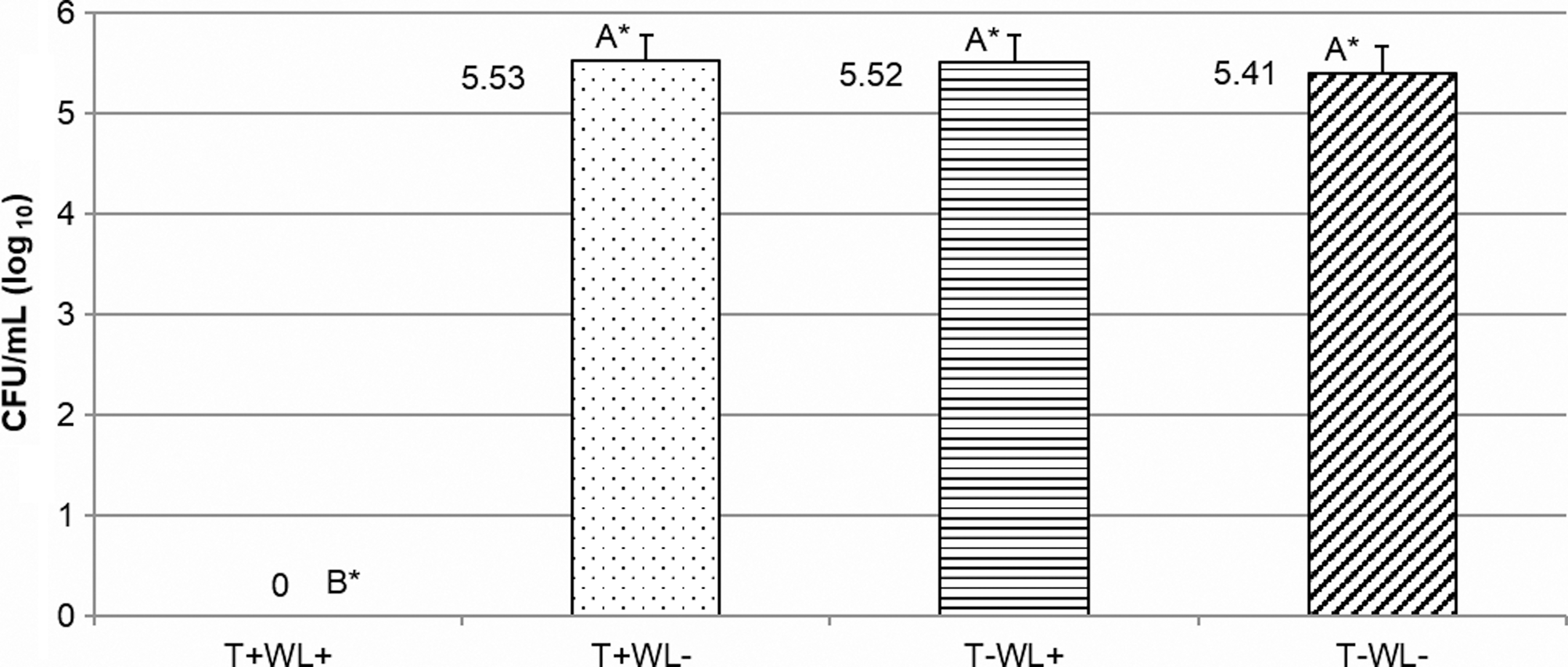
Treatment effects considering toluidine blue at 25 μM (T) and white light at 42 J/cm2 (WL) (PACT group: T+WL+), with T and without WL (photosensitizer [PS] group: T+WL−), without T and with WL (Light group: T−WL+), and without T and without WL (control group: T−WL−) on the viability of Streptococcus mutans suspensions. *Data followed by different letters differ statistically (p<0.05).
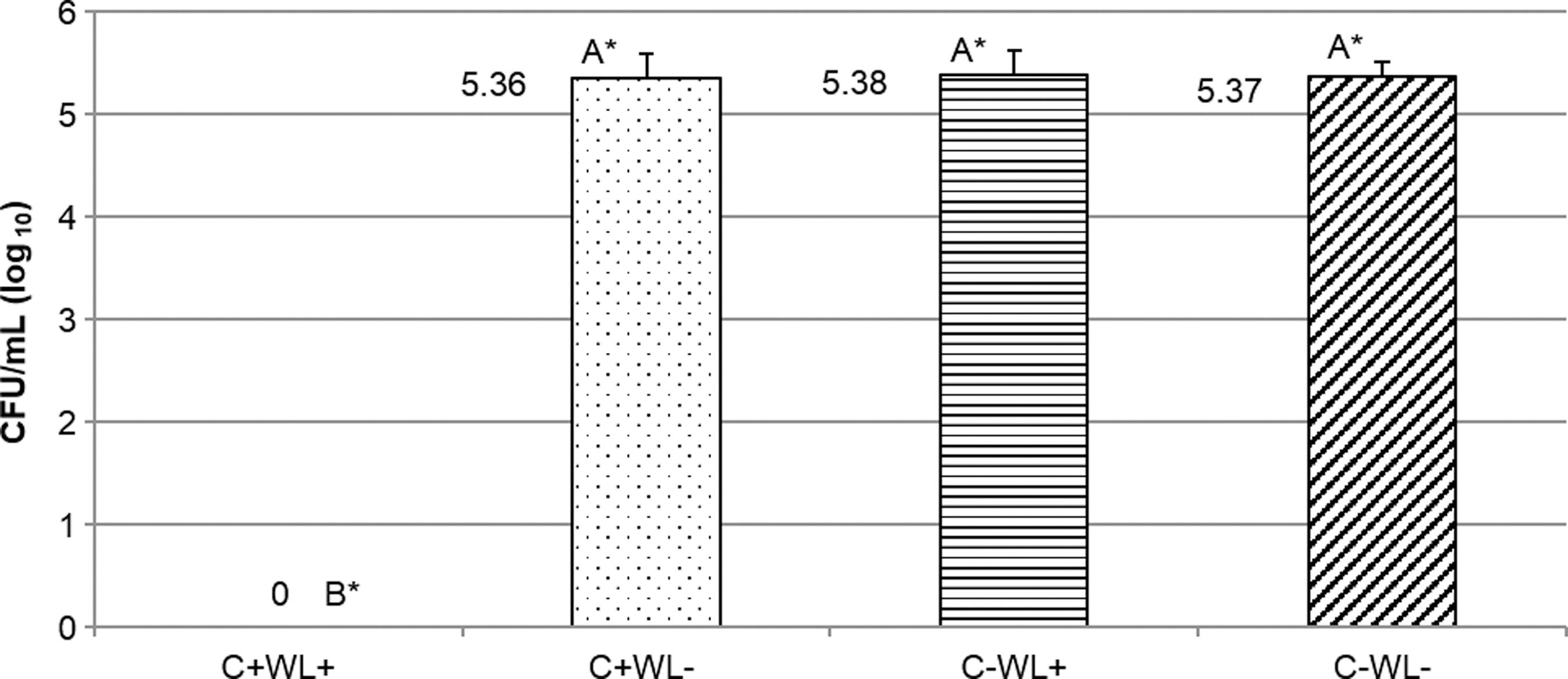
Treatment effects considering curcumin at 0.75 _M (C) and white light at 42 J/cm2 (WL) (PACT group: C+WL+), with C and without WL (PS group: C+WL−), without C and with WL (Light group: C−WL+), and without C and without L (control group: C−WL−) on the viability of Streptococcus mutans suspensions.*Data followed by different letters differ statistically (p<0.05).
Discussion
The present study investigated the effects of a new broad visible light (white light) at high power intensity to activate two different photosensitizers. The results showed that low concentrations of curcumin and toluidine blue exposed to a short white light illumination time (C+WL+ and T+WL+) caused a lethal photosensitization (∼ 99.9%) of S. mutans in suspension, which was different from the control groups (C−WL− and T−WL−). Light illumination alone (C−WL+ and T−WL+) did not produce bacterial reduction (p>0.05), which might indicate that there is no thermal effect. These results reaffirm that PACT intervention is an alternative option as an antimicrobial treatment in the dental field, and corroborates previous reports that achieved good outcomes when this procedure was tested in in vitro and in vivo approaches over cariogenic microorganisms. 5,9,28 Even though optimal results were achieved with this antimicrobial purpose, the illumination time used for these above-cited investigations 5,9,28 was much longer than the light exposure time proposed for this study.
In this investigation, a 5.41 log10 (±0.26) and 5.37 log10 (±0.22) bacterial reduction was achieved when toluidine blue and curcumin, respectively, were exposed to only 12.2 sec (42 J/cm2) of a novel source of white light. This is not the first investigation to use white light; 23 however, the time in the presence of curcumin and toluidine blue was much shorter than the ones reported in the literature referent to the white light. 23 –25 The current light source used (LumaCare™ system) presents high power density (3410 mW/cm2) provided by the current tested light (white light), and it is highly absorbed by the substrates because of its broad spectrum with a low percentage of reflection, allowing a more relevant interaction with a superficial tissue layer; short illumination times are known to be more efficient in reactive oxygen species (ROS) generation allied to a low photobleaching rate (photodegradation of PS). 13
The results based on the use of white light here are in agreement with previous reports that achieved a notable photokilling rate using the same broad spectrum. Wood et al. 24 verified that erythrosine (a dental plaque disclosing agent) at 22 μM showed a bactericidal efficacy of 1–2 log10 of S. mutans biofilms when irradiated for 15 min using a 400 W white light source. In this same year, Metcalf et al., 12 using this same light source and PS cited previously, fractionated the light dose (6.75 J/cm2) into five intervals of 1 min each, which achieved 3 log10 of bacterial viable S. mutans biofilm reduction. Still, further fractionation of the light dose into 10×30 sec doses separated by 2 min recovery periods resulted in 3.7 log10 of cell killing, an improvement of 1.7 log10 compared with the continuous irradiation protocol. They concluded that short illumination times followed by dark periods were able to redistribute the PS and avoid photobleaching, allowing the equilibrium of the PS concentration gradient. In the present study, we were able to achieve a remarkable photokilling rate of S. mutans suspensions after an extremely short exposure time, 12.2 sec, and by low concentrations of toluidine blue and curcumin, which can indicate that PACT mediated by both PS illuminated by this novel source of white light is a promising intervention against oral microorganisms. The next step is to study these therapies against this bacterium organized as biofilm.
The correlation between ROS and efficacy of the PACT have been questioned by previous reports. 4,13 It is expected that light sources that provide energy near PS peak absorption are able to generate more oxygen singlet and free radicals. However, even though in the present investigation we used a broad-visible light and two different PS, which are absorbed by lights that cover the red and blue spectrum, the data show a lethal photoinactivation (∼99.9%) of S. mutans after PACT mediated by the white light. This source of light presents a central wavelength of 550 nm (±150 nm) and a high power intensity delivered by only 12.2 sec. The absorption peak of curcumin and toluidine blue are close to 430 nm and 636 nm, respectively, so that the white light most likely covered both peaks (400–700 nm). Furthermore, other compounds in addition to ROS could be responsible for the effect of photodynamic application. The effect of photodynamic process is dependent upon the conjugation of PS and target cells, as ROS present high reactivity and limited diffusion power. 12 S. mutans are gram-positive bacteria that present a thick membrane that is relatively permeable, and is intimately associated with lipoteichoic and negatively charged teichuronic acids. 29 This membrane displays a relatively high degree of porosity, as various macromolecules, such as glycopeptides and polysaccharides with a molecular weight in the 30,000–60,000 range, were found to readily diffuse to the inner plasma membrane. 29 Thus, in this class of bacteria, the outer wall does not act as a permeability barrier for the most commonly used PS. Curcumin binds preferentially to lipid membrane and some cell proteins, whereas toluidine blue, a positively charged cationic phenotiazinum dye, acts to membrane gram-positive peptidoglycan cell walls, generating strand breaks in the organism's nucleic acid, with resultant genetic mutation and photodamage. These interactions are the result of molecular weight (toluidine blue, 305.83 g mol−1 and curcumin, 368.38 g mol−1) that allow the diffusion of PS to outer walls of studied micro-organisms allied to capacity of solubility. 5,24 In this experiment, curcumin was dissolved in 0.25 % DMSO to increase its solubility, because of its high hydrophobicity, a property not presented by toluidine blue. A vehicle control was used to monitor the effect of 0.25% DMSO in the cells, demonstrating no toxic effect using the DMSO at this concentration. Even though no toxicity was verified (data not shown), DMSO is not an optimal vehicle for in vivo application, because it increases membrane permeability to the drugs and may cause tissue damage and systemic effects. 13,20 Therefore, further studies need to investigate different vehicles to improve curcumin solubility in water. Moreover, low concentrations of dyes were able to produce high photodynamic effects. 5,14 Some authors have shown that high concentrations could serve as a “barrier” to light difusion, because of optical quenching by excess dye in solution. 5,30 Factors related to the type of microorganism and its organization (suspension or biofilm), concentration of dyes and type of light source, could be directly related to PACT effectiveness.
Conclusions
This present investigation is the first report to demonstrate the high bactericidal rate at a short illumination time of a noncoherent broad-spectrum visible light in the presence of low concentrations of curcumin and toluidine blue. It gives the basis for future studies aiming to apply this approach to reduce bacteria-related oral infections. The parameters tested are feasible for the dental practice, as this approach uses a visible light in a very short exposure time. Apart from PS limitations, such as staining potential (toluidine blue) and hydrophobicity (curcumin), further investigations on biofilms, and more information regarding side effects on host cells are needed to ensure the potential of this new light source to be clinically used.
Footnotes
Acknowledgments
The authors thank the Brazilian Government Agency – CAPES Foundation grant BEX #8485/11-9 (scholarship to Marco Aurelio Paschoal).
Author Disclosure Statement
No competing financial interests exist.