




Abstract
Introduction
T
Previous studies have shown that ultimate success in tooth whitening depends upon the concentration of the tooth bleaching gel, the light source, and the total treatment duration. 8,9 For more effective tooth whitening, different light sources may be used, such as quartz–tungsten–halogen lamps, lasers, plasma lamps, or light-emitting diodes (LEDs). When the bleaching agent is activated under the influence of light, some amount of light is absorbed and the resulting energy is converted into heat. Light sources may cause photothermal effects that are then associated with the chemical effect of the bleaching materials. Some authors consider this a major mechanism of action of all light-activated bleaching procedures. 10,11 Others consider light as the catalyst that increases the absorption of the gel and accelerates the chemical oxidation process. 8,12
Conclusions of the present literature regarding the effectiveness of photoactivator devices are limited by contradictory results. To further examine this issue, the purpose of this study was to evaluate the optical effect of experimental bleaching procedures activated by following light sources: LED light with center wavelength of 405 nm (LED405), white organic LED with wavelength of 400–760 nm (OLED) and femtosecond laser with center wavelength of 770 nm during application of 10% CP, 16% CP, 30% CP, 25% HP, or 38% HP, which could promote faster and more intense tooth bleaching without the excessive temperature rise. 13
Null hypotheses were: (1) The optical effect of bleaching is not affected by experimental light activation. (2) The optical effect of bleaching is not affected by type of bleaching agent.
Materials and Methods
Specimen preparation
For better objectivity, instead of using human or bovine teeth, the research was conducted on 200 pastilles made of hydroxylapatite powder (Hydroxyapatite for analysis, ACROS Organics Co., USA). Each pastille contained 400 mg of hydroxylapatite powder weighted in the scale (Mettler PM200, Switzerland) and compressed (Universal GP1, Croatia) under the pressure of 20 bars. The pastilles were dried in the dry sterilizer (Instrumentaria, Croatia) at a temperature of 150°C for 2 h to obtain the strength. Each was 10 mm in diameter and 2 mm thick. 14 They were absolutely equal, had the same red–green–blue (RGB) index before dyeing in tea (coefficient of variation <1%) and there was no other parameter apart from the bleaching process that could affect the outcome.
Pastilles were immersed in the tea solution for 8 h to create stained specimens (Fig. 1). Solution was made from 2 g of green tea (Cedevita, Croatia) boiled in 100 mL of distilled water for 5 min. 15 After drying, pastilles were randomly divided into four groups specified by the type of light source: LED405, OLED, and femtosecond laser (Fig. 2) or its absence (control group), each consisting of 50 pastilles. The distance between the light source and pastille was set to 10 mm. Each group was divided in five subgroups (n=10) specified by the applied bleaching gel: 10% CP, 16% CP, 30% CP, 25% HP, and 38% HP (Table 1). Bleaching gels were applied to the tooth surface in a layer ∼2 mm thick. Pastilles were bleached for 30 min.

Immersion of the pastilles in green tea.
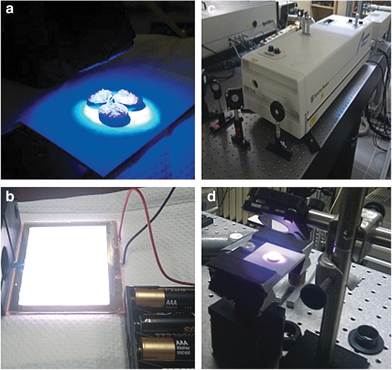
Experimental light activation.
Experimental light sources
Basic data on experimental light sources included in this study are presented in Table 2. Light-emitting diode (LED) is a semiconductor light source used for general lighting as well as for activation of tooth bleaching. 11,12 OLEDs are primarily used for general illumination and have never been tested in light-activated bleaching. They consist of an organic LED in which the emissive electroluminescent layer is a film of organic compound that emits light in response to an electric current. 16 Femtosecond pulse laser is a laser that emits pulses with a duration of 100 fs with average power of 1W. The whole system of femtosecond laser used in this study consists of a pump laser at 532 nm (Millenia, Spectra Physics, USA) and Tsunami oscillator (Ti:sapphire laser) which generates femtosecond pulses. The role of the pump laser is to excite the crystal Ti:sapphire in the oscillator, which ultimately generates femtosecond pulses.
LED, light-emitting diode; OLED, organic light-emitting diode.
Color change measurements
Instruments such as spectrophotometers and colorimeters are used to measure the colors of various materials and have shown potential of an accurate and reliable determination of the color of teeth. 17 –21 Ultraviolet-visible–near-infrared (UV/Vis/NIR) spectrophotometers are very precise, but also expensive, complex, and difficult to use for measuring the color of teeth in vivo. RGB-based colorimeters are widely used for color determination in dentistry, especially if the samples have flat surfaces like the pastilles used in this study. Three-dimensional color spaces based on retinal sensitivity: International Commission on Illumination (CIE) RGB, CIE XYZ, and CIE L*a*b* have been proposed for color analysis, with XYZ and L*a*b* derived from RGB color space. For more accurate and reliable determination of the color change, both colorimetric and spectrophotometric measurements were performed, respectively. The color of the pastilles was determined using RGB colorimeter (PCE RGB2 Color Analyzer, Germany). In order to facilitate data interpretation, RGB index was formed as the sum of colorimetric values R+G+B. Furthermore, reflectance spectra and optical properties of each pastille were assessed in the 300–800 nm wavelength range using UV/Vis/NIR spectroscopy (UV 3600, Shimadzu, Japan) equipped with an integrating sphere. A black rectangular cardboard segment (4×4 cm) with a central orifice of 10 mm in diameter was used to position the specimens in front of the sphere holder. Data were recorded with a computer connected to the spectrophotometer, obtaining a graph of light reflectance percentage per nanometer by using UV Probe software (Shimadzu, Japan).
Data analysis
Data were not multivariately normal and required nonparametric methods for the analysis of repeated measurements. Normality was checked by the Shapiro–Wilk test and the normal probability plot. Reported descriptive statistics included medians and interquartile range. Friedman test was applied to assess the differences among values of RGB index observed at the baseline, after immersion in tea and after further tooth bleaching procedures. Pairwise comparisons of repeated measurements were performed by the Wilcoxon signed rank test. Furthermore, effects of different tooth bleaching procedures, analyzed by the increase in the value of RGB index of dyed pastilles, were compared by the Kruskal–Wallis test and exact Wilcoxon rank sum test, for pairwise comparisons.
The significance level was set at 0.05. p Values were adjusted for multiple comparisons according to the Benjamini–Hochberg method, controlling the false discovery rate. Analysis was performed using SAS System, version 8.2 (SAS Institute Inc., NC, USA).
Results
RGB analysis
The baseline median (interquartile range) value of RGB index of hydroxylapatite pastilles was 3050 (3032–3063). A significant reduction was observed after dyeing in tea (p<0.001), dropping the median value of RGB index to 752 (736–814). After a further 30 min bleaching process, the values of the RGB index increased significantly during all bleaching treatments (p<0.001), but remained below the baseline values (p<0.001).
Bleaching with OLED had similar effect on the increase in the value of RGB index to that of bleaching without the light source, regardless of applied bleaching agent (Fig. 3). Application of bleaching agent with a lower concentration of peroxide–10% CP and 16% CP–led to a significantly smaller increase in the value of the RGB index (median of ∼600) than application of 30% CP, 25% HP, or 38% HP (median of ∼1500; p<0.001 for all comparisons between these two groups). Differences within the groups were not statistically significant (Fig. 4).
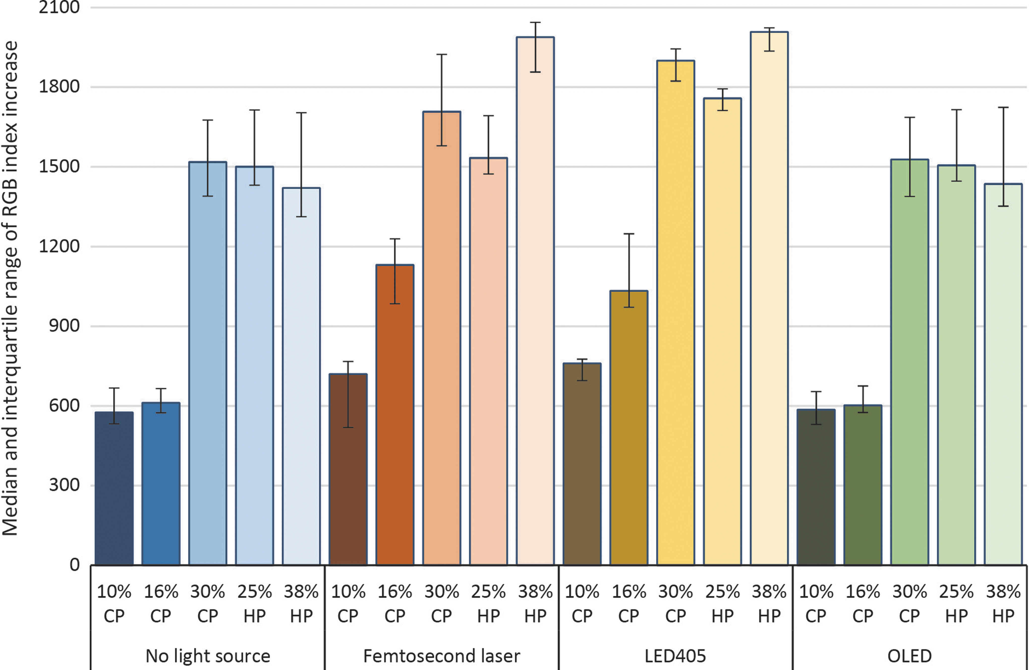
Median (interquartile range) increase in the red–green–blue (RGB) index of dyed pastilles after different bleaching treatments. LED405, light-emitting diode with a center wavelength of 405 nm; OLED, organic LED; CP, carbamide peroxide; HP, hydrogen peroxide.
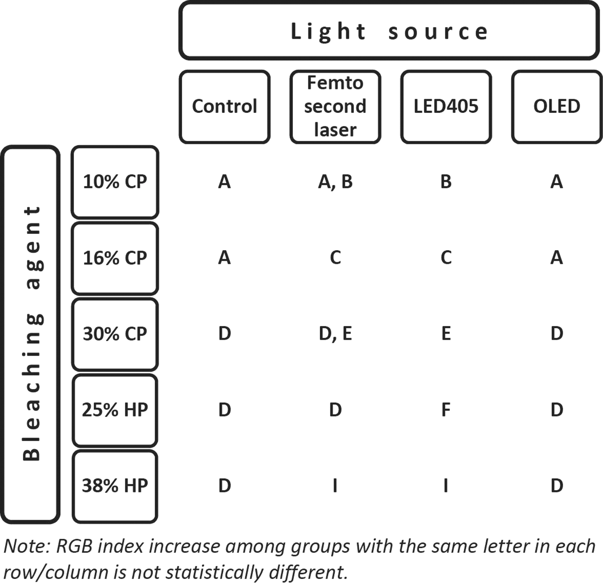
Results summary: significant differences in the increase of red–green–blue (RGB) index among light sources (presented by different letters in a row) and bleaching agents (presented by different letters in a column). LED405, light-emitting diode with a center wavelength of 405 nm; OLED, organic LED; CP, carbamide peroxide; HP, hydrogen peroxide.
Compared with bleaching with OLED and without the light source, LED405 induced a significantly larger increase in the value of the RGB index (p=0.029 for application of 25% HP; p<0.001 for application of other gels; same for OLED and no light source) as did the combination of femtosecond laser with 16% CP (p<0.001) and 38% HP (p<0.001) (Fig. 4). During treatments with LED405 and femtosecond laser, the effect of the bleaching agent was dependent on the concentration of peroxide. Application of 38% HP demonstrated the largest increase in the value of RGB index (median of 2008 for LED405 and 1988 for femtosecond laser), followed by 30% CP (1900 and 1708), 25% HP (1758 and 1534), 16% CP (1033 and 1131), and 10% CP (760 and 720) (Fig. 3). Differences between all gels were highly significant during treatment with LED405 (p=0.010 for comparison of 30% CP and 38% HP; p<0.001 for all other gel comparisons) (Fig. 4). The difference between 30% CP and 25% HP was not significant during treatment with femtosecond laser (p=0.064), in contrast to comparisons of other gels (p=0.011 for comparison of 30% CP and 38% HP; p<0.001 for all other gel comparisons). LED405 and femtosecond laser demonstrated a statistically significant difference in the increase of RGB index only when 25% HP gel was used (p=0.005), with the greater increase produced by the former.
Spectroscopic measurements
The results of light reflection, for all bleaching gels and light sources, measured with UV/Vis/NIR spectrophotometer are presented in Fig. 5. Spectroscopic measurements showed similar results to RGB analysis. The largest whitening effect was observed during bleaching with LED405, and the smallest was observed during bleaching without the light activation. Treatments with OLED were rated relatively better than in RGB analysis, producing similar or even greater improvements in color than femtosecond laser. Spectroscopic analysis of bleaching effects of different gels generally underlined conclusions made in RGB analysis: a higher concentration of peroxide in the bleaching agent enhanced the bleaching effect. However, some discrepancy was observed during treatment with LED405. Application of HP gels revealed a relatively underestimated effect in comparison with the CP gels. when contrasted to the results of RGB analysis.
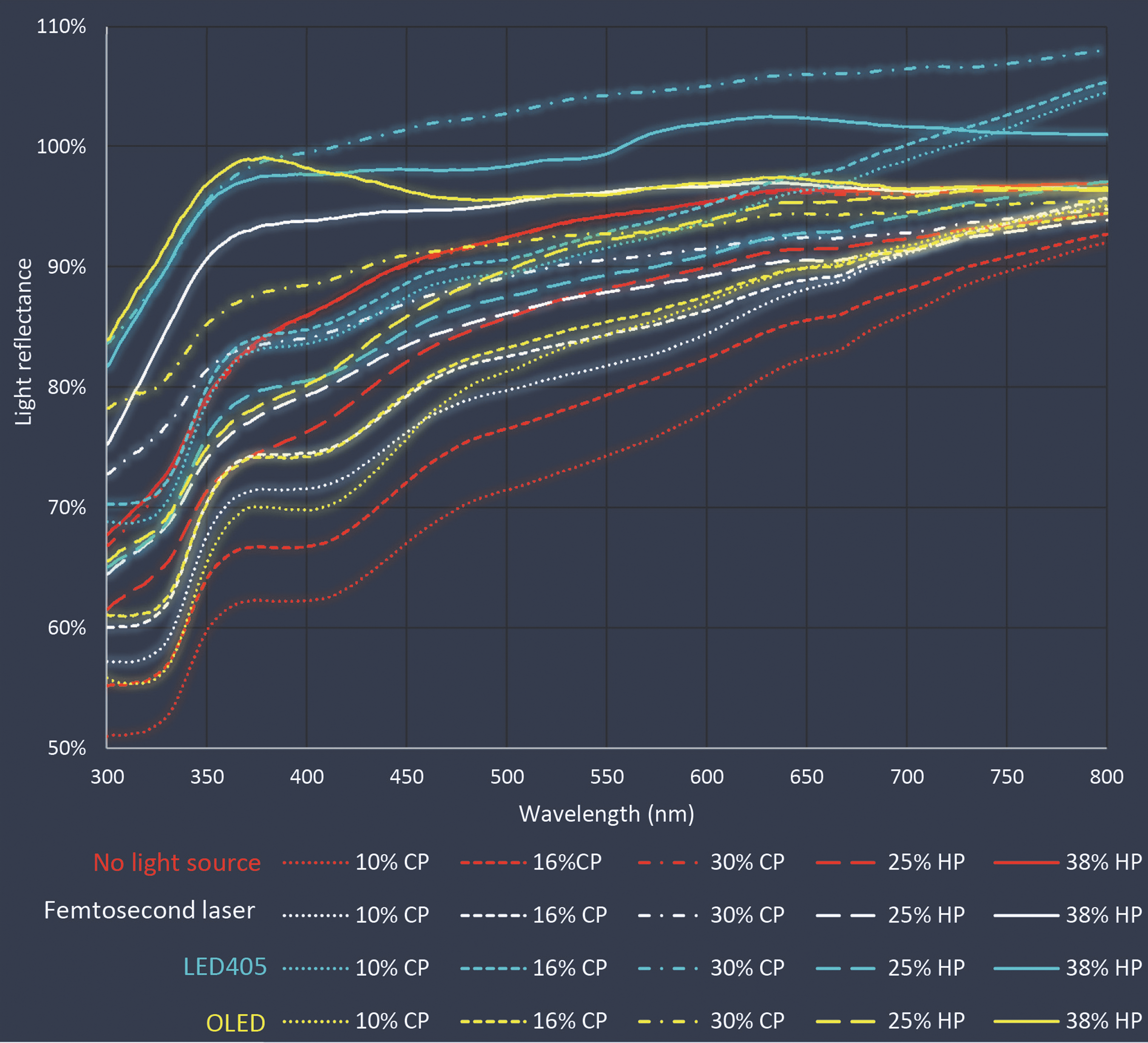
Ultraviolet-visible–near-infrared (UV/Vis/NIR) spectrum after different bleaching treatments. LED405, light-emitting diode with a center wavelength of 405 nm; OLED, organic LED; CP, carbamide peroxide; HP, hydrogen peroxide.
Discussion
Various lights used to accelerate power bleaching procedures have been investigated previously. 9,12,22,23 When light comes in the contact with the surface of tooth, one amount of light is reflected as a result of refractory index and the tooth surface. Reflected or transmitted color is a result of interaction with pigmented molecules and the amount of absorbed light. The wavelength of the absorbed light is the opposite of visible wavelength or the tooth color. 22
The light sources used for tooth bleaching mainly lead to excessive heat, which could improve the effect of bleaching gel. According to Goldstein and Garber, the most efficient temperature for heating the gel is between 46°C and 60°C. 24 Li et al., 25 Tavares et al., 26 Luk et al., 27 and Wong et al. 28 showed that the use of halogen lamps or LED 29 in combination with bleaching gels leads to a significant improvement in the tooth color compared with bleaching without light activation. On the other hand, Papathanasiou et al. 30 and Hein et al. 22 showed that the use of halogen lamps in conjunction with the bleaching gel does not lead to bleaching improvements. Ploeger et al. 31 concluded that the use of light does not contribute to the decomposition of hydrogen peroxide to the extent that would be visible in clinical practice.
This in vitro study analyzed the color change of hydroxylapatite pastilles during simulated tooth bleaching procedures using experimental light sources: LED405, OLED, and femtosecond laser. Results of colorimetric and spectrophotometric analyses revealed positive effect of the experimental light-activated bleaching on the values of RGB index and light reflectance. Therefore, hypothesis 1, that light activation does not lead to improvement in color, was rejected.
The possible effect of LED405, with a wavelength near UV, can be explained by the equation H2O2+hν→2HO• (h=Planck's constant) in which light of a specific frequency, ν, is absorbed, resulting in bond fission of H2O2 into two hydroxyl radicals. This type of energy can only be provided by high-frequency light, corresponding to a wavelength of 400 nm and lower (UV) which makes its use in the oral cavity difficult. 29 Improved performance of LED light source in combination with bleaching gels still remains unproven. 32,33 The results of our study suggest that application of LED405 enhances the bleaching effect. Colorimetric measurements showed significant improvement in the color of hydroxylapatite pastilles after 30 min of bleaching with LED405, compared with bleaching without light activation, regardless of applied bleaching gel. Furthermore, LED405 was the best rated among tested light sources in both colorimetric and spectrophotometric analysis. Spectrophotometric measurements showed the best reflection in the visible part of the spectrum closer to the brighter colors, that is, white, during bleaching with LED405.
The mechanism of laser systems for bleaching purposes depends upon the power, wavelength of the radiation, and pump mode. 34 Wavelengths with a high absorption coefficient in water and tooth mineral are readily absorbed at the tooth surface, where heat conversion takes place. These wavelengths (∼3000 nm) at the border between IR-B and IR-C, hardly penetrate deeper into dental hard tissue and, therefore, hardly pose a threat to the living pulp. 29 Lights in the red and NIR spectral range, like the one used in our study, act completely differently. The femtosecond laser had a central wavelength of ∼770 nm, which penetrates into biological tissue more easily. Thermal tissue damage during bleaching with femtosecond pulse laser can be minimized by appropriate choice of pulse duration and repetition rate, allowing for a sufficient “cool down” period between pulses. This modus is an important factor for efficacy and safety. 35 Studies have shown that NIR lasers could reduce pulp damage and relieve pain after the whitening process. 23 Some demonstrated no color improvement during bleaching with laser in combination with whitening gels, 29,36,37 only an adverse temperature increase in the pulp chamber. 27 On the other hand, Torres et al. have shown that the infrared diode laser can enhance the release of oxygen ions. 38 Wetter et al. suggested that the diode laser with 35% HP gel leads to better color improvement than the combination of gel and LED. 39 In our study, because of the small diameter of the laser beam, a system of lenses was created to increase the diameter and illuminate the entire surface of the pastille. Compared with bleaching without the light, the femtosecond laser showed significant improvement in the RGB index only when 16% CP and 38% HP were used, whereas spectrophotometric measurements showed optical improvement in combination with 10% CP, 16% CP, and 38% HP.
As no studies evaluating the bleaching effect of OLED were found in the available literature, our results cannot be compared with those of other authors. Colorimetric analysis of bleaching with OLED produced similar results to bleaching without the light source. However, spectrophotometric analysis indicated that OLED produces better results than bleaching with gel alone, and showed reflection in the visible part of the spectrum closer to the white color, similar to that of femtosecond laser or even slightly better.
The second null hypothesis, that optical effect of bleaching is not affected by type of bleaching agent, was rejected as well. The best bleaching results were achieved when using gels with higher concentrations of active ingredients, which led to a faster and stronger bleaching effect in a short time; however, the use of lower concentrations over time contributes to the color stability. 18
This study has some limitations. Research was conducted on pastilles of hydroxylapatite, which are different than human or bovine teeth in their composition, chemical structure, color, or physical feature. The pastilles were used as an experimental model to investigate the real and potential bleaching effect of new light sources. They served as experimental and uniform objects and were equal in their composition, size, shape, and color. These characteristics make pastilles a reliable choice for comparing the effects of different bleaching lights. On the other hand, human or bovine teeth are all different in their color, composition, size, and shape, and the true effect of the light source could be distorted by confounding. Furthermore, pastilles have a flat surface, and color change is easily measured with both RGB colorimeter and spectrophotometer.
We are not quite sure if the enhanced bleaching effect was the outcome of the heat produced by the light source or of the light output itself. Klaric et al. assessed the surface and pulp chamber temperatures during 30 min bleaching with LED405, OLED, and femtosecond laser, and concluded that generated temperature increase was below the critical threshold of 5.5°C for producing the thermal damage. 13 The improved color effect of light-activated bleaching may be attributed to the potential photochemical effect, such as photodissociation or photolysis, in which photons lead to electronic excitations and/or vibration in the molecule of hydrogen peroxide, cracking the certain chemical bonds. 29 Data on the mechanisms of action and the efficacy of light- and heat-activated dental bleaching are still very limited. Available studies do not allow for a final judgment regarding whether tooth bleaching can be increased or accelerated by additional light activation.
Conclusions
Bleaching procedures induced by light activation had a significant effect on the color change. The bleaching activation with LED405 and higher concentrations of peroxide in bleaching agents promoted better bleaching effect than other bleaching procedures. Application of activated bleaching procedures should be critically assessed, considering the possible physical, physiological, and pathophysiological implications. The patterns presented in this article provide a framework for ongoing further research to ascertain the effects of light sources on tooth bleaching, using different wavelengths and treatment durations, and different gel types and concentrations, as well as research on human or bovine teeth.
Footnotes
Acknowledgements
This study was supported by Croatian Science Foundation (Project 08/31 Evaluation of new bioactive materials and procedures in restorative dental medicine).
Author Disclosure Statement
No competing financial interests exist.