
Abstract
Introduction
N
Based on these clinical characteristics, a number of studies have evaluated the treatment effects on aging skin of different irradiating conditions of the therapeutic lasers. Chan et al. 10 assessed the efficacy of the Q-switched 1064nm Nd:YAG laser for skin rejuvenation, and found a slight association between laser toning with low fluence and mottled depigmentation. Trelles et al. 11 evaluated the use of long-pulsed 1064nm Nd:YAG laser with low energy density to remove periocular and perioral wrinkles. In their clinical study, Lee et al. 12 compared the treatment efficacy of Q-switched and long-pulsed 1064 nm Nd:YAG lasers on enlarged facial pores, and suggested a possible role for Nd:YAG lasers in pore size modulation. For treatment of facial rhytides, Crane et al. 13 evaluated the efficacy and safety of the 755 nm Alexandrite laser, according to different fluence conditions, with a fixed spot diameter. Tierney et al. 14 assessed the degree of improvement after a series of treatments with the 755 nm Alexandrite laser for hypertrophic, nodular, and macular port wine stains. To treat recalcitrant pigmentation after unsuccessful depigmentation therapy for vitiligo, Rao et al. 15 used the Q-switched 755 nm Alexandrite laser, and achieved excellent results, with no recurrence of pigment after a 1 year follow-up.
However, the existing approaches have two major limitations. First, current approaches produce mild and slight improvements for photoinduced aging, as they transmit a therapeutic laser with a low fluence condition. Second, most existing studies employed the 755 nm Alexandrite laser to remove unwanted hair and tattoos, because of the relatively low penetration depth. Typically, the risk for harmful skin damage increases, whereas the beneficial thermal damage also increases; when the fluence rises, higher heat energies are delivered to the skin. Therefore, it is possible to provide more convincing treatment results, when the optimum irradiation conditions with relatively high fluence are established. Particularly, quantitative and histological evaluations are essential to prevent adverse sequelae after laser transmission. Using these optimum treatment conditions, the clinical applicability of the 755 nm Alexandrite laser can be drastically improved for skin rejuvenation.
The purpose of this study is to quantitatively and histologically compare degrees of epidermal/dermal tissue damages after irradiation with therapeutic lasers, to find ideal treatment conditions for skin rejuvenation under relatively high fluence conditions. For this purpose, we transmitted 1064 nm Nd:YAG and 755 nm Alexandrite lasers into pig skin, with different spot diameters and fluences, and estimated internal/external skin temperatures. We also histologically assessed epidermal/dermal changes for each laser condition.
Materials and Methods
Experimental subjects
Skin specimens were excised from the rear flanks of 6-month-old Yucatan mini pigs (Optifarm Solution, Korea), which have tissue properties most similar to human skin, within 24 h after animal euthanasia. 16,17 After designating a suitable region without any skin injury and scars, the hair was removed using electric clippers for more effective heat transfer. The size of skin specimens was set to 50 mm (W)×50 mm (H)×30 mm (D).
According to our previous study, 18,19 a total of three thermocouple slots were composed at depths of 2.5, 5.0, and 7.5 mm to measure dermal temperatures. Twenty-one gauge, 30 mm hypodermic needles (Korea Vaccine, Seoul, Korea) were employed to pierce the dermis layer of the pig skin, and were positioned parallel to the pig skin. We promptly inserted the thermocouple wires through the needles, until each extended 10 mm past its tip. The thermal diffusivity of each skin specimen was 0.82×10⁻⁷−0.6×10⁻⁷ m2/sec, within 71–100% of that for human skin (0.82×10⁻⁷−1.2×10⁻⁷ m2/sec). 20
Laser irradiation conditions
Long-pulsed 1064 nm Nd:YAG and 755 nm Alexandrite lasers were transmitted into the pig skin specimens and internal/external heat distributions were analyzed. To establish optimum irradiation conditions for skin rejuvenation under relatively high fluence, the fluence conditions were set to 26, 30, and 36 J/cm2, and the spot diameter conditions were 5, 8, and 10 mm at each fluence condition. The pulse duration was designated as 30 ms for all experiments. Based on these test conditions, we evaluated the treatment efficacy and safety of the therapeutic lasers, for a total of nine different irradiation conditions.
Temperature measurements
The epidermal temperatures of the pig skin specimen were measured using a model 876 commercial infrared (IR) camera (Testo, Lenzkirch, Germany). The IR camera was able to continuously and simultaneously capture the maximum and minimum temperatures, whereas the resolution of the thermal images was 640×480 pixels at a minimum focusing distance of 0.4 m. The thermal sensitivity was lower than 80 mK at 30°C, and the spectral range was within 8–14 μm. The temperature measuring ranges were manually switchable from the −20–100°C to the 0–280°C ranges, and measurement accuracies were±2°C and±2%, respectively. Thermal images were obtained every 0.01 sec and analyzed using AnalyzIR+1.2 software.
The dermis temperatures were measured at depths of 2.5, 5.0, and 7.5 mm by K-type thermocouple after injecting wire sensors into the thermocouple slots. The diameter of the employed thermocouple was 1.57 mm. The temperature measuring ranges were within −200–1200°C, and the error range was±0.75%. The temperature data were acquired using an NI PCI-611 data acquisition card (DAQ) (National Instruments, Austin, TX), by means of LabVIEW (National Instruments) software. We focused on the maximum temperatures of the epidermis and dermis layers, to compare the thermal limitations of skin damage for each treatment condition. Figure 1 shows the experimental setup for estimating internal/external skin temperatures.
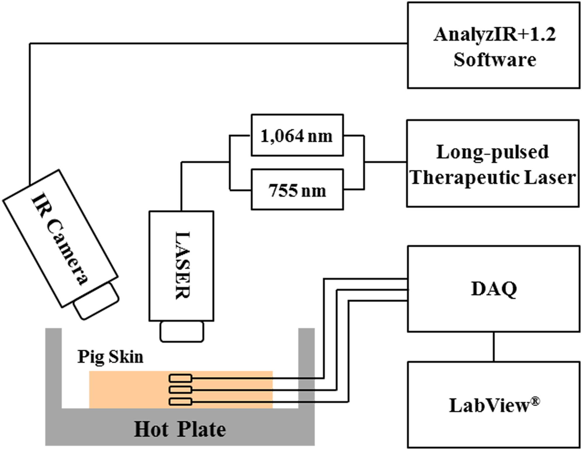
Experimental setup for estimating epidermal and dermal temperatures using the 1064 nm Nd:YAG and 755 nm Alexandrite lasers.
Histological evaluation
Routine punch biopsy specimens with a thickness and diameter of 8 mm were taken from the laser transmitted regions using a BP-80F biopsy punch (Kai Medical, Omera City, Japan), after irradiation of each laser three iterative times, to conduct histological examinations. 21 All biopsy specimens were fixed in 10% formalin, and embedded in paraffin. The samples were then sectioned to 5 μm in thickness and stained with hematoxylin/eosin (H&E) to evaluate epidermal/dermal changes. Histological images were taken of the slides at 100×magnification using a CH-2 calibrated light microscope (Olympus, Tokyo, Japan).
Data analyses
For statistical analyses, data were analyzed using the paired t test analysis via SPSS (Ver. 12.0 for Windows, Chicago, IL). A p value<0.5 was considered statistically significant.
Results
Comparison of the maximum dermal temperatures between the 1064 nm Nd:YAG and 755 nm Alexandrite lasers for the different transmitting conditions
The dermis temperatures were larger using the 1064 nm laser than using the 755 nm laser for all experimental conditions (p<0.05). The dermis temperatures continuously increased as the spot diameter and fluence increased for each penetration depth. The temperature increments also increased consistently as the spot diameter and fluence increased for all test conditions, and these increments were larger in the 1064 nm condition than in the 755 nm conditions (p<0.05). The dermis temperatures were far more strongly influenced by the spot diameter variations than by the fluence changes, irrespective of the laser types. Table 1 lists the epidermal and dermal temperatures for varying penetration depths between the 1064 and 755 nm lasers.
On the other hand, the dermis temperatures were decreased as the penetration depths deepened regardless of the laser types, and the temperature decrements drastically increased as the fluence and spot diameter increased (p<0.05). The temperature decrements at 10 mm and 36 J/cm2 between the penetration depths of 2.5 and 7.5 mm were ∼4.85 and 6.13 times larger than those with the condition of 5 mm and 26 J/cm2 for each laser, respectively. In contrast, the temperature differences between therapeutic lasers markedly diminished as the penetration depths deepened. Although the dermis temperatures were largely shown in the 1064 nm group at a penetration depth of 7.5 mm, the differences were quite small. Figure 2 compares the maximum dermis temperatures between the 1064 and 755 nm lasers, for the penetration depth of 2.5 mm.
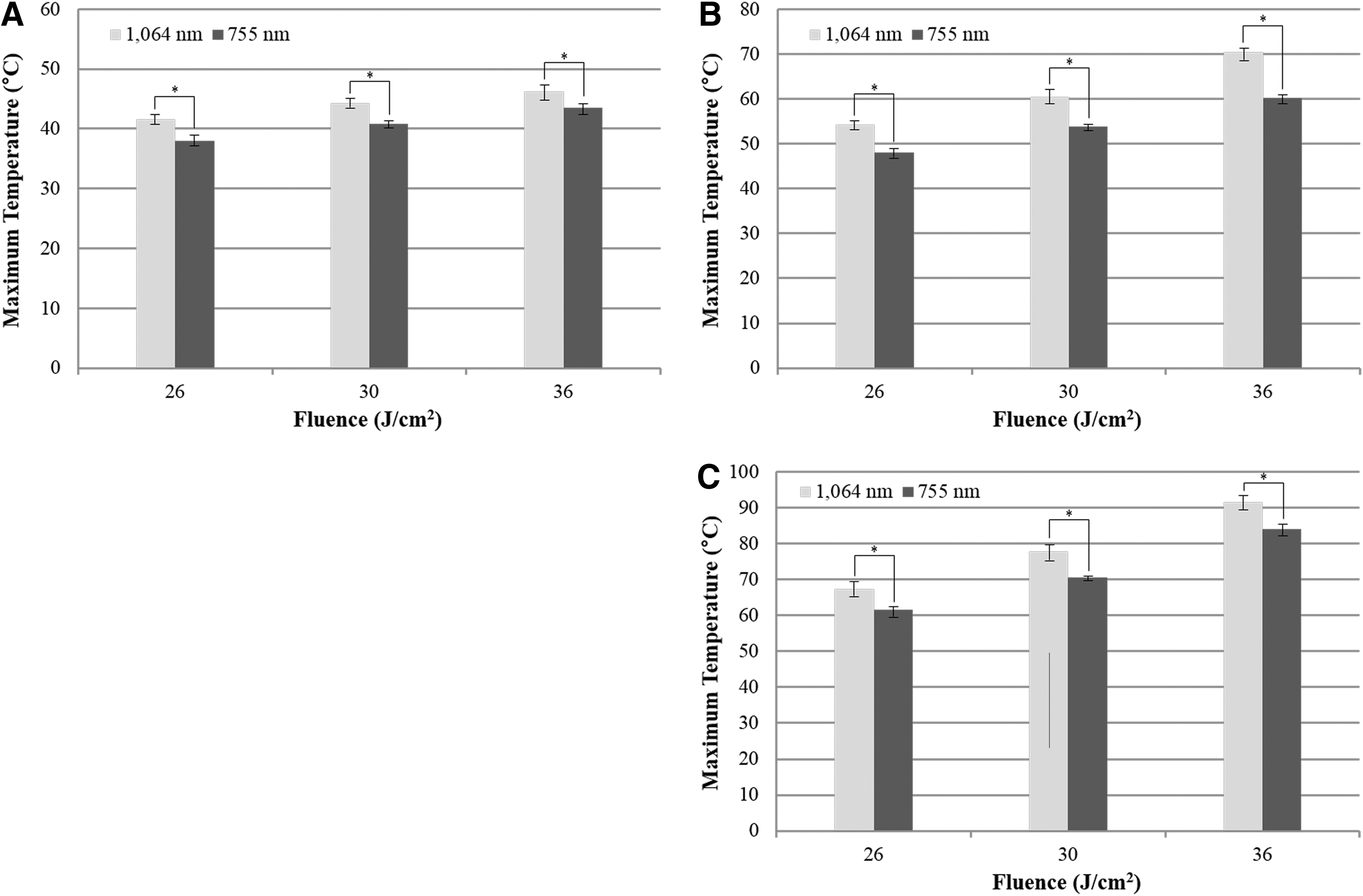
Comparison of the maximum dermal temperatures at a depth of 2.5 mm between the 1064 nm Nd:YAG and 755 nm Alexandrite lasers according to the different fluence and spot diameter conditions.
Comparison of the maximum epidermal temperatures between the 1064 nm Nd:YAG and the 755 nm Alexandrite lasers according to the different fluence and spot diameter conditions
The epidermis temperatures were larger in the 755 nm laser groups than in the 1064 nm groups for all experimental conditions, whereas the temperature increments were also slightly larger in the 755 nm condition (p<0.05). The epidermis temperatures continuously increased as the spot diameter and fluence increased. However, the surface temperatures were influenced more seriously by fluence changes, rather than by the spot diameter variations, unlike the results from the dermis layer. Moreover, the temperature variations of the epidermal layers were relatively small in comparison with those of the dermal layers, for all irradiation conditions. The maximum temperature differences were >50°C at a depth of 2.5 mm, whereas the differences were <10°C on the epidermal layers. Figure 3 depicts the maximum epidermis temperatures between the 1064 and 755 nm lasers, for the different irradiation conditions.
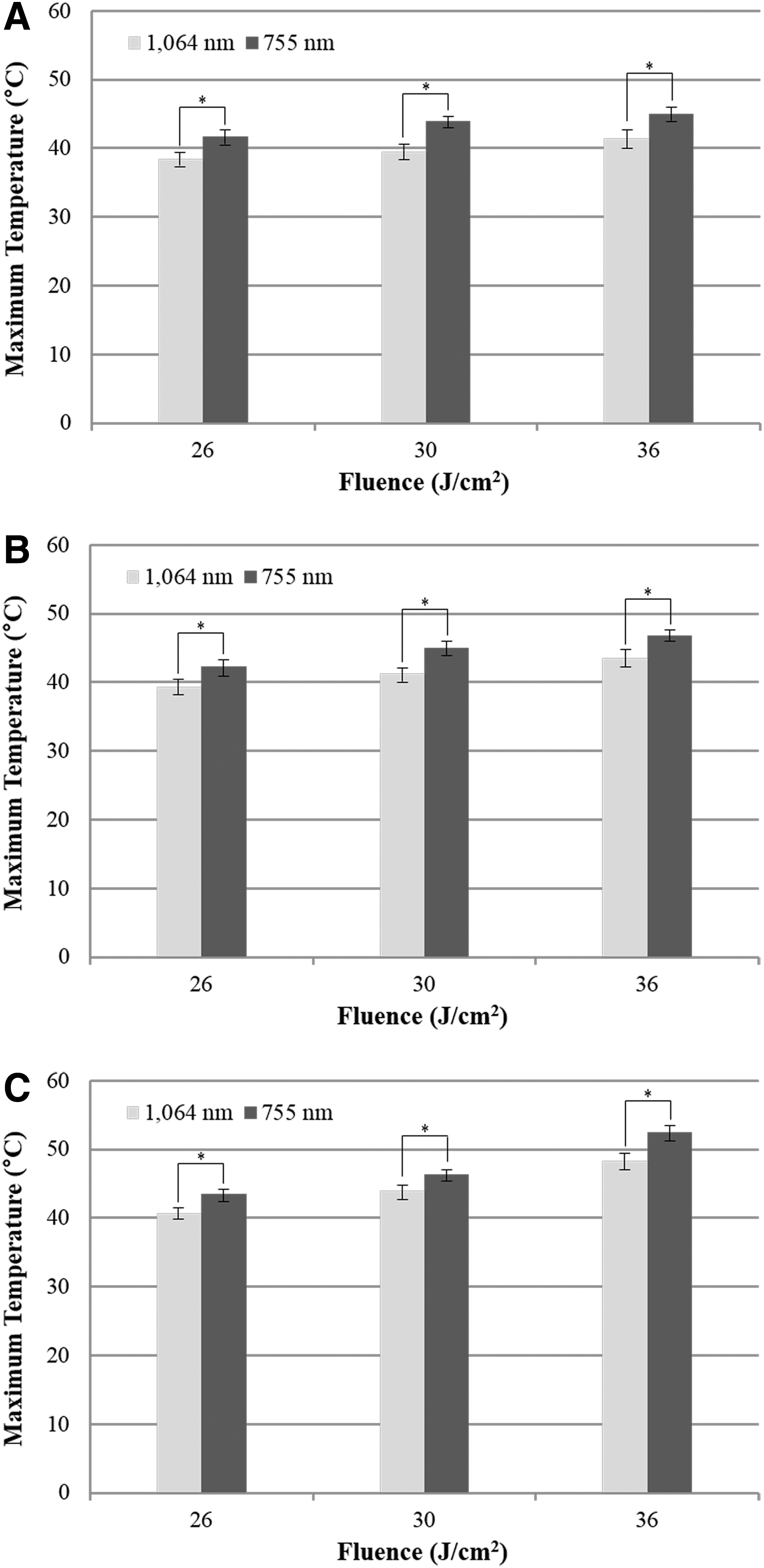
Comparison of the maximum epidermal temperatures between the 1064 nm Nd:YAG and 755 nm Alexandrite lasers for the different fluence and spot diameter conditions.
Histology
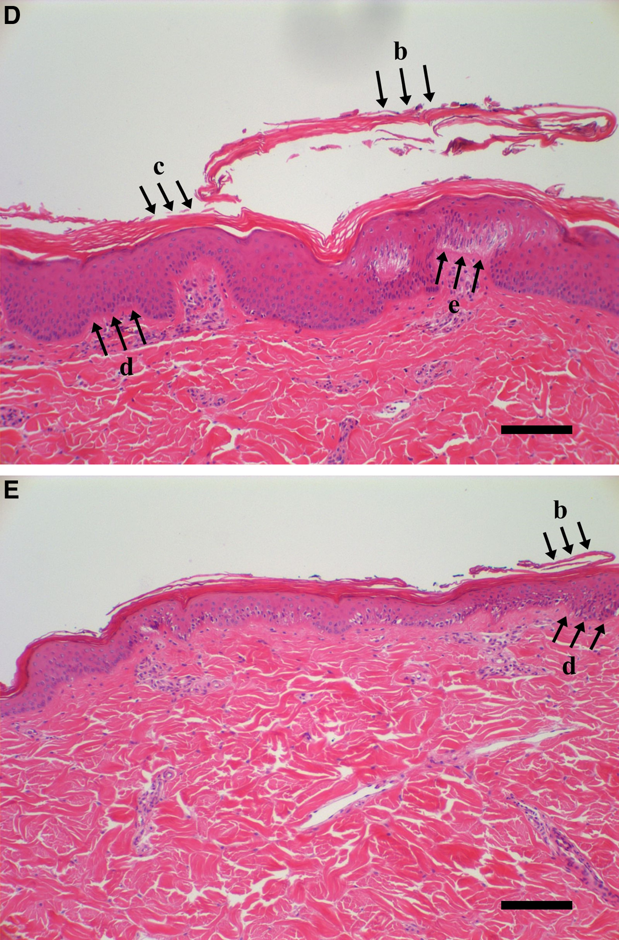
Figure 4a shows histological sections of an unirradiated control specimen. Control specimens did not include any thermal injuries on the skin surface, and our skin specimens were composed of epidermis, dermis, and dermal appendages. At a spot diameter of 5 mm, both the 1064 and 755 nm lasers caused only slight deformations of fibers on the dermis layers for all fluence conditions, and serious tissue damage did not occur. At a spot diameter of 8 mm, more fibrous tissues were formed in the dermis layers as the fluence increased. Particularly, the 1064 and 755 nm lasers induced the most active stimulation of fibroblast activity and collagen reformation with the fluence conditions of 30 and 36 J/cm2, respectively, without any serious injuries (Fig. 4b,c). In the case of the 10 mm spot diameter, each laser provided clinically significant changes only for the fluence condition of 26 J/cm2. However, the irradiation conditions of 8 mm with 36 J/cm2, 10 mm with 30 J/cm2, and 10 mm with 36 J/cm2 for the 1064 nm laser, as well as 10 mm with 30 J/cm2 and 10 mm with 36 J/cm2 for the 755nm laser, where the dermis temperatures rose >70°C, caused significant tissue damage to the epidermal and dermal layers. The fluence condition of 36 J/cm2 caused serious epidermal damages, such as epidermolysis and cornification, accompanied by the collapse of basal layers and epidermal necrosis (Fig. 4d, e). Figure 4 demonstrates the representative histological changes of the pig skin specimens, after laser irradiation.
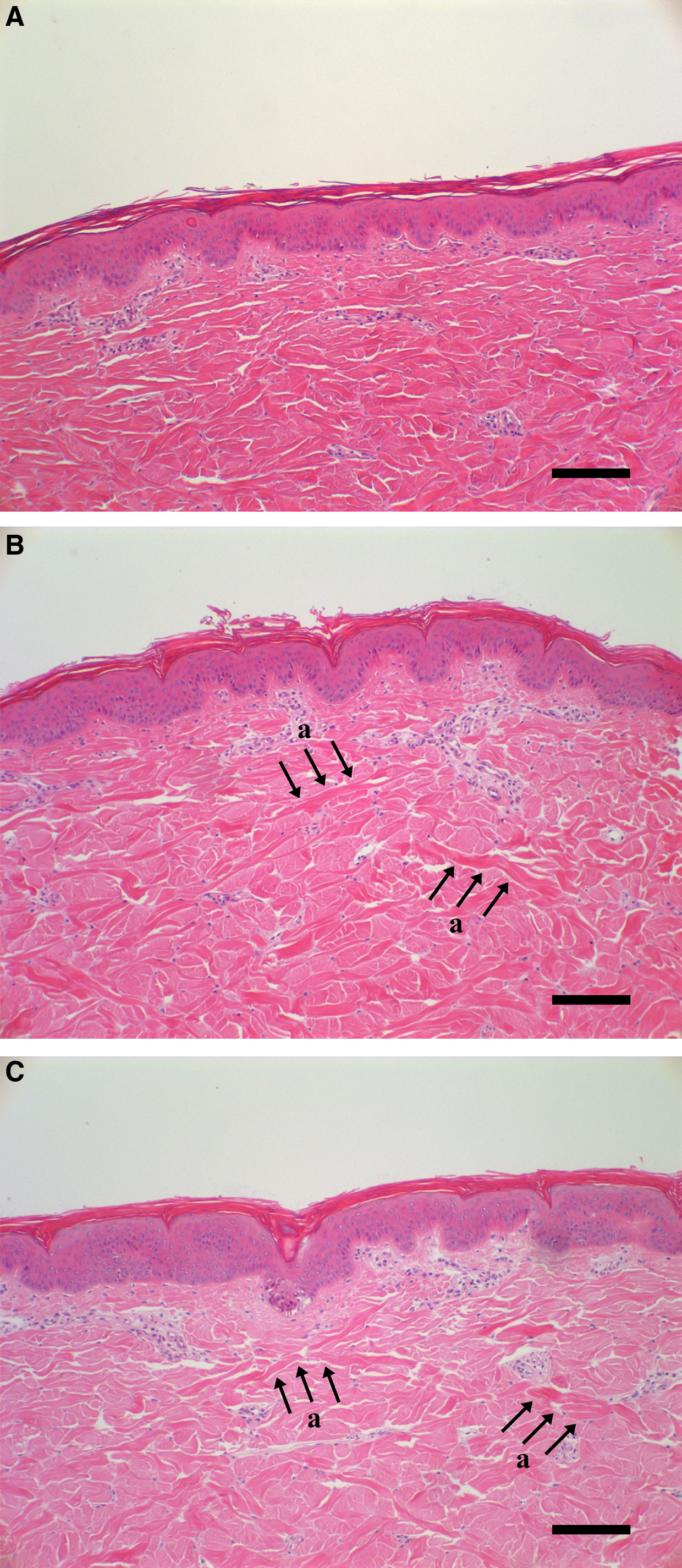
Representative hematoxylin and eosin stain (H&E) of pig skin specimens after the two laser irradiation modalities.
Discussion
The NSR technique delivers useful packets of photothermal damages to the lower dermis through the epidermis without any significant injuries, encouraging wound healing, and resulting in collagen regeneration and remodeling. 22 Therefore, to locate optimum treatment conditions for skin rejuvenation, it is essential to quantitatively and histologically evaluate the epidermal/dermal changes simultaneously after the laser transmissions. In our study, we transmitted the long-pulsed 1064 nm Nd:YAG and 755 nm Alexandrite lasers into pig skin samples, and evaluated degrees of epidermal/dermal tissue damages for the various transmitting conditions.
Analyses of the maximum temperatures between the epidermis and dermis layers revealed totally different characteristics for the temperature variations. The epidermal temperatures and its increments were larger in the 755 nm laser groups than in the 1064 nm groups, whereas the opposite results were obtained on the dermis layers. These findings are mainly the result of the different penetration characteristics of the two lasers. The 755 nm laser penetrates more shallowly than the 1064 nm laser because of its inherent nature. Therefore, the heat energies are focused on a relatively low depth. On the other hand, the 1064 nm laser concentrates heat energy more deeply under the same irradiation conditions, because of the relatively high penetrability. 23,24 Therefore, the epidermal temperatures were more strongly influenced by the 755 nm laser, whereas the 1064 nm laser induced larger temperature variations in dermis layer. Moreover, the epidermal temperatures were influenced more by fluences than by spot diameters, whereas the dermis temperatures were altered more greatly by spot diameter changes. Fluence changes do not significantly alter the penetration depth, whereas the epidermal temperatures are strongly influenced, as fluence indicates the energy density delivered per unit area. The increase of the spot diameter greatly influences the penetration depth, because the unit area where the heat energies are concentrated is expanded. 25 Therefore, the heat energies are transmitted more deeply and the dermis temperature alters significantly as the spot diameter increases. These characteristics are responsible for the particular temperature variations between the epidermis and dermis layers.
In their experimental study, Leach et al. 26 reported 47°C as the limiting temperature for visible changes of the epidermis, and 50–55°C as the threshold for irreversible injuries. Moreover, a dermal temperature within 60–70°C can provide tighter skin by inducing beneficial thermal damages on the dermis layers, and promoting collagen regeneration. 27 –29 However, permanent damages can occur when the dermal temperature increases >80°C. 30 In our experiments, all irradiation conditions except for the condition of 10 mm and 36 J/cm2 showed epidermal temperatures <47°C, and did not cause any serious tissue damage. On the other hand, only the conditions of 8 mm with 30 J/cm2 and 10 mm with 26 J/cm2 for the 1064 nm laser group, and the conditions of 8 mm with 36 J/cm2 and 10 mm with 26 J/cm2 for the 755 nm laser group corresponded to the aforementioned ideal dermis temperature conditions. The irradiation condition of 10 mm and 36 J/cm2 was inappropriate for skin rejuvenation, as the epidermal and dermal temperatures were increased >47°C and 80°C, respectively.
These experimental results were very similar to the histological results for all experimental conditions. The treatment condition of 8 mm with 30 J/cm2 and 10 mm with 26 J/cm2 for the 1064 nm group, as well as 8 mm with 36 J/cm2 and 10 mm with 26 cm2 for the 755 nm group, provided histologically meaningful results. In addition, the condition of 10 mm and 36 J/cm2, which elevated the epidermal temperatures >80°C, produced irreversible damage on the dermis layers. It was noteworthy that the 1064 nm laser group typically showed more active fibrous formations than did the 755 nm laser group for the same transmitting conditions. Moreover, the condition of 10 mm and 36 J/cm2 caused the collapse of basal layers as well as epidermal necrosis under the 1064 nm laser condition, whereas the basal layers were not collapsed under the 755 nm laser condition because of the relatively low internal temperatures. Therefore, the 1064 nm laser could lead to more serious tissue injuries during laser treatment, although more beneficial treatment results could be obtained because of a deeper penetration capacity. According to these quantitative and histological results, we selected the irradiating conditions of 8 mm with 30 J/cm2 and 10 mm with 26 J/cm2 for the 1064 nm Nd:YAG laser, and the conditions of 8 mm with 36 J/cm2 and 10 mm with 26 J/cm2 for the 755 nm Alexandrite laser as being the ideal treatment conditions for skin rejuvenation. We also found the potential clinical usefulness of the Nd:YAG and Alexandrite laser treatments, using relatively high fluences, for skin aging.
In this study, no cooling procedures were involved during laser transmissions, as we only focused on tissue damage caused by the therapeutic lasers, without any external conditions. Typically, the cooling procedures can minimize serious tissue damage on the epidermal layers, thereby improving treatment safety. However, the treatment effects can be degraded even under the same treatment conditions, as the epidermal and dermal temperatures drastically decrease. These aspects indicate that larger fluence values are available in laser treatment. Therefore, our optimum treatment ranges can be expanded, when cooling procedures are employed before and after laser transmissions.
Conclusions
The results of the present study showed that the 1064 nm Nd:YAG and 755 nm Alexandrite lasers could provide reliable thermal damage to the dermis without any serious epidermal injuries, under relatively high fluence conditions. Moreover, the 1064 nm laser provided more active fibrous formations than did the 755 nm laser, whereas higher risks for tissue damages simultaneously occurred. Based on these experimental results, we selected the optimum treatment conditions for skin rejuvenation as 8 mm with 30 J/cm2 and 10 mm with 26 J/cm2 for the 1064 nm laser, and 8 mm with 36 J/cm2 and 10 mm with 26 J/cm2 for the 755 nm laser, respectively.
In a future study, we will perform in vivo experiments with more reliable testing materials, involving hairless mice. Furthermore, we will evaluate the degrees of tissue damage through cytological examinations.
Footnotes
Acknowledgments
This study was supported by a grant of the Korea Healthcare Technology R&D Project, Ministry of Health & Welfare, Republic of Korea (A102058).
Author Disclosure Statement
No competing financial interests exist