
Abstract
Introduction
O
Photodynamic therapy (PDT) is a noninvasive treatment that has been used in anticancer therapy. PDT requires uptake of a photosensitizer by cells, and irradiation of the cells with light, which, although nontoxic individually, is cytotoxic toward target cells in combination. The photosensitizer generates reactive oxygen species after activation with light energy, thereby inducing cell death. Previous reports have shown that PDT with acridine orange derivatives, but not with chlorophyll as the photosynthesizer, can inhibit the proliferation of osteosarcoma cells 4 –6 and diminish the size of osteocarcinoma tumors. Pheophorbide A is a chlorophyll derivative whose peak absorption maxima are at wavelengths of 410 and 670 nm in red light. 7 In previous experiments, a light source of ∼670 nm was selected to activate this photosensitizer. PDT with pheophorbide A has been shown to be highly cytotoxic toward cancer cells, inhibiting the proliferation of various cancer cell lines such as hepatocellular carcinoma, 8,9 bladder cancer, 10 oral and laryngeal cancer, 11 and pancreatic cancer. 12 The mechanism of cell death by PDT is dependent upon the combination of photosensitizer, light dose, and cell type. 13 PDT with pheophorbide A has been reported to induce apoptosis in a hepatoma cell line R-HepG2, 14 a rabbit liver VX2 tumor model, 8 and a uterine carcinosarcoma. 15
However, pheophorbide A is a hydrophobic agent, which militates against its clinical use. In contrast, Na-pheophorbide A is water soluble and is taken up more rapidly into cells than is pheophorbide A. 16 Recently, PDT with Na-pheophorbide A as the photosensitizer was shown to be effective as a bactericidal treatment. 17 –19 However, there have been no reports regarding the cytocidal effects of PDT with Na-pheophorbide A toward cancer cells. The purpose of this study was to investigate how effective PDT with Na-pheophorbide A is as a cytotoxic treatment of osteosarcoma cells in vitro. Furthermore, to determine whether necrosis or apoptosis is involved in the growth inhibition of osteosarcoma cells by PDT, we also analyzed apoptotic cells after PDT using terminal deoxynucleotidyl transferase-mediated dUTP nick end labeling (TUNEL) staining and measurement of caspase activity.
Materials and Methods
Cell culture
The human osteosarcoma cell line HuO9 20,21 was purchased from the Japanese Collection of Research Bioresources (Osaka, Japan). The cells were maintained in Roswell Park Memorial Institute (RPMI) 1640 medium (Gibco, Grand Island, NY) supplemented with 10% fetal calf serum (FCS) (Nippon Bio-supp. Center, Tokyo, Japan) at 37°C in a humidified atmosphere with 5% CO2. Cells were subcultured at a split ratio of 1:4 every 7 days, using 0.25% trypsin plus 0.02% ethylenediaminetetraacetic acid (EDTA) in phosphate-buffered saline (PBS), and were then maintained as described.
Photosensitizer
Na-pheophorbide A (Chlorophyll Research Institute, Yamanashi, Japan), a chlorophyll derivative, was used as the photosensitizer. Na-pheophorbide A (20 mg) was dissolved in 10 mL of distilled water and was then diluted with 90 mL of PBS and stored at 4°C. The concentration of this solution was 280 μmol/L. For each experiment, the stored solution was diluted with PBS to make concentrations ranging from 1.4 to 28 μmol/L.
Laser source
A Panalas 664UP (Panasonic institute, Yokohama, Japan) with a laser diode of 664 nm wave length and 150 mW output power was used. Power densities of 50, 75, 100, 150, and 200 mW were selected. Laser irradiation energy was calculated using the following formulae:
Cells were irradiated by exposure of the surface of a cell monolayer within the well of a cell plate to radiation at doses ranging from 0.1 to 10 J/cm2 in a dark environment using a quartz optic fiber. The laser irradiation mode was a continuous wave. An experimental setting photo is shown in Fig. 1A.
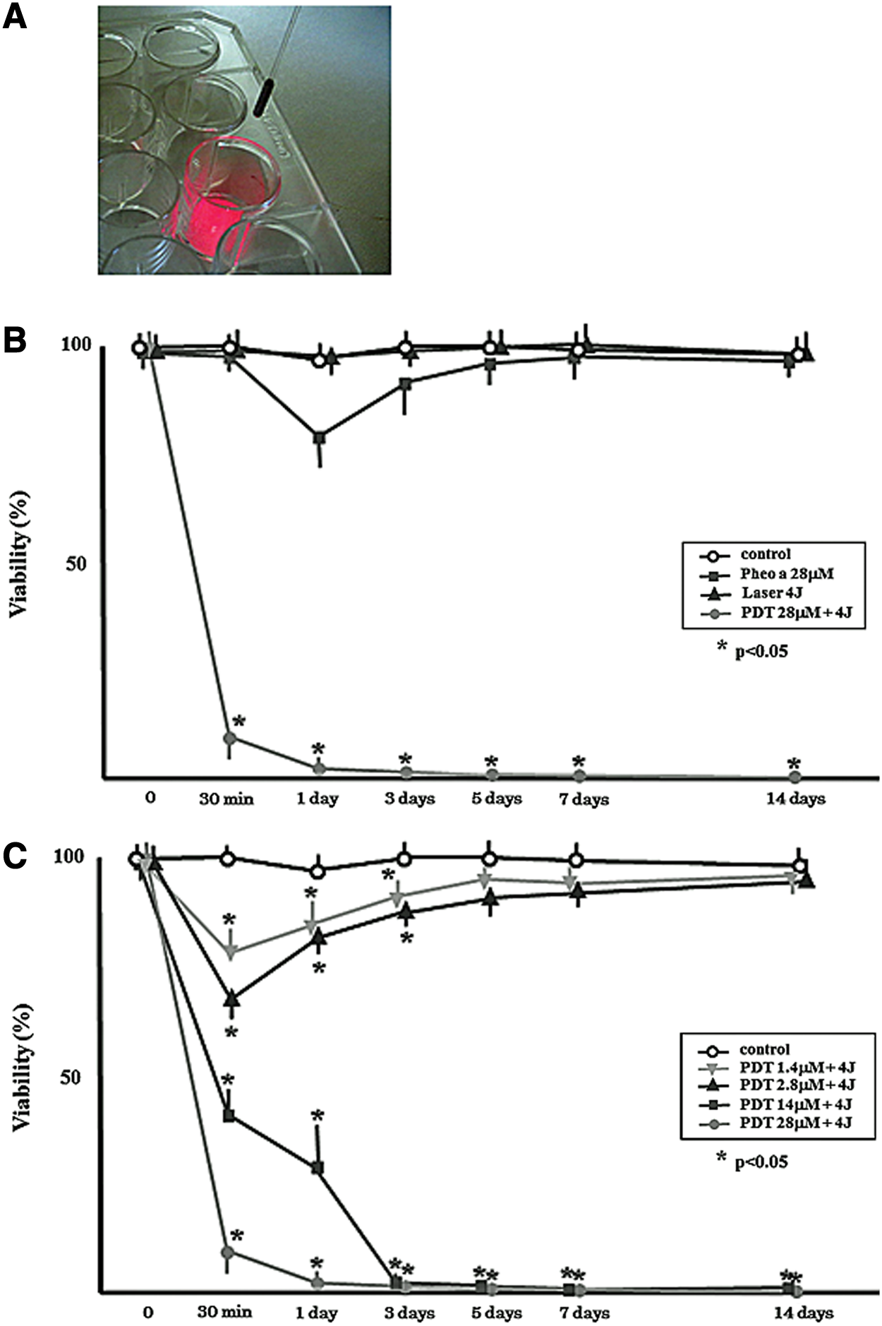
Effect of photodynamic therapy (PDT) treatment with Na-pheophorbide on Hu09 cell viability. An experimental setting is shown in
Intracellular uptake of Na-pheophorbide A into Hu09 cells
Cells were inoculated on a Chamber Slide (IWAKI5722-004, SANSYO, Tokyo, Japan) at a density of 1×10³ cells/mL and were cultured at 37°C in a humidified atmosphere with 5% CO2 for 2 days. After the Hu09 cells had attached and were growing on the base of the chamber, the medium was removed and the cells were washed twice with 1 mL of PBS. Growth medium and 28 μmol/L of Na-pheophorbide A in a volume of 1 mL were added to the chamber and were incubated with the cells. Morphological change of the cells and uptake of Na-pheophorbide A into the cells was evaluated using fluorescence microscopy (BZ-8000, KEYENCE, Osaka, Japan) after incubation for 60 min or 24 h.
TUNEL assay
Double stranded DNA fragmentation, which is characteristic of apoptosis, was assayed by TUNEL staining using an ApoTag in situ apoptosis detection kit (INTERGEN, Purchase, NY) according to the manufacturer's instructions. Briefly, cells on slides were sequentially fixed in 1% paraformaldehyde; quenched in 2% hydrogen peroxide; rinsed in PBS, pH 7.4; and incubated in 1× Equilibration Buffer (INTERGEN) for 10–15 sec and then with terminal deoxynucleotidyl transferase (TdT) for 1 h at 37 °C. The slides were then blocked with Stop/Wash Buffer (INTERGEN) and incubated with peroxidase-conjugated anti-digoxigenin antibody for 30 min at room temperature. Finally, staining was visualized using diaminobenzidine (DAB) (Sigma, St Louis, MO) and counterstaining with methyl green. The TUNEL assay was performed 24 h after PDT with 28 μmol/L of Na-pheophorbide A and 4 J/cm2 of irradiation energy.
Effects of PDT on cell viability
Viable cells in the control group, the Na-pheophorbide A-only treated group, the laser irradiation-only group, and the PDT (Na-pheophorbide A treatment and irradiation) group were detected using the trypan blue dye exclusion staining method. For each group, cells (1×10⁴/well) were seeded in a 24-well culture plate (Falcon 3047, Becton Dickinson, NJ) and were cultured at 37°C in a humidified atmosphere of 5% CO2 for 5 days. The medium was then removed and the cells were washed with 1 mL of PBS. For the PDT group, the cells were then incubated for 30 min with 0.5 ml of 1.4, 2.8, 14, or 28 μmol/L of a Na-pheophorbide A solution in a dark environment. For the Na-pheophorbide a group, cells were incubated with 0.5 mL of 28 μmol/L of a Na-pheophorbide A solution, similarly to the PDT group. After incubation, the solution was removed from the well plate and the cells were washed twice with 1 mL of PBS. For the PDT and laser-irradiation groups, cells were irradiated using 4 J/cm2 of irradiation energy per well. For all groups, following removal of the solution, the cells were washed twice with 1 mL of PBS followed by the addition of 0.5 mL of 0.25% trypsin and 0.02% EDTA to each well, and incubated at 37 °C for 3 min to remove the Hu09 cells. Viable Hu09 cells that excluded the trypan blue dye were counted. Cell viability was evaluated at 30 min and at 1, 3, 5, 7, and 14 days after PDT. Four different wells were prepared for each time point, and experiments were performed in triplicate.
Cell viability was also measured using the MTS [3-(4,5-dimethylthiazol-2-yl)-5-(3-carboxymethoxyphenyl)-2-(4-sulfophenyl)-2H-tetrazolium] assay. Cells (1×10⁴/well) were cultured in 96 well plates (Falcon 3072, Becton Dickinson, NJ) in 100 μL of culture medium at 37°C in a humidified atmosphere of 5% CO2 for 3 days. The medium was then removed and the cells were washed twice with 100 μL of PBS. The cells were then incubated with 1.4, 2.8, 14, or 28 μmol/L of Na-pheophorbide A in culture medium for 30 min in a dark environment followed by exposure to laser irradiation at various power densities (0.4–10 J). Following irradiation, 10 μL of the tetrazolium compound MTS and the electron coupling reagent [phenazine methosulfate (PMS)] were added to the wells, and the cells were incubated for 3 h at 37°C in a humidified atmosphere of 5% CO2. Absorption was measured at 490 nm using a microplate reader (MPR-A41, TOSOH, Tokyo, Japan). In some experiments, laser irradiation was repeated three times after pretreatment of the cells with 28 μmol/L of Na-pheophorbide A. Four different wells were prepared for each group, and experiments were performed in triplicate.
Caspase activity assay
HuO9 cells (1×10⁶/well in 5 mL of culture medium) were incubated on a six-well plate for 72 h in a humidified atmosphere of 5% CO2 at 37°C, and cells were processed with the above-described treatment of the different groups (PDT, Na-pheophorbide A only, and laser only groups). After 2 and 24 h, the activation status of several members of the caspase family during PDT-induced cytotoxicity was assessed. The activation of caspases-3, -8, and -9 was studied using an APOPCYTO™ Colorimetric Assay Kit (MBL, Nagoya, Japan). Cells were harvested using 0.25% trypsin and 0.02% EDTA. Cells were resuspended in ice-cold Cell Lysis Buffer (50 μL) and were incubated on ice for 10 min. Cellular debris was precipitated by centrifugation of the cell lysates at 10,000g for 5 min at 4°C. Supernatants (cell extracts) were transferred to new microcentrifuge tubes and put on ice. Reaction buffer (50 μL) containing 10 mM dithiothreitol (DTT) and 50 μL of cell lysate was added to each well of a 96-well microplate. Wells in which 50 μL of Cell Lysis Buffer were added without cell lysates were also prepared to measure blank absorption. Caspase-3, -8, and -9 substrates in a volume of 5 μL were added to each well. The plate, which was covered with a plate sealer, and the cells, were incubated at 37°C for 1–2 h. One hundred μL of p-nitroanilide (pNA) standards were added to an empty well. The absorbance in each well was measured at 405 nm, and the specific activity of caspases -3, -8, and -9 in each sample was calculated.
Statistics
Statistical evaluation was performed using ANOVA with SPSS. Results are presented as means±standard deviation (SD).
Results
Intracellular uptake of Na-pheophorbide A
The intracellular localization of Na-pheophorbide A that was taken up into the HuO9 cells was examined using a fluorescence microscope. After 60 min of exposure to 28 μmol/L of Na-pheophorbide A, the Hu09 cells displayed a clear pattern of intracellular fluorescence, which was detected in cytoplasmic compartments but not within the nuclei (Fig. 2B). No cellular morphological change was observed under phase-contrast microscopy (Fig. 2A). After 24 h incubation, morphological changes of the cells including slight shrinkage of the cells and some loss of cell–cell contact became apparent as shown in Fig. 2C. The intracellular fluorescence intensity of cells that had taken up Na-pheophorbide A was greater after 24 h incubation with Na-pheophorbide A than after 60 min incubation (Fig. 2D).
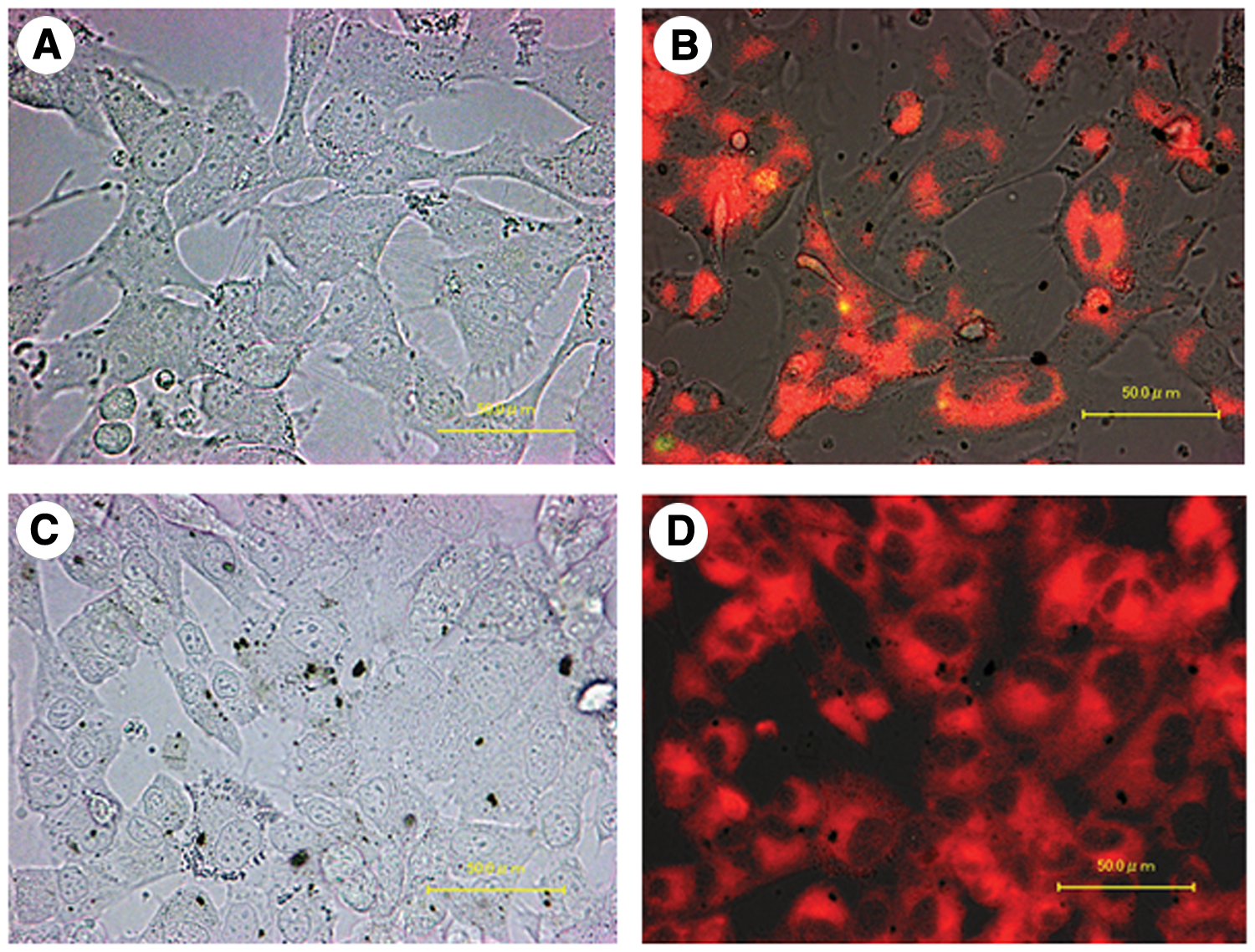
Uptake of Na-pheophorbide A into Hu09 cells The morphological changes and intracellular uptake of Hu09 cells with 28 μmol/L of Na-pheophorbide A were photographed by a phase-contrast microscope and fluorescence microscopy. After 60 min of treatment with only 28 μmol/L of Na-pheophorbide A, there were no morphological changes
Cytotoxic effect of PDT with Na-pheophorbide a on HuO9 cells
We tested the cytotoxic effect of PDT with Na-pheophorbide A on HuO9 cells by comparison of the viability of PDT with Na-pheophorbide A-treated cells with the viability of control, Na-pheophorbide A only-, and laser only-treated groups of cells. We first performed differential staining to determine the number of living and dead cells following treatment. Trypan blue staining was used for this purpose, because this staining made it possible to visually observe individual cells. Based on trypan blue staining, the viability of the Hu09 cells in the PDT group showed a significant decrease after 30 min of treatment compared with the other groups. Cell viability in the PDT group did not recover over a period of 14 days, indicating that PDT with Na-pheophorbide A was cytotoxic for these cells. In the Na-pheophorbide A only-group, cell viability temporarily decreased 1 day after treatment, but cell viability had recovered to the same level as that of the control group after 5 days of culture (Fig. 1B). The observed damage to some Hu09 cells in this assay caused by exposure to 28 μmol/L of Na-pheophorbide A without laser irradiation was consistent with the morphological damage observed in Fig. 2C, except that in the trypan blue assay, the surviving cells then exhibited regrowth. Decreased cell viability in the PDT with Na-pheophorbide A group was dose dependent at early time points (Fig. 1C). However, after 3 days of treatment, PDT with 14 μmol/L Na-pheophorbide A and 4 J irradiation was as effective in decreasing cell viability as PDT with 28 μmol/L Na-pheophorbide A and 4 J irradiation (Fig. 1C).
We further investigated cell viability of treated cells using the MTS method and various PDT conditions, including changes in the concentration of Na-pheophorbide A, in the degree of laser energy, and in the number of irradiation times. The effect of changes in the concentration of Na-pheophorbide A was determined by comparison of cell viability in the PDT- (PDT with 28 or 2.8 μmol/L of Na-pheophorbide A) Na-pheophorbide A- (28 μmol/L Na-pheophorbide A), and laser irradiated-only groups at 1, 2, and 4 days following treatment. Cells in the PDT group treated with 28 μmol/L of Na-pheophorbide A showed a significant decrease in absorbance at 490 nm (a measure of cell viability in the MTS assay) at every time point compared with the control group (group that did not receive any treatment), indicating a decrease in living Hu09 cells after PDT. A slight but significant decrease in absorbance compared with control was also observed in cells in the PDT group treated with 2.8 μmol/L of Na-pheophorbide A and in the Na-pheophorbide A-only group (28 μmol/L) (Fig. 3A). The effect of changes in the degree of laser energy in PDT on cell viability was assayed using laser energies ranging from 0.4 to 10 J/cm2 and Na- pheophorbide A (28 μmol/L). On day 4, the 10 J/cm2 PDT group displayed a very large decrease in absorbance compared with the 0.4 J/cm2 and the 1 J/cm2 PDT groups on days 1 and 4 (Fig. 3B). Absorbance was higher in the 0.4 and 1 J/cm2 PDT groups after 4 days of treatment than after 1 day of treatment, suggesting the regrowth of surviving cells (Fig. 3B). Variation in the number of times that the cells were irradiated indicated that three irradiations resulted in a significant decrease in the absorbance level not only of the PDT group pretreated with 28 μmol/L of Na-pheophorbide A, but also of the PDT group pretreated with 2.8 μmol/L of Na-pheophorbide A (Fig. 3C). These data indicated that repeated irradiation suppressed Hu09 cell proliferation even when the cells were pretreated with a low concentration of Na-pheophorbide A.
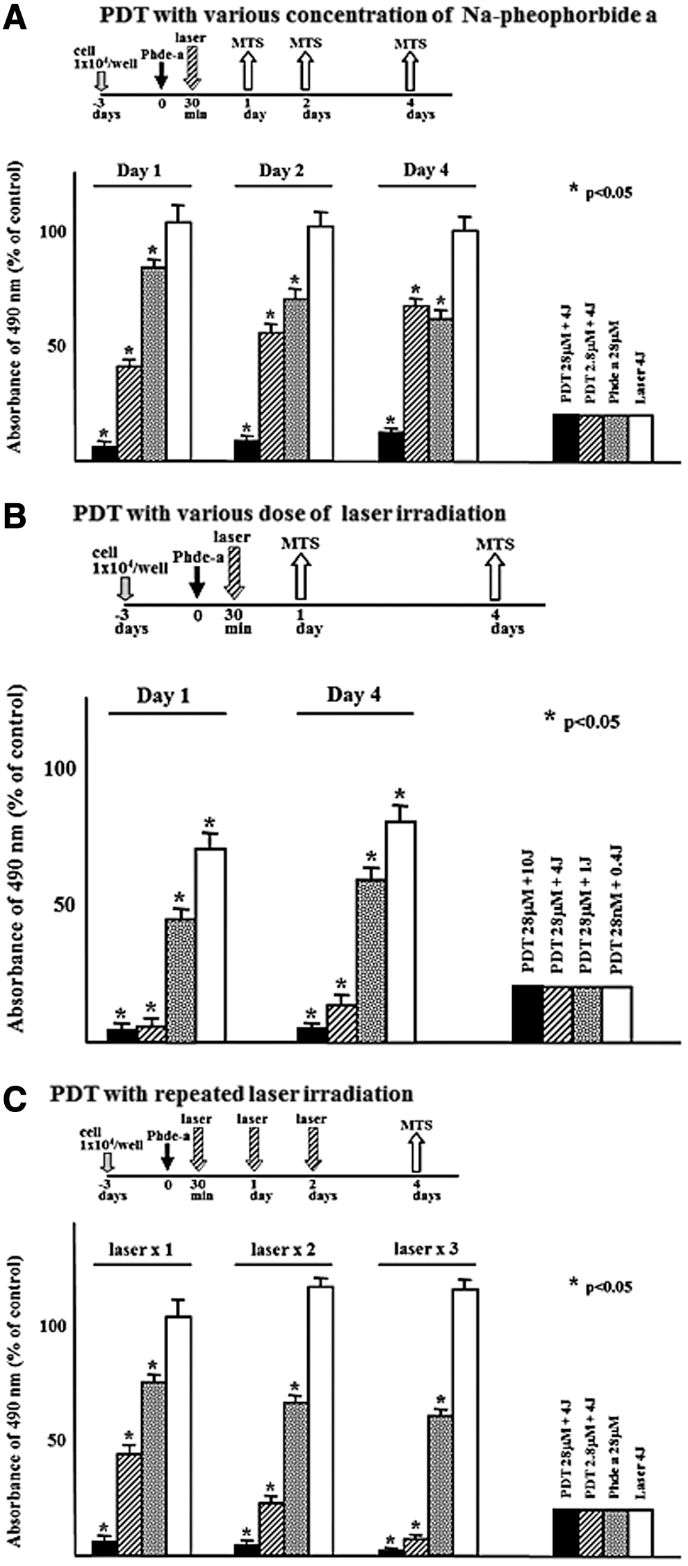
Cytotoxic activity of photodynamic therapy (PDT) with Na-pheophorbide A under various conditions. Cell viability of cells treated with PDT using various concentrations of Na-pheophorbide A
Analysis of apoptotic cells after PDT by TUNEL staining
For analysis of apoptotic cells, TUNEL staining was performed 24 h after PDT with 28 μmol/L of Na-pheophorbide A and 4 J/cm2 irradiation. Figure 4 shows the TUNEL positive cells in the control and PDT-treated cells. This result indicated that PDT treatment induced apoptosis in most of the Hu09 cells.
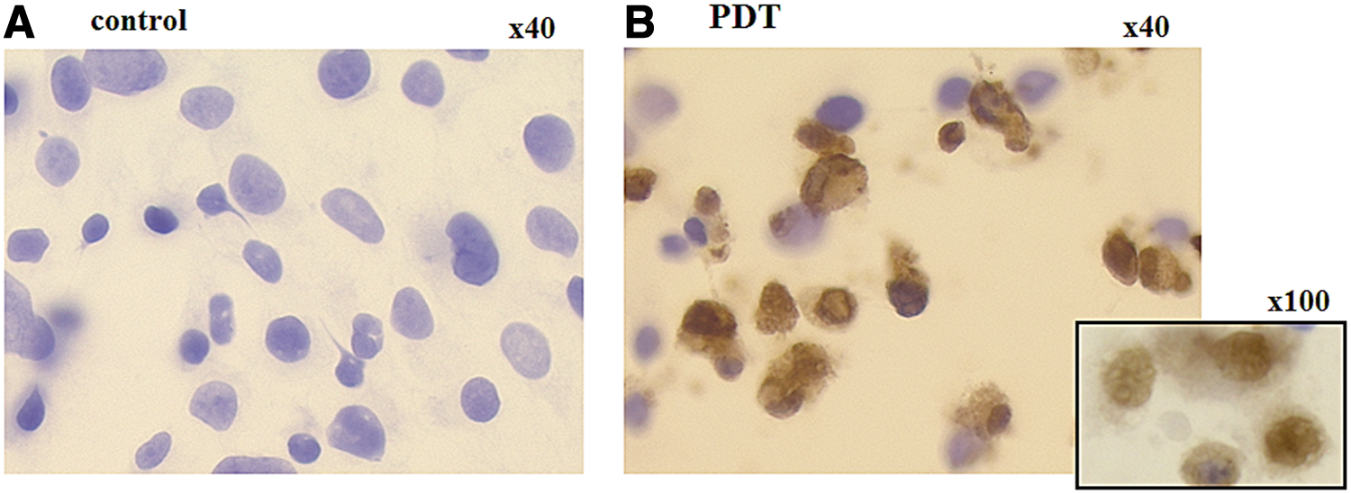
Terminal deoxynucleotidyl transferase-mediated dUTP nick end labeling (TUNEL) staining of Hu09 cells after photodynamiuc therapy (PDT). Apoptosis in Hu09 cells following PDT was analyzed by comparison of TUNEL staining of control cells
Effect of PDT on the activation of caspases-3, -8, and -9
The activity of caspases -3, -8, and -9, which is another measure of apoptosis, was assayed in the PDT (with 28 μmol/L of Na-pheophorbide A and 4 J/cm2 irradiation) and control groups of cells. All three caspases exhibited a significant increase in activity, as measured by absorbance at 405 nm, at 2 h after PDT treatment, compared with control cells. These caspase activities had returned to the control level by 24 h (Fig. 4A). However, at 24 h after PDT, most of the Hu09 cells had died (Fig. 1), which would explain why caspase activity was not detected at this time point. The effect of altering the concentration of Na-pheophorbide (1.4, 2.8, 14, and 28 μmol/L) in PDT with 4 J/cm2 irradiation energy on caspase activity is shown in Fig. 5B. All Na-pheophorbide A concentrations tested resulted in significantly higher activation of caspases -3, -8, and -9 compared with the control and laser-only groups. PDT with high concentrations of Na-pheophorbide A (28 and 14 μmol/L) resulted in higher caspase activities than PDT with lower Na-pheophorbide A concentrations (Fig. 5B). In addition, all densities of laser energy tested (0.4, 1, 4, and 10 J/cm2) in PDT with 14 μmol/L Na-pheophorbide A resulted in significantly higher caspase activities than those of the control group, and caspase activities increased as the degree of laser energy increased (Fig. 5C).

Effect of photodynamic therapy (PDT) treatment on the activity of caspases -3, -8, and -9. Activation of caspases -3, -8, and -9 after PDT treatment with Na-pheophorbide A of HuO9 cells was assayed using the APOPCYTO™ Colorimetric Assay. Activity is expressed as absorbance at 405 nm per mg protein. Caspase activity was assayed as follows.
Discussion
This study demonstrated, using Trypan blue staining and MTS assays, that PDT with Na-pheophorbide a inhibited the viability of osteosarcoma (Hu09) cells. The cytotoxic effects of PDT on Hu09 cells were clearly demonstrated when cells were pretreated with >14 μmol/L of Na-pheophorbide A, and >4 J/cm2 of laser irradiation (Fig. 3A, B). Interestingly, repeated irradiation showed high cytocidal activity even following a single pretreatment of cells with a low concentration of Na-pheophorbide A before laser irradiation (Fig. 3C). Therefore, repeated irradiation has very high cytocidal activity for these cells. Most of the Hu09 cells displayed positive TUNEL staining at 24 h after PDT. Furthermore the activity of caspases in both the intrinsic (caspases -9, -3) and the extrinsic (caspase 8) apoptotic pathways increased after PDT with Na-pheophorbide A. The appearance of TUNEL positive cells and the increased activity of caspases suggested that PDT induces apoptotic changes in the Hu09 osteosarcoma cells. These findings suggest that the cytocidal activity of PDT with Na-pheophorbide A toward osteosarcoma cells may be mediated through the induction of apoptosis in these cells. Strong cytocidal activity of PDT was observed when PDT was performed with 14 μmol/L Na-pheophorbide A and 4 J irradiation. Further studies are required to determine the best pre-incubation time before irradiation, and the best light source for clinical use.
Photodynamic therapy is a minimally invasive treatment for oncological and non-oncological diseases. 22 In our previous study, PDT with Na-pheophorbide A was effective for the treatment of osteomyelitis 19 and septic arthritis 7,18 in rat models. Pheophorbide A is a photosensitizer made from chlorophyll that is derived from a natural source. There have been several studies regarding the application of PDT with chlorophyll for the treatment of cancer in mammals in vitro and in vivo. 9,12,15,17,23 –25 However, the chlorophyll derivative pheophorbide A is not suitable for clinical use, because of its hydrophobic nature. 16 We therefore focused the present study on PDT with Na-pheophorbide A, which is a water-soluble photosensitizer in which Na has been added to the pheophorbide A structure.
To our knowledge, this is the first study that shows effective cytocidal activity of PDT with Na-pheophorbide A toward osteosarcoma cells in vitro. Acridine orange, another photosensitizer, has been used clinically for the treatment of musculoskeletal sarcoma patients before surgery, to reduce tumor size. 4 –6 Because acridine and its derivatives are DNA- and RNA- binding compounds, when using acridine, there is the possibility of carcinogenesis as a result of interference with normal DNA function. 26 It is believed that Na-pheophorbide A is a safer photosensitizer than acridine orange, because it is not expected to produce such carcinogenetic effects.
In our study, fluorescence microscopy analyses indicated that a clear pattern of intracellular fluorescence, resulting from uptake of Na-pheophorbide A, was observed at an early time point in the Hu09 cells (after 60 min of Na-pheophorbide A treatment), and that this fluorescence persisted for at least 24 h. Fluorescence was detected in cellular cytoplasmic compartments, but not within the nuclei. In a recent study of pheophorbide A uptake into a number of different cells, this photosensitizer was observed in the cytoplasm but not within the nuclei. 24 Conversely, Na-pheophorbide A becomes incorporated into the nuclei, mitochondria, and lysosomes of human oral mucosa cells. 7 It is possible that the subcellular location of the photosensitizer differs, depending upon the quality of the cell. The pattern of uptake of Na-pheophorbide A into the osteosarcoma cell line (Hu09), and the morphological changes induced in these cells following uptake in this study, were similar to those reported for Jurkat leukemia cells. 23 Furthermore, in our study, a loss of connection between osteosarcoma cells was observed after 24 h of Na-pheophorbide A uptake compared with the cells following uptake for 60 min. In addition, in the study of Jurkat leukemia cells, a small amount of membrane blebbing was observed in the photosensitizer-only group. 23 The combined data indicate that a high concentration of photosensitizer can be cytotoxic for target cells in the absence of light energy.
The toxic effect of a high concentration of photosensitizer in the absence of light irradiation was further shown in our study. Therefore, the trypan blue staining assay, regarding intracellular uptake, indicated that cell viability was temporarily, and nonsignificantly, decreased in the Na-pheophorbide A only group (28 μmol/L without irradiation) compared with the control group (Fig. 1B). Furthermore, in the MTS assay, the Na-pheophorbide A only group (28 μmol/L) displayed higher toxicity compared with the laser irradiation only group (Fig. 3A,C). In addition, cytotoxicity was induced by changing the energy of laser irradiation and the number of irradiations, even with pretreatment solutions that contained low concentrations of Na-pheophorbide A. Such low concentration pretreatment solutions should be considered in PDT treatment in order to reduce damage to normal cells.
It was suggested in a recent review that the main mechanism of cell death induction by PDT was induction of a cellular apoptotic response. 16 Apoptotic caspases are classified into extrinsic and intrinsic or mitochondrial pathways. In the extrinsic pathway, caspase 8 stimulation by the death receptor leads to the proteolytic activation of the main effector caspases -3 and -7. In the intrinsic or mitochondrial pathway, caspase 9 is activated by apoptosome-led cytochrome c or procaspase 9, and subsequently activates caspases -3 and -7. 27 The caspase activity assays of our study indicated that Na-pheophorbide A mediated PDT induced apoptosis of HuO9 cells via activation of both the intrinsic and the extrinsic pathways. It has been reported that reactive oxygen species are major factors in the induction of apoptosis via intrinsic pathways leading to caspase -9 and -3 activation. 28 It is known that after the photosensitizer absorbs specific wavelengths of visible light, it generates reactive oxygen species in cellular organelles. 27 It is, therefore, possible that after the uptake of Na-pheophorbide into mitochondria, it might generate reactive oxygen species at the time of irradiation that activates caspase 9. Using Mito Tracker® Green, Tang et al. showed that pheophorbide A was located in the mitochondria. They further showed that PDT with Na-pheophorbide A probably induced apoptosis via the mitochondrial-mediated pathway in human uterine carcinoma cells. 15 Panzarini et al. reported that PDT induced cell death by stimulation of Fas and Fas ligand expression. 29 These observations suggest that the mechanism by which PDT induces apoptosis is complicated, and varies according to the type of photosensor used, and the light source.
Although further studies are required to analyze the mechanism responsible for the apoptotic activities of PDT, the results of the current study indicate that PDT with Na-pheophorbide A might provide a new therapeutic tool for the treatment of osteosarcoma.
Conclusions
Our study demonstrated a cytocidal effect of PDT with Na-pheophorbide A on osteosarcoma (Hu09) cells, which was accompanied by the induction of apoptosis via intrinsic and extrinsic pathways.
Footnotes
Author Disclosure Statement
No competing financial interests exist.