
Abstract
Introduction
I
In the context of laser therapy, photodynamic therapy (PDT) assumes that the interaction of light with a suitable wavelength with photosensitizer (PS) and oxygen results in reactive oxygen species (ROS) capable of inducing cell unviability. 6,7 This highlights that the antimicrobial activity of ROS results primarily from the electronic excitation of the dye by light, followed by reaction mechanisms from its excited state. 8 The singlet oxygen ( 1 O2), which is a key intermediate in such reactions, may induce oxidative processes and damage essential cellular components such as the plasma membrane, or irreversibly alter metabolic activities, resulting in the death of various microorganisms. 9,10 Therefore, the PS absorption band should be resonant with radiation emitted, well absorbing the wavelength. 2,11
Malachite green (MG) has been widely reported as a PS in antimicrobial photodynamic chemotherapy (APCT), having an absorption band in the red region of the electromagnetic spectrum (630–700 nm). 11 –13 Its molecule is cationic and belongs to the family of triphenylmethane (TPM), with three benzene rings. TPMs have shown great efficacy in the in vitro inactivation of Gram-positive bacteria and facultative anaerobic bacteria, as part of the composition of various PS. 14 However, phenyl derivatives can also be present in plant extracts from various plant species. 15 –17
Shinopsis brasiliensis Engl., a plant from the Brazilian semiarid region, has effective antimicrobial activity related to the high amount of tannin, flavonoids, and polyphenols. 18 These secondary metabolites, derived from the plant metabolism with known bactericidal action 18,19 , can also contain PS structures, 20 such as those presented in significant PS such as blue methylene, 21 which optimize their antimicrobial properties with appropriate laser light.
Therefore, the aim of this study was to evaluate the effect of PDT associated with the use of MG and extracts of S. brasiliensis Engl., as potential photosensitizing agents on bacteria involved in several human infections.
Materials and Methods
Plant sample and extracts preparation
Leaves and bark of S. brasiliensis Engl. were analyzed. Plant samples were collected in the region of Borborema-PB. The exsiccate was deposited in the Professor Jayme Coelho de Morais herbarium, belonging to the Center for Agricultural Sciences, Federal University of Paraíba, under the number EAN-14049.
The leaves and barks of S. brasiliensis Engl. were subjected to the drying process in an oven with renovation and air circulation at 40°C. Then, the material was triturated in vertical rotor mill with a particle size ∼10 mesh. The maceration process for obtaining the extracts used a hydroalcoholic solution at 30:70 water: ethanol (v/v) as solvent. The hydroalcoholic extracts were dried by nebulization with a spray dryer device, LabPlant, using Aerosil 200® as a pharmacotechnical stabilizer. The material atomization inside the chamber was performed by employing a peristaltic pump with injector needle of 0.5 mm. The input temperature was 150°C and the output temperature was 75°–80°C, under a power flow of 3.3 mL min−1. After nebulization, dilutions were made at 500, 400, 300, 200, 100, 50, and 1 mg of samples in 1 mL of dimethyl sulfoxide (DMSO) 10%. The microbial analyses were performed using each of the dilutions prepared from leaf and bark separately.
Fourier transform infrared spectroscopy (FT-IR)
FT-IR spectra of herbal medicines were recorded on a Perkin-Elmer Model 1600 apparatus using KBr stressed discs in the range of 4000–500 cm−1.
Laser source
The source was a red-laser emitting Theralase (DMC®) Indium–Gallium–Aluminium–Phosphide (InGaAlP) active medium, with a maximum output power of 100 mW, 660 nm wavelength, continuous emission, and dosimetry 4 J/cm2. The laser was applied using a fang attached to a universal gripper support, ensuring the standardization of the experiments and a distance of 2 cm from the tip of the laser and the disk center, which contained the test substance. The application was made in a timely manner (34 sec on each disk tested).
Antimicrobial activity
Standard strains from the American Type Culture Collection (ATCC) were used: E. coli (25922), S. aureus (25923), Pseudomonas aeruginosa (27853) and Enterococcus faecalis (29212).
To obtain a bacterial inoculum, strains were individually inoculated into test tubes containing 5 mL of 0.9% sterile saline solution. The bacterial suspension produced was standardized by a spectrophotometer at 625 nm wavelength to obtain 85% transmittance, which is equivalent to 106 colony-forming units (CFU)/mL.
The antimicrobial activity was performed by disk diffusion method 11 using sterile paper discs, 6 mm in diameter. The discs were impregnated with 20 μL of each extract dilution and MG dye at 0.01–0.1 mg.mL−1 which were arranged in a petri dish onto the surface of Mueller–Hinton agar, previously seeded with the bacterial suspension. The MG was included as control. Assays were divided into groups (Table 1).
PDT, photodynamic therapy.
The groups laser and bark extract (B+L+), laser and leaf extract (F+L+), and laser and MG (M+L+) received PDT with low-intensity laser irradiation. The Theralase red light (DMC, São Paulo, SP) was activated for 34 sec at the specifications listed. The tip of the optical probe was at a distance of 1 cm from the disc impregnated with the sample. In group laser only (L+) only a sterile disk, without impregnation of any substance, was treated with the laser.
After these procedures, plates were placed in oven at 37°C for 24 h. The biological effect of PDT, isolated application of low-intensity laser therapy, and the use of extracts and the MG dye in different bacterial species were measured in millimeters by measuring the diameter of inhibition halos produced. Assays were performed in quadruplicate, and statistically analyzed by independent samples Mann–Whitney U test (SPSS/PC, ver. 10.0, Chicago, IL), p values<0.05 were considered significant.
Results
Inhibition zones were observed in all groups, except for control group L+ that did not present any inhibition halo, showing that only the application of low-intensity laser (660 nm) does not inhibit bacterial growth. The dried extract from the bark of S. brasiliensis showed inhibitory activity in all bacteria, with laser increasing the produced halos, with the exception of E. coli, in which the presence of an inhibition halo was visualized only at the highest concentration studied (500 mg.mL−1) (Table 2). P. aeruginosa showed an inhibition zone at all concentrations of the extract of the bark. Although there was significant variation between the B+L+ and bark extract only (B+L−) groups at concentrations of 50 and 300 mg.mL−1 (p>0.05), there were statistically significant variations between these groups (p=0.029) at the other concentrations. The leaf extract of S. brasiliensis also showed an inhibition halo in all the studied bacteria, as is shown in Table 3. The inhibition halo was greater in the group with laser application, showing positive results for E. coli and P. aeruginosa.
Mean±standard deviations, throughout.
p Value<0.05 was statistically significant in the independent samples Mann–Whitney U test.
S.a., Staphylococcus aureus; E.c., Escherichia coli; P.a., Pseudomonas aeruginosa; E.f., Enterococcus faecalis; -, no inhibition zone; B+L +, laser+bark extract; B+L−: bark extract only.
Mean±standard deviations, throughout.
p Value <0.05 was statistically significant, independent samples Mann–Whitney U test.
S.a., Staphylococcus aureus; E.c., Escherichia coli; P.a., Pseudomonas aeruginosa; E.f., Enterococcus faecalis; −, no inhibition zone; F+L+, laser+ leaf extract; F+L-, leaf extract only.
Table 4 shows that 0.01% MG was effective only in E. faecalis in groups M+L+ and MG only (M+L−), with a statistically significant difference between the groups (p=0.029), although the concentration of 0.1% was effective in almost all strains tested, except for S. aureus, because there was no inhibitory activity in the group where the laser was not applied.
Mean±standard deviations, throughout.
p Value<0.05 was statistically significant, independent samples Mann–Whitney U test.
S.a., Staphylococcus aureus; E.c., Escherichia coli; P.a., Pseudomonas aeruginosa; E.f., Enterococcus faecalis; −, no inhibition zone; M+L+, laser+malachite green; M+L−, malachite green only.
The FT-IR analysis (Fig. 1) shows typical infrared absorption of phenolic compounds such as the stretching band in the ranges 1702–1685 cm−1 (carbonyl – peak 5), 1607–1516 cm−1 (C=C bonds in aromatic rings – peak 4), and 788–674 cm−1 (aromatic C-H bonds – peak 1). Peaks between 1087 and 1307 cm−1 correspond to the axial deformation in C-O bonds (peak 3) in alcohols, ethers, carboxylic acids, and esters, indicating the presence of a wide variety of metabolites, such as tannins and flavonoids. Between 800 and 900 cm−1 (peak 2) stretching of the C-O group and deformation of the O-H group in carboxylic acids occurs. In 2974–2847 cm−1, the characteristic of stretching C-H associated with aromatic rings is attributed to the nature of organic compounds in the extract (peak 6). And peaks in the range 3000–3400 cm−1 are associated with OH stretching frequency (peak 7).
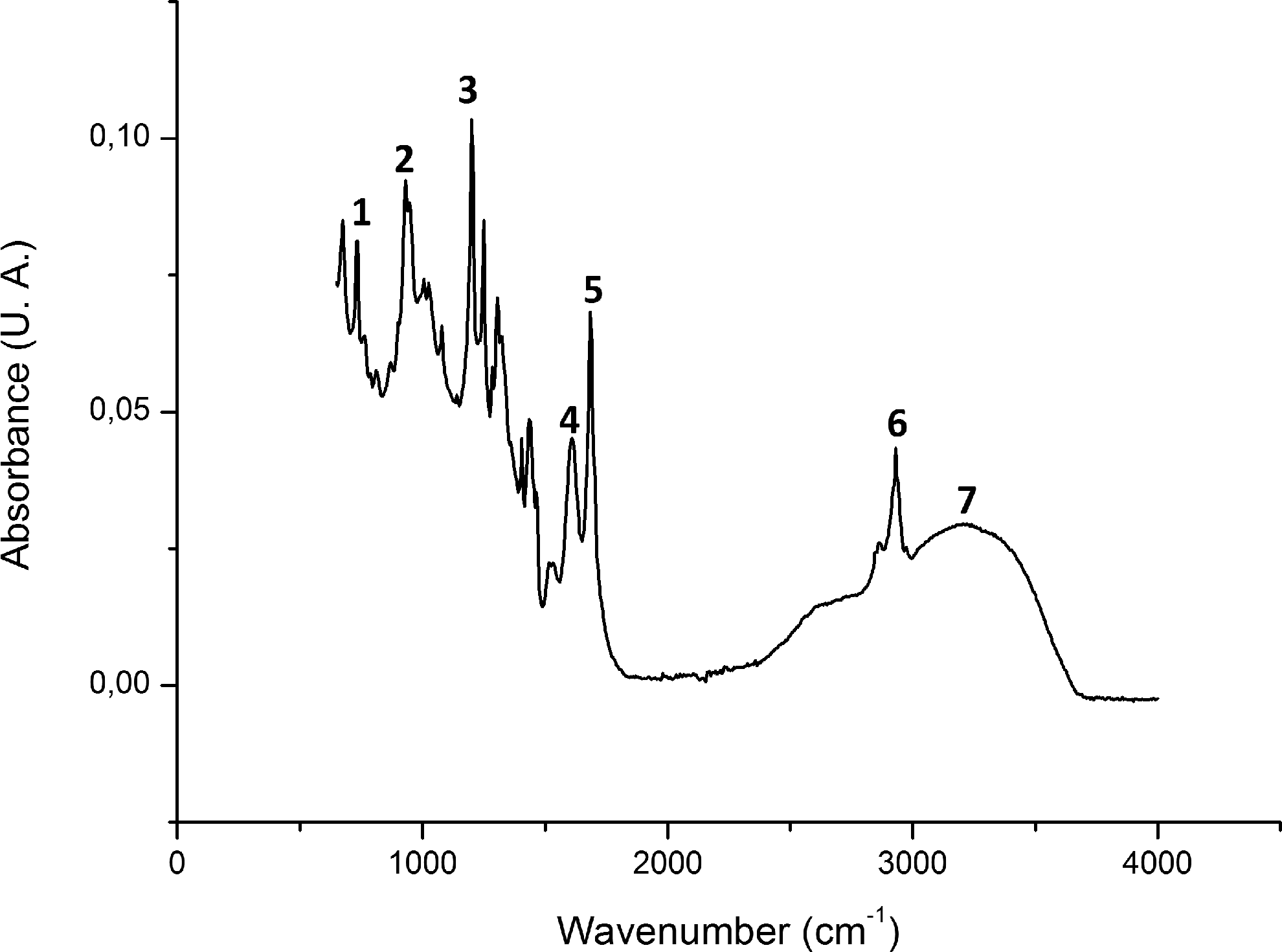
Fourier transform infrared spectroscopy (FT-IR) spectra of nebulized extract of Schinopsis brasiliensis Engl.
Discussion
As the results suggest, S. aureus was the most sensitive pathogen to PDT, with MG showing variation for 7.81±0.21 (p=0.029) when the laser was applied to the MG at 0.1 mg.mL−1, compared with the control group M+L− with no activity. In a similar study, in which when this PS was used at concentrations ranging between 37.5 and 3000 mM arranged on organized biofilms of E. coli and S. aureus, there was variation of bacteria by reducing the percentage of CFU/mL converted to logarithmic form. This study also demonstrated a higher sensitivity of S. aureus, with a decrease from 5.48 to 3.84 CFU/mL (log10) when the PS was applied to 3000 mm, compared with the control group with no PS application. 1
S. aureus is one of the most frequently identified pathogens in association with healthcare, particularly hospital-acquired pneumonia associated with a mechanical ventilator, 22,23 in addition to skin infections such as atopic dermatitis 24 of which numerous subtypes are identified as being resistant to methicillin, the standard choice of antibiotic, 3 PDT effectiveness in in vitro tests using MG shows a new perspective in the development of essential therapies to control this pathogen.
This process can be investigated by measurements made with FT-IR, which suggests a possible mechanisms of interaction of the laser with the cells. 14 MG is a basic dye with affinity for anionic polyelectrolytes, including lipids, glycolipids, and phospholipids that are present in cell membranes of bacteria, which destabilizes them. To increase the binding of these PS with bacteria, isothiocyanate can be used with the aim of increasing the affinity of this group with proteins, in particular amino acids with SH- and NH2-free groups. With FT-IR, it is evident that this PS forms complexes with the structure of cell wall of bacteria, in which photoproducts are generated with absorption in the 300–400 nm spectra, such as ketones that degrade cell structures. 6,25,26
Therefore, it was understood that MG is naturally toxic to many species of bacteria, because the concentration of 0.1 mg.mL−1 produced zones of inhibition of 10.02±0.61 mm for E. coli, and of 9.33±0.28 mm for P. aeruginosa in the M+L− group. However, when applying the red laser, photoproducts can be produced and further damage is triggered, with an increased inhibition zone to 10.60±0.45 mm (p>0.05) for E. coli, and 11.41±0.22 mm (p=0.029) for P. aeruginosa, in the M+L+ group at 0.1 mg.mL−1.
Regarding the use of nebulized extracts of S. brasiliensis Engl., some herbal considerations may explain its application in isolation, and combined with the laser, showing its photodynamic character as an important antibacterial agent.
S. brasiliensis belongs to the Anacaridaceae 27 family, and phenolic compounds, tannins, and flavonoids have been reported as key components of their plant extracts with high antimicrobial activity. 18,27 The mechanism of antimicrobial action of tannins may be related to their complexation with metal ions, reducing the availability of essential ions to bacterial metabolism. 28 Flavonoids such as 2,3-flavones and hexamethoxy flavones are agents capable of forming complexes with soluble proteins, and the cell wall of bacteria and may disrupt bacterial membranes. 29,30
Therefore, such inhibitory mechanisms of bacterial metabolism explain why nebulized extracts obtained from bark and leaf were effective on all bacteria evaluated even without applying the laser, as shown by the B+;L− and leaf extract only (F+L-) groups. Therefore, it can be noted that even without applying the laser, leaf extracts produced inhibition zones of 9.45±0.49 mm at 50 mg.mL−1 to E. faecalis, demonstrating greater efficacy of this extract when compared with groups B+L- and F+L+. Studies have shown that E. faecalis is related to urinary nosocomial infections, being also frequent in endocarditis and endodontic infections in the dental context, and with the presence of numerous species resistant to vancomycin being recorded. 31 –33 Therefore, we highlight the importance of this study, which points to new perspectives in the treatment of these infections.
The most important observations can be made regarding groups B+L+ and F+L+. In these groups, an increased inhibition zone for all bacteria tested at various concentrations was noted, in nebulized extracts of both barks and leaves. In group B+L+, S. aureus was the most sensitive to variations from 14.78±0.24 to 19.90±0.48 mm (p=0.029) at 300 mg.mL−1 when the laser at 660 nm was applied; and to variations from 18.55±0.37 to 24.55±0.36 mm (p=0.029) at 500 mg.mL−1.The same occurred in group F+L+, in which a variation of 2.2 mm with the red laser was observed, starting with a halo of 15.30±0.18 mm in the group F+L- to a halo of 17.50±0.45 mm (p=0.029) in the F+L+ group.
The literature has not reported why plant extracts properly irradiated with laser light can increase their antimicrobial activity, as was evidenced in testing by a larger zone of inhibition. And there has been no study with nebulized extracts from this perspective: as potential photosensitizing agents in PDT. However, research published by Fernandes et al. 20 may encourage some hypotheses. The aforementioned study showed a thermal characterization of various plant extracts of the semiarid northeast, including S. brasiliensis using FT-IR, the same method used to find photochromic compounds in dyes such as MG. 14
The use of FT-IR spectroscopy for herbal preparations focuses on the identification and assessment of the stability of the functional groups contained in the chemical constituents. The functional group identification is based on the FT-IR peaks attributed to the stretching and bending vibrations. 34 The antimicrobial activity of S. brasiliensis has been reported by Saraiva et al. 35 and Silva et al., 19 which was attributed to the presence of phenolic compounds, mainly tannins and flavonoids. Phytochemical studies also reported the presence of a new alkyl phenol, (methyl 6-eicosanyl-2-hydroxy-4-methoxybenzoate) and an unusual steroid (5α,8α -epidioxyergosta-6,22-dien-3-β-ol). 36
In the analysis using FT-IR, several absorption peaks of the radiation by organic molecules was observed (Fig. 1), present in flavonoids, tannins, phenolic compounds, and aromatic radicals, which were converted into molecular energy vibration. 37 –39 The vibrational spectrum appears as a series of bands, because each change of level of vibrational energy corresponds to a number of changes in the levels of rotational energy. 40 In Fig. 1, we observed absorption peaks corresponding to the laser wavelength employed between 600 and 800 cm−1. Absorption in these wavelengths is related to angular deformations outside the planes in C-H bonds of aromatic rings in phenol/aromatic compounds. 35,40
The presence of aromatic groups in various PS, such as MG and methylene blue perhaps point to the photodynamic character of nebulized extracts of S. brasiliensis, as many reactions in aromatic chains are reported in oxyreductive processes triggered by electronic excitation derived from the energy provided by the laser, producing irreversible cell damage. Therefore, such compounds are used in the synthesis of photosensitive agents and the formulation of new PS. 41,42 The extracts obtained from the bark and leaves of S. brasiliensis Engl. can be considered a mixture of PS with variable composition, with tests being needed to isolate and characterize the main active substance responsible for the photodynamic process.
Conclusions
Favorable results for the use of MG and nebulized extracts of S. brasiliensis Engl were found in PDT as antibacterial agents against all the tested pathogens. S. aureus proved to be the most sensitive microorganism, with group B+L+ being the most effective, showing a halo of inhibition of 24.55±0.36 mm. Further studies should be conducted so that photosensitizers obtained from medicinal plants are identified and validated as a new therapeutic application against bacterial infections.
Footnotes
Author Disclosure Statement
No competing financial interests exist.