
Abstract
Introduction
E
It is well known that exposure of cells to IR results in the formation of reactive oxygen species (ROS). ROS are also produced during the metabolism of oxygen. However, the physiological levels of ROS are important for the growth of cells, adaptation to stress, processes of DNA repair, and other physiological processes. 5,6 When the levels of ROS increase, for example after the influence of IR, they can damage biomolecules in cells, which leads to the effects described earlier.
Currently, there is considerable interest in therapeutic lasers in research and medicine. We have focused on therapeutic lasers, because of their well-known biostimulative, anti-inflammatory effect in in vivo and in vitro experiments. 7 –10 It is known that the one of the cellular targets of the laser light is mitochondria. They contain many photoacceptor molecules, which may be stimulated by laser light and activate cascading reactions in the cell. 6 Mitochondria are the energy stations of the cell involved in redox processes and in the formation of ROS. Many years of experimentation have shown that single low-level laser treatment increases the catalase concentration, 11 –13 superoxide dismutase activity, 13 –16 and concentration of glutathione. 17 Also low-level laser irradiation leads to a decrease of lipid peroxidation, 11,14,16 and malondialdehyde levels. 16,18 In cells activated by oxidative stress, low-level laser treatment reduces ROS 15,17,19 and increases mitochondrial membrane potential. 19,20 Total oxidative status is likewise decreased after low-level laser treatment. 21 This apparenty provides a suitable basis for radioprotective feature of laser light. Accordingly, irradiation of therapeutic laser is possible regulative method of cellular radiosensitization which make cells more protective against changes in the extracellular environment.
For our experiment, we chose a 940 nm therapeutic laser, which may generate radiation on power up to 5 W. It is a newly developed laser and its intracellular action is not clearly understood. Present experimental evidence indicates that in in vivo experiments, the 940 nm laser increases the amount of cell proliferation or the amount of cell differentiation in a power dependent manner. 22 In an in vivo experiment on rats, neither the antioxidative nor the oxidative status were changed in serum after a 6-day treatment of a mucoperiosteum wound lesion by this laser. 23 The number of inflammatory cells on day 1 and the mitotic activity of fibroblasts on day 7 and 14 are significantly increased after the end of laser therapy. 21,23 On the other hand, several studies indicate that a 940 nm laser has no impact on collagen synthesis or vascularization 21,23 However, after 940 nm laser treatment, insulin-like growth factor, vascular-endothelial growth factor, and transforming growth factor-beta mRNA are increased. 24
The possible radioprotective effect of laser may be widely used in oncology and radiology when an irradiated organism must be protected from ionizing radiation. For example, it is a well-known fact that radiation therapy is associated with risk of hematopoietic changes in oncology patients. We hypothesized that the 940 nm laser has radioprotective features for the life span and blood/hematopoietic cells of mice. To test this hypothesis, we selected parameters of the life span of gamma-irradiated mice and also absolute cell counts and proportions of peripheral blood cells as characteristics of organism sensitivity to IR.
Materials and Methods
Experimental animals
Two-month-old C57BL/6 female mice (Velaz, s.r.o., Únětice, Czech Republic) with an initial body weight of 20±1 g were used for this study. Animals were housed in standard vivarium caging under a standard condition (temperature, pressure) and a 12/12 light/dark cycle. Food and water were provided ad libitum. Mice were acclimated for 1 week before use. All experiments were approved by The Ethical Committee of the Faculty of Military Health Sciences in Hradec Kralove and by The Ethical Committee of the Ministry of Defence of the Czech Republic.
Groups
Following an acclimation period, mice were randomized to 11 experimental groups (Table 1). Mice in the first, control group were in their cages all the time. In other groups, mice were exposed to a single laser and/or gamma irradiation (for more details, see Table 1). Study design is considered in greater detail on Fig. 1 and in Table 2.
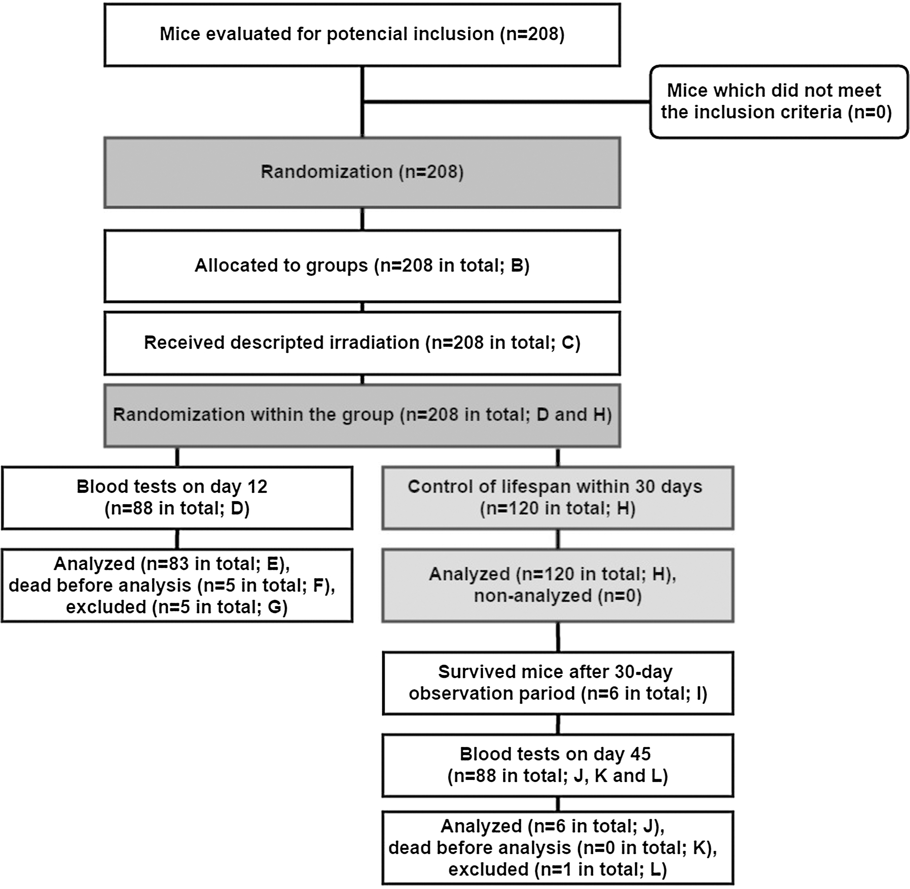
Study design. Letters in parenthesis refer to Table 2, where a detailed description of the study design may be found.
Other parameters of laser were: power output, 5 W; power density, 0.44 W/cm2; frequency, 20 Hz. For more details, see section on laser irradiation and Table 3.
Other parameters of gamma radiation were: dose, 8.7 Gy; dose rate, 1.3 Gy/min. For more details, see subchapter on gamma irradiation.
Number of mice is summarized from two analogic studies: pilot study and actual experiment.
Total number of mice allocated to blood test analysis on day 12.
Mice were excluded from statistical analysis, because of zero absolute counts of blood cells.
Statistical analysis was not performed because the insufficient number of mice.
Bold text indicates two groups in which mice survived after 30-day observation period.
Gamma irradiation
Unanesthetized mice from experimental groups were individually immobilized in special cages, which were placed in the circular layout (called a “carousel”). Mice were exposed to whole body irradiation at a single lethal dose of 8.7 Gy using a 60Co gamma source (Chisotron, Chirana, Czech Republic) at a dose rate of 1.3 Gy/min. Subsequently, mice were immediately placed in their cages.
Laser irradiation
The experiments were conducted with the 940 nm diode laser (Biolase, Inc., USA). Each mouse was immobilized in the dorsal position. Immobilized mice were irradiated in pulse wave mode at different fluences (Table 3). Laser was applied perpendicularly in a scanning manner to an unshaved abdominal skin surface, with the distance maintained at 0.5 cm. Before the beginning of the experiment, the laser was calibrated using a radiometer (Rm-6600A Universal Radiometer, LaserProbe, Berlin, Germany). The used laser is a Class IV laser product according to safety.
Life span
One group of mice was placed in other cages, and was used for control of the life span within 30 days after the influence of gamma and/or 940 nm laser irradiation.
Peripheral blood
The blood was collected intracardially into a heparin-containing solution (final concentration 20 UI/mL, Zentiva, Prague, Czech Republic) with the animal under ether anesthesia on day 12 of the experiment. Total white blood cells (WBCs), red blood cells (RBCs), lymphocytes, monocytes, and neutrophils were determined on the Sysmex XE-2100 hematological analyzer (Sysmex-Toa, Japan).
After the end of 30 days, life span study blood of mice who had survived was also analyzed by the method described earlier. A blood test on those mice was performed on day 45.
Statistical analysis
In statistical analysis, life span data are expressed as the mean±standard error. Life span was analyzed by Kaplan–Meier procedure (log rank test statistics) with a p value<0.05.
In a statistical analysis of difference in blood among groups, data were expressed as the mean±standard deviation. Statistical significance was evaluated by Mann–Whitney U test. The results were considered statistically significant when the p value was<0.05.
Results
The experiment provided confirmation of alteration of the life span of mice, the blood cell counts, and percentages by the 940 nm laser treatment (5 W, 20 Hz, 3 J/cm2, and 18 J/cm2) following the gamma radiation (p<0.05). Laser with those parameters prolongs the life span of mice. In the gamma-laser3 group, absolute counts of white blood cells were higher than in the gamma group, which was based on higher absolute counts of lymphocytes and neutrophils (p<0.05). In the same group, there was a statistically higher percentage of monocytes than in the gamma group (p<0.05). In the gamma-laser18 group, only percentages of lymphocytes and monocytes were significantly different than in the gamma group (p<0.05). An unexpected result was that after laser treatment, the blood cell count and percentage in healthy, non-gamma-irradiated mice were lower than those in the control group. A more precise description of results is as follows.
Life span of mice treated with 940 nm laser and/or gamma radiation
As can be seen in Table 4 and Fig. 2, in the gamma-laser3 and gamma-laser18 groups. the life span of mice was longer than in the Gamma group and other groups. In Table 4, we summarized the means for survival time for all groups and found that 50% of mice in the gamma-laser3 group and 9.1% of mice in the gamma-laser18 group had survived to the 30th day after gamma irradiation, but only in the first one was the difference in survival compared with the gamma group significant (p=0.007 vs. 0.875). Meanwhile, survival in the gamma-laser3 group was significantly different than in other gamma-irradiated groups, except the gamma-laser18 group, in which p=0.23 in comparison with the gamma group.

Effect of 940 nm laser on the life span of mice during a 30-day period. Day 0 is day of gamma irradiation (8.7 Gy). The results are expressed as survival functions. The control and laser-irradiated groups were omitted from the graph for the sake of a better illustration.
The control and laser-irradiated groups were omitted from the graph (Fig. 2) for the sake of illustration. All (100%) of the mice in these groups were alive during the experiment.
Changes in WBC count in mice treated with 940 nm laser and gamma radiation on day 12
Among gamma-irradiated mice, only in three groups was the count of WBCs significantly higher than in the gamma group (Fig. 3). These were the gamma-laser3, laser3-gamma and laser12-gamma groups (p<0.05). The surprising thing was that in the laser3 and laser18 groups the count of WBCs was decreased in comparison with the control group (p<0.05). In the laser12 group, the count of WBCs was lower than in the control group as well (p=0.056).
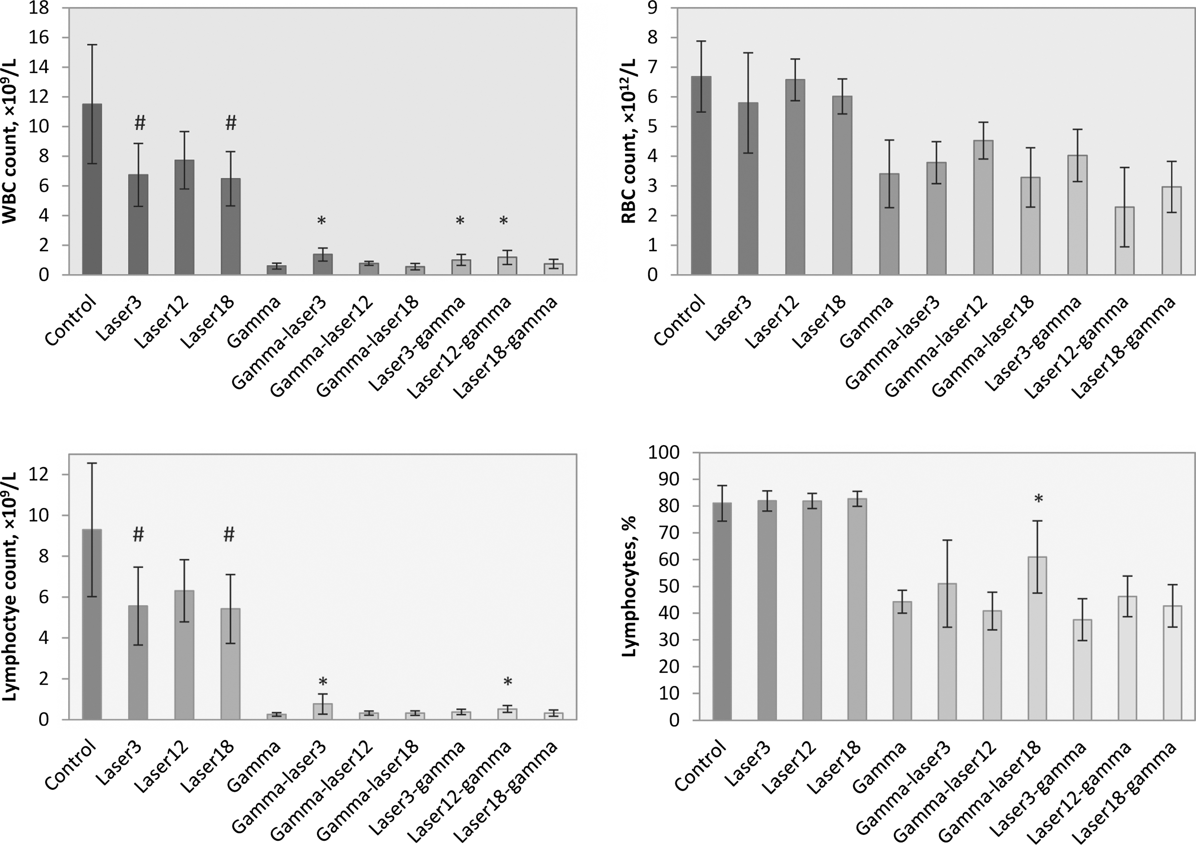
Effect of 940 nm laser on counts/percentages of white blood cells (WBC), red blood cells (RBC), and lymphocytes in mice on the 12th day after gamma irradiation. The results are expressed as mean±SD. * and # indicate significant differences between the marked group and the gamma and control groups (p<0.05), respectively.
Changes in absolute RBC count in mice treated with 940 nm laser and gamma radiation on day 12
Significant changes were not found between gamma-irradiated and non-gamma-irradiated mice (Fig. 3). Furthermore, there was no significant difference in RBC count between the laser groups and the control group, or among groups with gamma-irradiated mice (p<0.05). However, in the gamma-laser12 group, the RBC count was higher than in the gamma group (p=0.056).
Changes in absolute lymphocyte count and percentage in mice treated with 940 nm laser and gamma radiation on day 12
On day 12, lymphocyte count and percentage were altered by 940-nm laser in mice treated with or without gamma-radiation (Fig. 3). It has been observed that in the gamma-laser3 and laser12-gamma groups, the count of lymphocytes was significantly higher than in the gamma group (p<0.05). Meanwhile only the gamma-laser18 group had a higher percentage of lymphocytes than the gamma group (p<0.05). These results were accompanied by significantly lower counts of lymphocytes in the laser3 and laser18 groups than in the control group, and the percentage of lymphocytes as in the control group (p<0.05).
Changes in absolute monocyte count and percentage in mice treated with 940 nm laser and gamma radiation on day 12
Figure 4 shows changes in monocyte count and percentage altered by 940 nm laser in mice treated with or without gamma radiation. A twofold lower percentage of monocytes was registered in the gamma-laser3 and gamma-laser18 groups than in the gamma group (p<0.05). Meanwhile, among these two groups also a lower, but not a significant, count of monocytes was found in the gamma-laser18 group (p=0.073). In the gamma-laser12, laser3-gamma, and laser18-gamma groups, the count of monocytes was significantly higher than in the gamma group (p<0.05). On day 12, counts of monocytes were significantly lower in the laser3 and laser18 groups than in the control group, but there were not changes in percentage of monocytes in those groups (p<0.05).

Effect of 940 nm laser on counts/percentages of monocytes and neutrophils in mice on 12th day after gamma irradiation. The results are expressed as mean±SD. * and # indicate significant differences between the marked group and the gamma and control groups (p<0.05), respectively.
Changes in absolute neutrophil count and percentage in mice treated with 940 nm laser and gamma radiation on day 12
As can be seen on Fig. 4, the count of neutrophils was significantly higher in the gamma-laser3, laser12-gamma, and laser18-gamma groups (p<0.05). Conversely, in the gamma-laser12 and laser12 gamma groups, the percentage of neutrophils was different than in the gamma group (p<0.05). In first group it was lower, and in the second it was higher. On day 12 in the laser3 and laser18 groups, the count of neutrophils was decreased in comparison with the control group (p<0.05).
Changes in life span, blood cell counts, and percentages in mice treated with 940 nm laser on day 45
As can be seen in Table 2, mice survived to day 45 only in the gamma-laser3 group (five mice) and the gamma-laser18 group (one mouse). One mouse from the first group was excluded from statistical analysis, because of zero values for some blood cell counts and percentages.
In Fig. 5 it can be seen that on day 45 no significant changes were perceived in five experimental parameters between the gamma-laser3 group and the control group. There were counts of RBCs (p=0.17), monocytes (p=0.17), neutrophils (p=0.262), and percentages of neutrophils (p=0.078). The results in the gamma-laser18 group are interesting, and are worth a more careful look in future experiments.
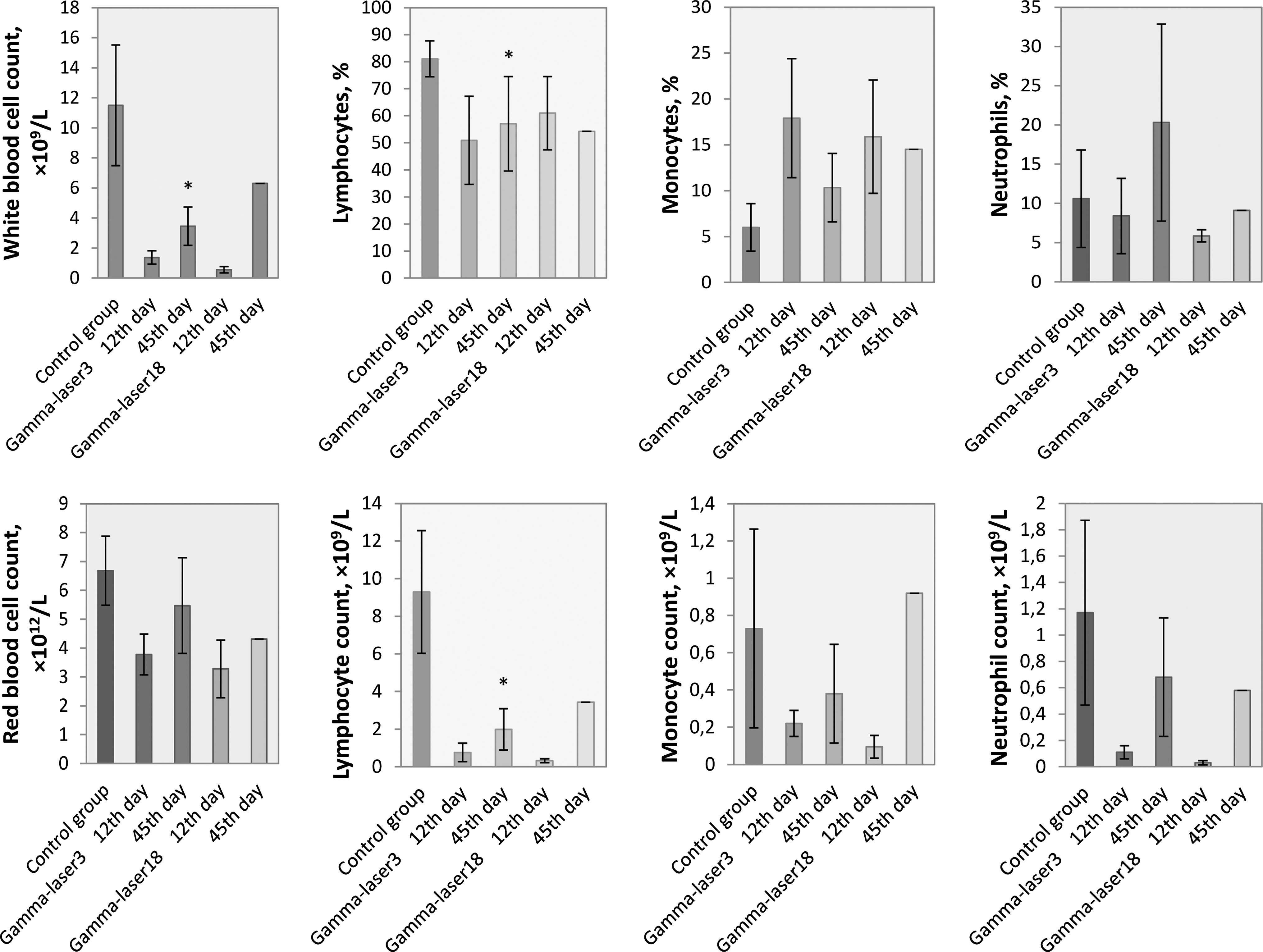
Effect of 940 nm laser on counts/percentages of WBCs, RBCs, lymphocytes, monocytes, and neutrophils in mice on the 12th and 45th day after gamma irradiation. The results are expressed as mean±SD. The results for the gamma-laser18 group on day 45 are expressed as they were measured in the blood of one mouse that survived. Statistically significant differences between the gamma-laser3 and control groups are indicated by * (p<0.05).
Discussion
This study shows that the 940 nm laser (3 J/cm2, 20 Hz, 5 W) has a radioprotective effect in mice treated with laser after gamma irradiation. Its presence has been demonstrated in two ways. First, we found that laser with these parameters utilized 24 h after exposure to a lethal dose of gamma radiation (8.7 Gy) prolonged the life span of mice (p<0.05). Second, the findings that counts of WBCs (p=0.001), lymphocytes (p=0.004), and neutrophils (p=0.004) were respectively 2.32, 2.99, and 3.33 times higher on day 12 in this group than in the gamma group. Moreover, on day 45 after gamma irradiation, some signs of hematopoiesis repair were found in blood. There were no significant differences in counts of RBCs (p=0.17), monocytes (p=0.17), neutrophils (p=0.262), and percentage of neutrophils (p=0.078) between this group and the control group.
The lifespan of mice depends not only on dose and type of radiation, but also on the age and strain of the mice. 25 For example, the well-known median lethal dose or LD50/30 in female mice lies within the range of 5.5–7.8 Gy, 25 and for the mice that we used in actual study is equal to 6.7±0.06 Gy. 25 From biological point of view, there is a close connection between the survivability of irradiated organisms and the general state of their body organs. Certainly one of the most important among these is blood, because the dose of ionizing radiation of 8.7 Gy is lethal because of the dramatic reduction of blood cells. 1,25 At a dose of 8.7 Gy, the gamma-irradiated mice have hematopoietic syndrome with a drop in the number of blood cells (aplastic anemia). The main reason for death in such mice is infections (decrease of white blood cells), inner bleeding (decrease of platelets), and anemia (decrease of red blood cells). The question is why have mice irradiated with laser after gamma radiation survived? It is conceivable that the reason for their survival is higher counts of blood cells. It is useful to look at the blood in more detail.
Referring again to Figs. 3 and 4, we can see what the difference is between the gamma-laser3 group and other groups. WBC count in this group was 232% higher (p<0.05) than in the gamma group, which was attributed to higher counts of lymphocytes and neutrophils in this group than in the gamma group (p<0.05). It is a well-known fact that the sensitivity of cells depends on the rate of proliferation and degree of differentiation of cells. 25 Laser irradiating at a wavelength of 940 nm increases proliferation and differentiation of cells, 21 –23,26 the same as other near-infrared lasers. 27 Therapeutic lasers at the near-infrared region of irradiation are known as a source of ROS, which result in proliferation, mitotic activity, and reparative processes in cells by activation of a cascade of reaction. 6,27,28 Probably, after gamma radiation, 940 nm laser at a low dose of 3 J/cm2 leads to indirect mitotic activation of stem and progenitor cells, which leads to significantly higher counts and percentage of blood cells in that group than in the gamma-irradiated groups. Laser at a higher energy fluence leads to activation of only differentiation, 22 which may not play crucial role in the survivability of mice.
Present experimental evidence indicates, however, that not only in these groups do such changes exist. Also, we found changes in the laser12-gamma group. In the laser12-gamma group, the count of WBCs was 199% higher than in the gamma group (p<0.05), as were higher counts of lymphocytes and neutrophils (p<0.05) (Figs. 3 and 4). This is in remarkable contrast to data of the life span of mice, because the mice in that group had died first, as can be seen in Table 4. Before beginning a more detailed analysis of these results, it is helpful to consider the influence of only 940 nm laser on mice.
Figures 3 and 4 clearly illustrate the basic differences between the laser groups and the control group. We have no a priori reason to expect that laser irradiation should significantly decrease the count of blood cells. This reduction is not dose dependent, because in the laser3 and laser18 groups, reduction of WBCs and their different lines was more intensive (p<0.05) than in the laser12 group. Sharma et al. observed similar dependence after irradiation of neurons with an 810 nm laser at different energy fluences. 28 At low fluences (up to 3 J/cm2) laser treatment had a beneficial effect on neurons by increasing ROS and nitric oxice (NO) production, whereas at high fluence (30 J/cm2) laser treatment had the opposite effect. Increase of ROS and NO levels was so high that laser light damaged neurons. It is of some interest to observe that in Sharma's study, energy fluence at 10 J/cm2 caused reduction of ROS and NO production. It is not improbable that a similar mechanism of laser influence underlies our results.
The study of radioprotective features of laser light in in vivo experiments has not been adequately elucidated in the literature. However, radioprotective features of 650 nm laser radiation were analyzed by Voskanyan et al. 29,30 Voskyanyan et al. used a 0.7 mW laser module with a fluence of 1 mJ/cm2 for treatment of mouse backs, and with a fluence on 2×1 mJ/cm2 for treatment of mouse backs and abdomens. In the experiment they used a 60Co source for gamma radiation with dose power of 1.8 Gy/min (total dose 3 Gy). Mice were irradiated with gamma prior to laser irradiation, and the time interval between the two irradiations did not exceed 30 min. Voskanyan et al. analyzed lymphocyte count 24 and 72 h after irradiation. Counts of lymphocytes were also doubly decreased than those in intact mice 24 h after irradiation, and were not significantly increased 72 h after irradiation. It is especially interesting to note that 24 h after irradiation in the gamma-irradiated groups, the lymphocyte count depended on the fluence of laser irradiation (lower in gamma-irradiated mice and higher in mice irradiated with gamma and laser irradiation of 2×1 mJ/cm2), but 72 h after irradiation the lymphocyte count was higher in gamma-irradiated mice than that in mice irradiated with gamma and laser irradiation. Our study and the studies of Voskanyan et al. had showed also that indirect laser irradiation may lead to stimulation of hematopoiesis in bone marrow (the count of karyocytes in bone marrow was also analyzed by Voskanyan 29 ). In a similar study, Voskanyan et al. showed a beneficial reparative effect of 650 nm laser on hematopoiesis and mitotic activity of bone marrow. 30
It will be useful to reconsider the indirect influence of 940 nm laser on hematopoiesis in future studies. We used laser at higher levels of fluence and intensity than are used in other research; 29,30 therefore, the results may be entirely different. Also laser with these parameters produces a rise in temperature in irradiated tissues as is described by the developers of the 940 nm laser used in our study.
Conclusions
In conclusion, we have shown that 940 nm laser with fluence on 3 J/cm2 has radioprotective features on gamma-irradiated mice. In essence, this tells us that this type of laser can be used at some stage for therapy of irradiated organisms. We believe that this finding may be intensively used in various disciplines such as radiobiology and oncology.
Footnotes
Acknowledgments
This work was supported in the form of a student grant by Czech Technical University in Prague (SGS11/144/OHK4/2T/17) and by the Ministry of Defence of the Czech Republic by the grant Health Problems of Weapons of Mass Destruction (A long-term organization development plan 1011).
Author Disclosure Statement
No competing financial interests exists.