
Abstract
Introduction
T
The osseointegrated implants used for this purpose have many disadvantages, such as requirement for operation, expensiveness, and the long duration need for osseointegration. 1 Kanomi, in 1997, designed the mini-implant for orthodontics. 2 Factors that may increase the need for a mini screw are uncooperative patients, undesirable extraoral appliances, and the inadequacy of the dental elements and the surrounding bone. 3 In addition, these screws have some advantages such as easy applicability, immediate loading, and time saving. 4,5 However, the most common clinical challenge is the early loss of the mini screw. Primary stability is essential to be able to apply force to the mini screws.
It has been noted that the majority of the losses of mini screws result from primary stability failure. 6 This problem can be solved by longer and thicker implant usage in prosthodontic treatment; 7 however, this method is not conducive to the use of orthodontic mini screws, because of the placement regions. Therefore, clinicians are trying to develop alternative choices to increase the stability.
Many processes that have been shown to be stimulated by low-level laser therapy (LLLT) are cell proliferation, 8,9 collagen and protein syntheses, 10 wound healing, 11 –13 differentiation of bone and cartilage cells, 14,15 and cell regeneration. 16
LLLT speeds up the blood flow, improves the mechanism of the revitalization processes, reduces the risk of infection, boosts the metabolic activities, and accelerates the healing of the damaged tissue. 16 Although there are many research studies of LLLT applications in a variety of areas, no investigations were found concerning mini screw stability using various laser dose levels with different force level applications. Therefore, the aim of this study was to evaluate the effects of different laser doses and force levels on the stability of orthodontic mini screws used for anchorage, by histomorphometric analyses, including the mini screw and the bone tissue intact together. It was hypothesized that LLLT and force application would not significantly affect the orthodontic mini screw's stability.
Materials and Methods
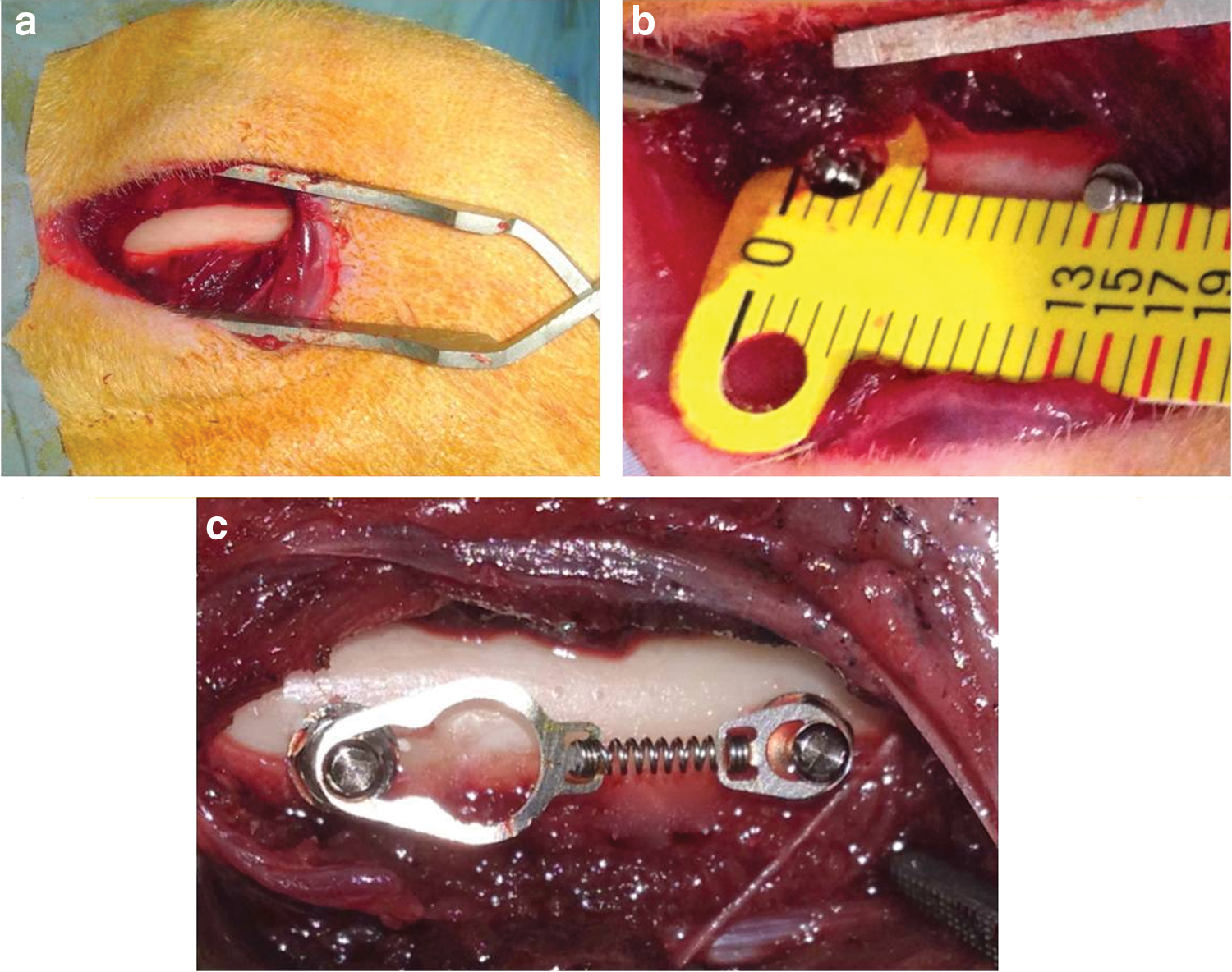
The protocol for this study was approved by the Experimental Animal Committee of Bezmialem Vakif University (30.11.2012/353). Seventeen 6-month-old male New Zealand white rabbits, weighing 3.0–3.5 kg, were used. A total of 68 cylindrical, self-drilling orthodontic mini screws (JeilMed, Seoul, Korea) made of Ti6Al4V alloy with a diameter of 1.4 mm and length of 8 mm were included. All surgeries were performed under sterile conditions in a veterinary operating room. Rabbits were anesthetized using an intramuscular injection of ketamine hydrochloride (100 mg/kg) and xylazine (5 mg/kg). After that, the hair on the medial surfaces of the right and left fibulas was clipped, and the skin was cleaned with iodinate surgical soap. A 50 mm incision was made parallel to the longitudinal axis of the fibula, and the periosteum was stripped (Fig. 1a). Mini screws were placed into the first cortex of the fibula and their longitudinal axes were adjusted parallel to each other and perpendicular to the external cortical fibula without interfering with the secondary cortex (Fig. 1b). TAD coil spring gauge was used for determining the distance between the mini screws on each fibula (Fig. 1b). Two mini screws were placed in the randomly selected fibulas of each rabbit, and 150g of force was immediately applied using a nickel-titanium (Ni-Ti) closed-coil spring (TAD, GH Wire Company, Hanover, Germany; C2 size: medium, 15 mm) (Fig. 1c). The identification of the groups were as follows: in group 1 (n=8) no force and no laser were used; in group 2 (n=12) no force was used and laser dosage was 10 J/cm2; in group 3 (n=12) no force was used and laser dosage was 20 J/cm2); in group 4 (n=12) the force amount was 150g and no laser was used; in group 5 (n=12) the force amount was 150g and laser dosage was 10 J/cm2; and in group 6 (n=12) the force amount was 150g and laser dosage was 20 J/cm2.
All mini screws were inserted using an electronic torque meter (JeilMedical Corporation, ORTHONIA 111-ED-010, Seoul, Korea) (10 Ncm) by the same operator (M.G.). The tissues were then closed with absorbable sutures, and carprofen (4 mg/kg) was given for 3 days after surgery to minimize infection risks.
A GaAlAs diode laser device (Cheese dental laser; Wuhan Gigaa Optronics Technology Co. Ltd., Wuhan, China) was used in this study. This system operates in the near-infrared spectrum at a continuous wavelength of 810 nm and an output power of 0.3 W, and produces a spot size of ∼5.85 cm2. Treatment was initiated immediately after surgery and performed daily for 10 consecutive days. The application period per point was 195 or 390 sec, releasing an energy density of 10 or 20 J/cm2.
Four weeks post-surgery, all rabbits were euthanized with an intravenous overdose of sodium pentothal. The fibulas were dissected, and 68 bone blocks containing one mini screw were prepared, each with at least 2 mm of surrounding bone. Mini screws were prepared for histomorphometrical analyses. The specimens were fixed in 10% buffered formalin, dehydrated in increasing concentrations of ethanol (70–99%) over a period of 10 days, and embedded in methyl methacrylate (Technovit 7200 VLC; HeraeusKulzer, South Bend, IN). Fifty micrometer thick, undecalcified sections were prepared by use of a diamond-coated saw cutting and grinding system (Exakt, Norderstedt, Germany). Sections were stained with toluidine blue, and digital images were obtained with a digital camera attached to a light microscope (Olympus DP 70; Olympus, Tokyo, Japan) at a magnification rate of ×40. The percentages of bone to implant contact (BIC) at the lateral sides of the implants and cortical bone thickness (CBT) were calculated by image analysis software (ImageJ 1.33u; National Institutes of Health, Bethesda, MD). Because the surface at the bottom of the mini screws was a machined surface (implants were cut to a height of 6 mm during the manufacturing process), the apical surfaces were not included in the BIC calculations.
BIC values were calculated using the following equation
17
(Fig. 2):
CBT values were calculated by taking the average of measurements contacting the mini screw on both sides 18 (Fig. 2).
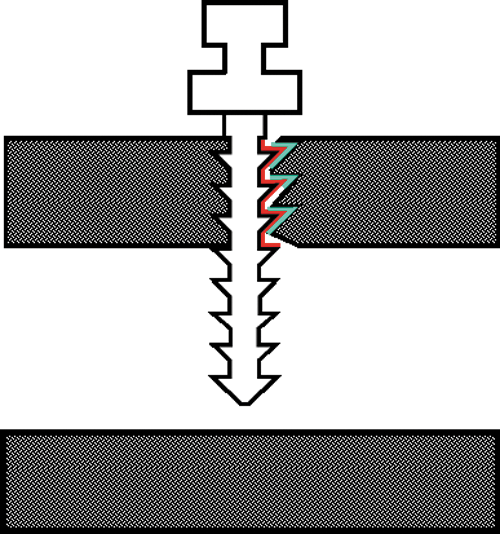
Measurement of bone to implant contact (BIC) and cortical bone thickness (CBT) values.
SPSS version 18.0 for Windows (SPSS Inc., Chicago, IL) was used for statistical analyses. One way ANOVA and Tukey's honest significant difference (HSD) multiple comparison tests were used for independent samples to compare quantitative measurements (p<0.05). Pearson correlation coefficient was used to evaluate the associations between the BIC and CBT values.
Results
After 4 weeks of the experimental period, the clinical observation results of all mini screws in the force-applied and non-applied groups were all successful and there was no evidence of mobility.
Images of the histological sections were used for histomorphometrical analyses (Fig. 3). More cortical bone tissues were detected throughout the insertion areas of the mini screw, and in some cortical regions, connective tissue was observed to be intertwined within the mini screw grooves.

Image of the histological sections according to Groups
The descriptive statistical results of groups and results of ANOVA are shown in Table 1. The highest BIC value was observed in group 6 (83.11±1.75). This was followed by, respectively, group 5 (72.70±2.04), group 3 (64.87±1.78), group 4 (57.18±1.42), group 2 (53.99±1.82), and group 1 (36.15±2.45).
BIC, bone to implant contact; CBT, cortical bone thickness.
There were significant differences in BIC values among all groups (F=67.51, p=0.001<0.05) (Table 1). The multiple comparison results of CBT values are shown in Table 2. There were significant differences between groups 1 and 2 (p=0.001<0.05), groups 1 and 3 (p=0.001<0.05), groups 1 and 4 (p=0.001<0.05), and group 1 and groups 5 and 6 (p=0.001<0.05). The values of group 1 were lower than those of the other groups.
p<0.001.
BIC, bone to implant contact.
The statistical evaluation of CBT values was determined according to the groups in Table 1. The highest CBT value was observed in group 3 (2.16±0.20). This was followed by, respectively, group 4 (2.03±0.25), group 2 (2.01±0.16), group 6 (1.99±0.22), group 5 (1.95±0.17) and group 1 (1.93±0.31). There were no significant differences in CBT values for any of the groups (p=0.982>0.05) (Table 1).
There were no statistically significant correlations between CBT and BIC values (r = −0.012, p=0.922).
Discussion
The results obtained in the present study demonstrate that LLLT of bone with a diode laser significantly improved the BIC values of orthodontics mini screws. Therefore, the first null hypothesis, which stated that diode laser application would not significantly affect the connection amount of mini screw to bone, was rejected.
When the groups, which had a similar laser application procedure and different force amounts, were compared, it was obvious that the force application positively affected the quantity of attachment. Therefore, because of the bone remodeling process, the second null hypothesis was also rejected.
In the present study, it was found that there was no significant difference in the CBT values for any of the groups. In addition, no substantial correlations were detected between CBT and BIC values. Therefore, the third null hypothesis, which declared that the CBT would not affect the osseointegration of the mini screw, was accepted within the conditions of this study.
In the literature, there are many different opinions about timing of animal euthanasia. Eighteen weeks of human bone metabolic process corresponds to 6 weeks of rabbit bone turnover. 19 This means that the rabbit metabolism is three times faster than the human metabolism. Previously, adequate time for osseointegration of the placed mini screws was reported to be 8 weeks. 20,21 Although it was reported that the lamellar bone formation and secondary remodeling events occurred in this period of time, 21 recently, it has been accepted that the critical time for osseointegration is 4 weeks, and that a longer waiting period for proper connection is pointless. 22,23 Therefore, in present study the time of euthanasia was determined to be 4 weeks.
A pilot study was performed just before the experimental stage of this research, in which the mini screws were placed in the tibia of the rabbits; 24 –26 however, unfortunately it was detected that the legs of the animals were broken because of the weak bone structure. It was thought that length of the mini screw could be the cause of the fracture, but as the most commonly used length of the mini screws is 8 mm in orthodontic practice, it was decided to change the experimental bone to the fibula, rather than changing the dimension of the mini screw. 27
It has been reported that the most suitable wavelength for biostimulation is 550–950 nm. 28 The laser types in this range are He-Ne and diode laser systems. For this study, the selected diode laser was in the infrared spectrum, having a high level of tissue penetration depth. 28
The force amount to be applied to mini screws is a controversial issue. There are many investigators reporting that it should be between 100 and 200g 29 and that it would not be successful if it was >200g. 3,30 –34 For this reason, the quantity of force was determined to be 150g.
When the previous studies were analyzed for determining the dose of LLLT, it was noticed that a consensus was absent concerning this issue. However, it is a fact beyond doubt that the dose of laser that is required for influencing the hard tissue should be more than the dose of laser that is required for stimulation of the soft tissue. 23 It is described as ranging from 4 to 10 J/cm2 for soft tissues. 35 –39 In light of this information, the laser doses selected were 10 and 20 J/cm2 in this investigation.
Although some researchers reported that there is no relationship between the cortical bone thickness and the stability of the mini screw, 40 –42 others reported an opposing argument. 43,44 According to the results of the present study, there was no statistically significant difference among the groups. This outcome may be associated with providing adequate stability of the mini screws for maintaining stability until the end of the experiment. The limitation of this study is that initial cortical bone thickness cannot be standardized. The limitation of this study is that the initial cortical bone thickness cannot be standardized since measuring cortical bone thickness on live animals is an extremely difficult issue. For the future, there is a need for research that compares clinically successful and unsuccessful mini screw groups, and, in addition, better standardization procedures are greatly needed for determining reliable impacts of these variables. Another limitation of this study is that the condition of experiment cannot be simulated in the mouth. The mini screws are placed in humans in an open oral environment. More accurate results can be obtained with human trials.
Conclusions
In this present research, clinical and histomorphometrical findings were evaluated, and the following results were obtained.
1. Utilizing orthodontic mini screws as anchorage devices is a reliable, effective, and easy method.
2. No mobility of the mini screws was noticed during the experimental period, which can be assumed as a success indicator.
3. The BIC values of groups receiving 20 J/cm2 laser doses were higher than those of the other groups. Therefore, LLLT may be an alternative method for increasing the stability of mini screws.
4. There was no correlation between CBT values and the BIC values for the stability of the orthodontic mini screw.
Footnotes
Author Disclosure Statement
No competing financial interests exist.