
Abstract
Introduction
D
Several methods have been suggested to increase the permeability of dentinal tubules, which include acid etching of the tooth surface, the use of irrigation solutions with ultrasonic activation, and the use of heat or light during the bleaching procedure. 5 The application of heat, light, or lasers is used to increase the temperature of a bleaching agent applied to the tooth surface. 6 Several sources such as curing lamps, LED lamps, and, since 1996, coherent light have been proposed to enhance the action of the bleaching gel. The first wavelengths utilized were the argon (480 nm) and CO2 (10,600 nm) lasers, but today the neodymium:yttrium-aluminum-garnet (Nd:YAG, 1064 nm), diode (810 and 980 nm), and the potassium-titanylphosphate (KTP, 532 nm) lasers are also used. 7
The Nd:YAG laser is a solid-state laser used in some dental and periodontal procedures such as apical surgery. It also melts and recrystallizes the superficial dentin, which consequently results in decreased apical leakage, superior cleaning, and smear layer removal. In addition, the Nd:YAG laser is recommended by many studies to accelerate the bleaching process. 8,9 There are two different wavelengths produced by surgical diode lasers. One uses aluminum-gallium-arsenide to emit ∼800 nm wavelengths, and the other uses indium-gallium-arsenide to emit 980 nm light energy. These lasers are used in the contact mode for rapid cutting, vaporizing, and bacterial reduction of tissue adjacent to the tooth structure, and are used in noncontact mode for deeper coagulation. 10 The diode laser is a type of continuous or pulsed wave laser that emits coherent well-collimated light. It penetrates deeply into dental hard tissues and achieves successful tooth whitening. 11,12
Although some studies investigated the efficacy of laser irradiation during intracoronal bleaching, no study has been conducted to investigate the changes in both color and enamel structure during intracoronal bleaching with laser irradiation. The aim of this study was to evaluate the color and enamel structure changes after intracoronal bleaching with sodium perborate under Nd:YAG and diode laser irradiation.
Materials and Methods
Preparation of samples
Thirty-six freshly extracted, single-rooted mandibular incisors without any caries were used. Standard endodontic access cavities were created using a 12 round diamond bur with a high-speed hand piece under water cooling. The working length was measured by inserting a #10 stainless steel file (VDW Antaeos, Munich, Germany) with a silicone stop until the tip of the file was observed at the level of the apical foramen. The root canals were prepared to size F3 with the ProTaper rotary system (DentsplyMaillefer, Ballaigues, Switzerland). Irrigation was performed with 2 mL of 2.5% sodium hypochloride after each filing. Following the final irrigation, root canals were dried with sterile paper points. The canals were filled with AH Plus (Dentsply De Trey, Konstanz, Germany) and gutta-percha (Diadent, Seoul, Korea) using a cold lateral condensation technique. After 2 weeks, the root fillings were removed 2 mm apical to the cementoenamel junction, and a 2-mm-thick layer of glass ionomer cement (GC Fuji II LC; GC Corporation, Tokyo, Japan) was applied.
Baseline color measurements and Raman spectroscopy detection
The baseline color values (L*, a*, b*) of each specimen were measured with a spectrophotometer (Easyshade,Vita, Bad Sackingeni, Germany). The spectrophotometer was calibrated with a standard white card before each group of specimens was measured. Measurements were repeated three times for each specimen before the mean values were calculated.
Baseline Raman spectra of each specimen were obtained before the application of the intracoronal bleaching agent. Spectra were recorded by a micro-Raman spectrometer (inVia Raman, Renishaw, United Kingdom) equipped with semiconductor laser diode (785 nm wavelength). Each spectrum was made under the following conditions: 100–3200 cm−1 range, 7000 ms integration time, five times average at room temperature, and the data were visualized on a computer. Similar to the methods used in previous studies, in the present study Raman absolute intensity (RAI), Raman relative intensity (RRI), and laser-induced fluorescence intensity (FI) at 960 cm−1 were defined and calculated from the Raman spectra. 13,14 RAI is the intensity of the Raman peak at 960 cm−1 before the spectrum baselines, and RRI is the intensity of the same peak after the spectrum baselines between 990 and 930 cm−1. FI is the difference between RA and RRI.
Bleaching and laser irradiation
All samples were randomly divided into three groups as follows: • Group 1: intracoronal bleaching with sodium perborate+NdYAG laser irradiation • Group 2: intracoronal bleaching with sodium perborate+diode laser irradiation • Group 3: intracoronal bleaching with sodium perborate without any laser irradiation
Sodium perborate and distilled water were mixed to form a thick paste and placed into the pulp chamber of all samples. In group 1, the NdYAG laser (Fotona Laser AT Fidelis Plus III) was applied for 30 sec with a 3.5 W power setting into the pulp chamber. In group 2, the 810 nm diode laser (AMD Picasso, Indianapolis, IN) with a standard hand piece and 400 mm fiber optic cable was applied for 30 sec in the continuous mode at the 1.5 W power setting into the pulp chamber. In group 3, no laser irradiation was applied. A cotton pellet was placed on the bleaching material to isolate it from the temporary filling. The access cavity was sealed with temporary filling material after the bleaching treatments of all samples. In total, three bleaching sessions were held in which the same bleaching protocols were applied after every 4 days. Therefore, specimens had been in contact with sodium perborate for 12 days during bleaching procedures. After the last bleaching session, a calcium hydroxide paste was applied to the pulp chambers for 7 days to neutralize the byproducts of the bleaching agents. The samples were restored with composite resin (Filtek Ultimate, 3M ESPE, St Paul, MN) in the pulp chamber.
The final tooth color was recorded with the same spectrophotometer as had been used in the baseline reading. ΔE is the total color difference, or the distance difference between the baseline and the final colors. The difference between color coordinates is calculated as ΔE*=((ΔL*) 2 +(Δa*) 2 +(Δb*) 2 )1/2.
Final Raman spectra from each specimen were obtained after the application of the bleaching agent. RRI and FI values were calculated from the final Raman spectrum. The baseline and final values were transformed to percentage values, the values of the baseline were considered as 100%, and the changed values were calculated as a percentage of the baseline.
Statistical analysis
Statistical analyses were performed with SPSS 18.0 software (SPSS Inc., Chicago, IL). Distribution of data was determined by the Shapiro–Wilks test. Continuous variables were expressed as mean±SD. deviation. The Anova or the Kruskal–Wallis test was used to determine the differences among the three groups. A p value of<0.05 was considered statistically significant for all tests.
Results
The statistical results of the color changes of the Nd:YAG, diode lasers and the control groups are presented in Table 1. There was a significant difference between the Nd:YAG laser group and the control group (p<0.05). The ΔE values in both the Nd:YAG and diode laser groups were higher than in the control group (Fig. 1). However, there was no significant difference found between the diode laser group and the control group or the diode laser group and the Nd:YAG laser group.
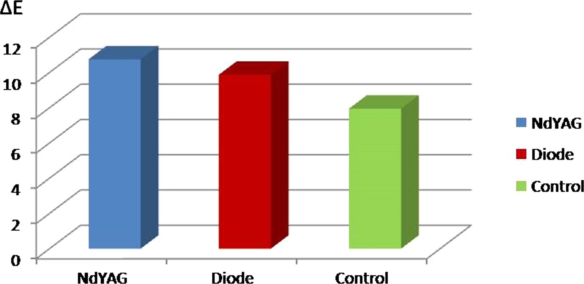
Color changes (ΔE) of groups after treatment.
Tables 2 and 3 showed the results of FI and RRI percentage changes in all groups. The Raman spectrum of enamel with RRI and FI calculation from the strongest band 960 cm−1, which arose from PO−3 are presented in Fig. 2. Figure 3 shows the percentage RRI and FI changes of groups after treatment. There was no significant difference among the groups in terms of RRI and FI percentage values (p>0.05).
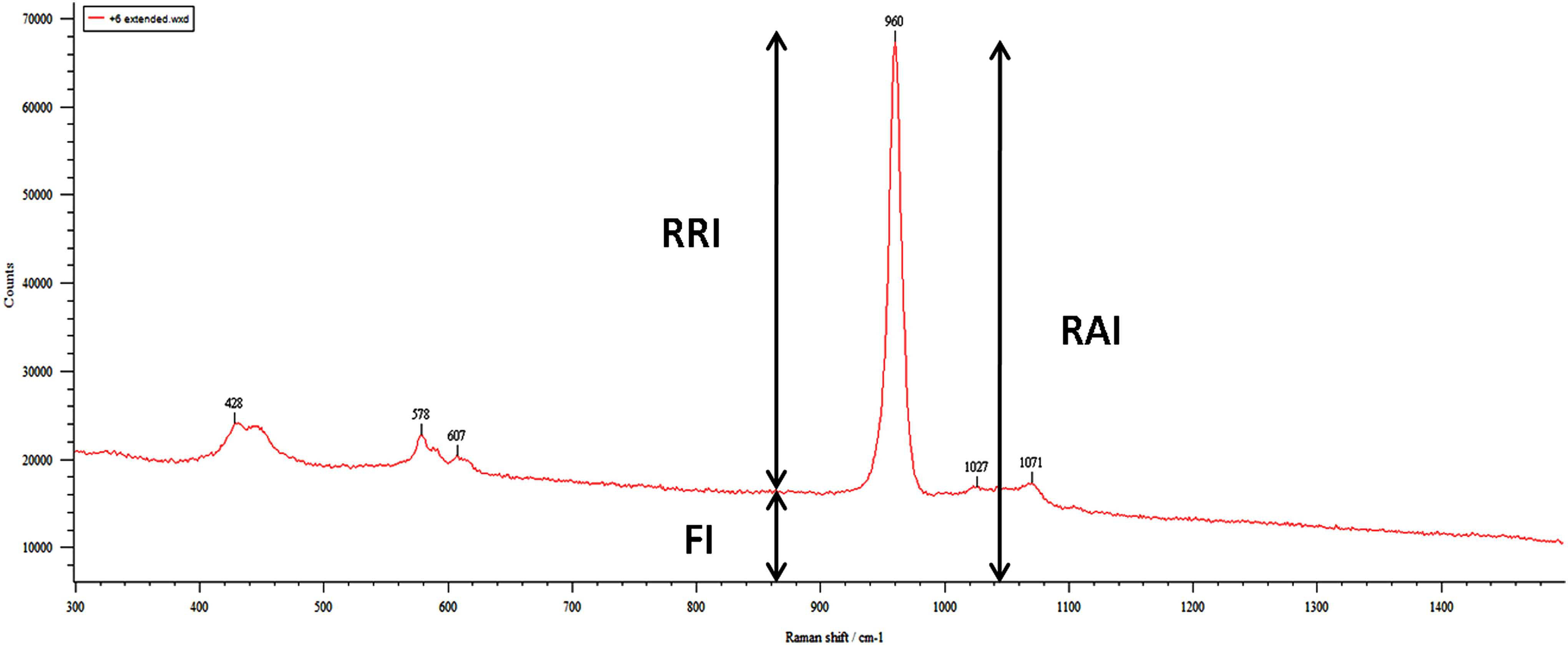
Raman spectrum of enamel with Raman relative intensity (RRI) and fluorescence intensity (FI) calculation.
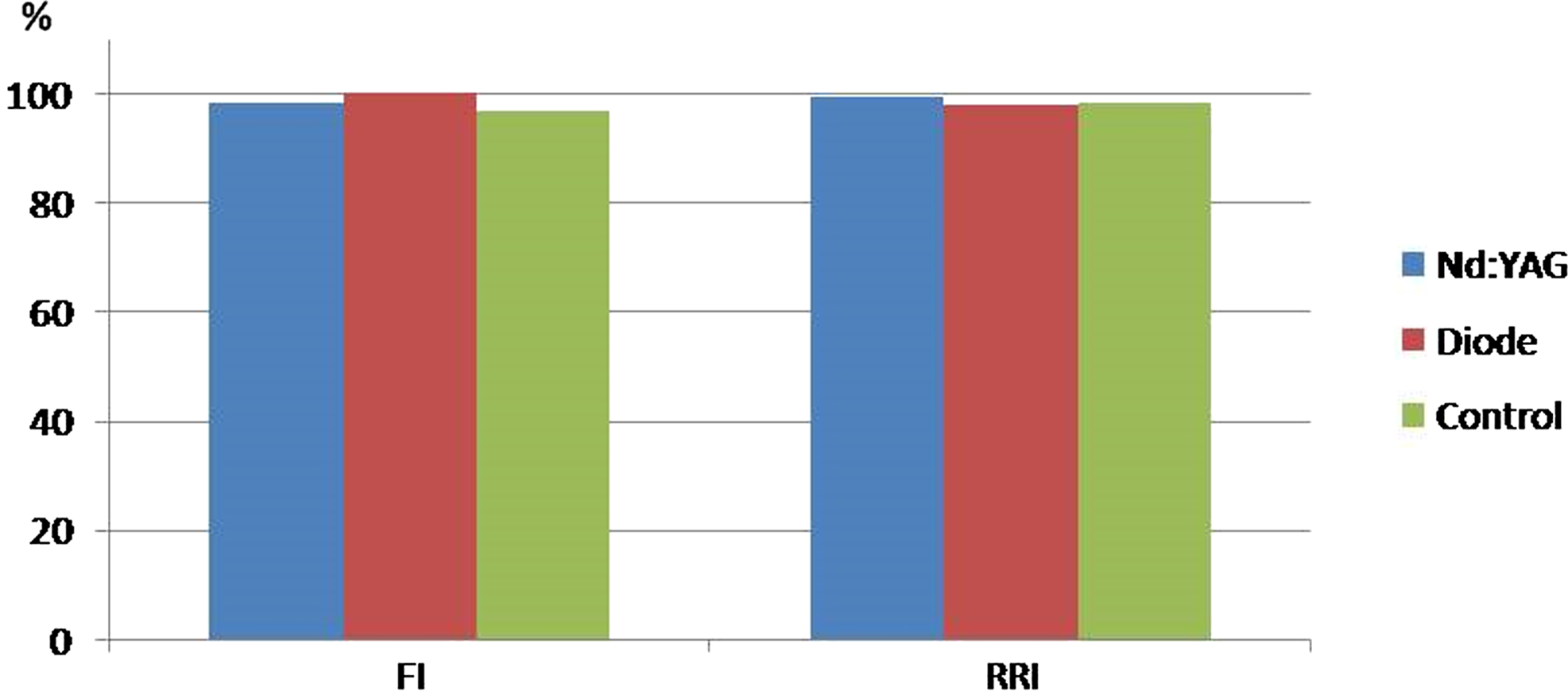
Percentage Raman relative intensity (RRI) and fluorescence intensity (FI) values of groups after treatment.
FI, fluorescence intensity.
RRI, Raman relative intensity.
Discussion
Intracoronal bleaching treatment with lasers has been investigated in previous studies. 5,15 However, limited data are available evaluating the effects of intracoronal bleaching on enamel structure. 16 In the present study, two commonly used lasers (NdYAG and diode lasers) were used in intracoronal bleaching with sodium perborate, and their effects on bleaching efficacy and the changes on enamel structure were investigated together.
A mixture of sodium perborate and distilled water was used as a bleaching agent. In the previous studies, sodium perborate was mixed with hydrogen peroxide, carbamide peroxide, or distilled water. 17,18 However, sodium perborate should be mixed with water rather than with hydrogen peroxide in order to prevent or minimize the occurrence of bleaching-related external root resorption. 2
The previous studies showed that sodium perborate was able to bleach nonvital teeth. 17 –19 All groups in the present study included sodium perborate in order to compare the efficacy of two laser systems. Ganesh et al. used a control group in which bleaching was done with distilled water, and reported no significant changes after 14 days. 19 The no laser group was accepted as the control group in the present study.
Activation by LED, the halogen lamp, or the walking bleach technique leads to similar increase in dentinal permeability. 20 Carrasco et al. reported that LEDs, the halogen lamp, or the walking bleach technique showed similar bleaching efficacy. 21 Another activation method in the bleaching procedure, laser irradiation, was used in the present study, and according to the results of the present study, laser activation increased bleaching efficacy. In the present study, Nd:YAG laser irradiation was found to be significantly effective in comparison with the control group. This result is incompatible with a previous in vivo study. 22 However, Strobl et al. evaluated the efficacy of the Nd:YAG laser in vital tooth bleaching. 22 To the knowledge of the authors, no study has ever mentioned Nd:YAG laser irradiation during devital bleaching.
There was no significant difference between the bleaching efficacy of the diode laser and the control group; however, the diode laser showed better results than the procedure used in the control group. Gontijo et al. reported that the use of a diode laser did not improve the bleaching result compared with light application with a halogen lamp. 23 Nevertheless, the differences between the diode laser and the Nd:YAG laser were close in comparison with the control group. The influence of different laser systems on bleaching efficacy was compared in several studies. 7,24 For example, Fornaini et al. compared the efficacy of the diode laser and the KTP laser during vital bleaching, and they reported that the diode laser demonstrated lower bleaching efficacy than the other laser system. 7 The present study is the first study comparing the efficacy of Nd:YAG and diode laser irradiation during intracoronal bleaching. Further studies with larger sample sizes are required to reach a conclusion regarding the efficacy of the diode laser and comparison with the Nd:YAG laser during intracoronal bleaching.
A spectrophotometer was used in this study to compare the bleaching efficacy of the laser systems. Quantitative and comparable data can be obtained by this method. It has been reported that a color difference of<1 ΔE is considered as a color match that cannot be identified by an independent observer. 25 The results of the present study indicated that in all groups, distinct color changes were recorded after the bleaching treatment. Furthermore, ΔE changes were>1 ΔE between the laser groups and the control group; therefore, it can be suggested that laser irradiation provides visible changes after intracoronal bleaching.
Raman spectrometry allows the molecular analysis of mineralized dental tissues. The Raman spectra of enamel represent the intensity of the signal according to the frequency, and allow comparative and quantitative analysis. 26 Some studies used Raman spectroscopy and scanning electron or atomic force microscopy together to analyze the structure of the enamel surface after tooth bleaching. 13,14 Although these studies investigated vital tooth bleaching, intracoronal bleaching was studied in the present study. Raman spectroscopy analysis was used alone, as bleaching gel was applied into the access cavity, and this examination method allowed analysis of subsurface enamel changes arising from intracoronal bleaching. 27 The concentration of the phosphate group is a good indicator for defining the extent of mineralization in the enamel structure. Moreover, the intensity of PO4 −3 in Raman spectroscopy is linearly proportional to the phosphate group concentration within the hydroxyapatite molecule. 13
RAI, RRI, and laser-induced FI were calculated for data analysis. FI and RRI methods were also used by previous studies. 13,14 According to the FI findings of the present study, close percentage changes were recorded in all groups. FI provides the information of organic matter. 28 However, no significant changes were observed in the diode laser and control groups. Limited changes were observed in RRI findings in all groups, which indicated that the phosphate group concentration in the enamel surface had scarcely changed.
Conclusions
Within the limitations of the present study, laser application, especially Nd:YAG laser irradiation, was able to increase the efficacy of intracoronal bleaching with sodium perborate. The results showed that irradiation with Nd:YAG laser is more effective than with diode laser, but further studies are required to draw conclusions regarding diode laser efficacy. Additionally, Raman spectroscopy analysis revealed that intracoronal bleaching with laser application did not affect enamel structure. The conclusion reached was that laser application is more effective than the bleaching procedure without laser irradiation and it is safe for the enamel surface during intracoronal bleaching with sodium perborate.
Footnotes
Author Disclosure Statment
No competing financial interests exist.